

Pediatric and Teen Ovarian Tissue Removed for Cryopreservation Contains Follicles Irrespective of Age, Disease Diagnosis, Treatment History, and Specimen Processing Methods
- PMID: 26697267
- PMCID: PMC4684654
- DOI: 10.1089/jayao.2015.0032
Pediatric and Teen Ovarian Tissue Removed for Cryopreservation Contains Follicles Irrespective of Age, Disease Diagnosis, Treatment History, and Specimen Processing Methods
Abstract
Purpose: Fertility preservation in a pediatric and teen female population is challenging because standard technologies of egg and embryo freezing may not be possible due to premenarcheal status. Ovarian tissue cryopreservation (OTC) with the intent of future ovarian tissue transplantation or in vitro follicle growth may be the only option to preserve fertility. The purpose of this study was to add to the general understanding of primordial follicle dynamics in young patients.
Methods: First, the unique infrastructure of the Oncofertility Consortium National Physicians Cooperative (OC-NPC) is described, which simultaneously drives clinical fertility preservation and basic research to explore and expand the reproductive options for those in need. Then, the OC-NPC research resource is used to perform a histological evaluation of ovarian tissue from 24 participants younger than 18 years of age.
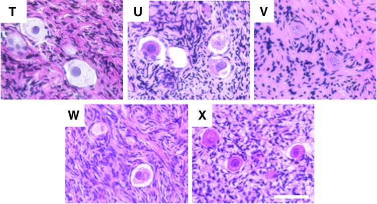
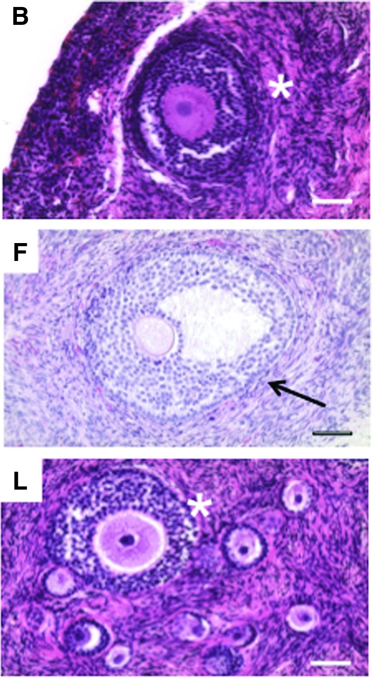
Results: Primordial follicles, which comprise the ovarian reserve, were observed in all participant tissues, irrespective of variables, including age, diagnosis, previous treatment history, tissue size, and tissue processing methods. Primordial follicles were present in ovarian tissue, even in participants who had a previous history of exposure to chemotherapy and/or radiation treatment regimens, which placed them at risk for iatrogenic infertility or premature ovarian failure.
Conclusion: Primordial follicles were observed in ovarian tissue from all participants examined, despite population and tissue heterogeneity. These results increase the understanding of human follicle dynamics and support OTC as a promising fertility preservation modality in the young female population. Future studies to evaluate follicle quality within these tissues are warranted.
Keywords: fertility preservation; follicle; oncofertility; ovarian tissue cryopreservation; ovary.
Figures


Similar articles
-
Impact of first-line cancer treatment on the follicle quality in cryopreserved ovarian samples from girls and young women.Hum Reprod. 2019 Sep 29;34(9):1674-1685. doi: 10.1093/humrep/dez125. Hum Reprod. 2019. PMID: 31411325 Free PMC article.
-
Successful fertility preservation following ovarian tissue vitrification in patients with primary ovarian insufficiency.Hum Reprod. 2015 Mar;30(3):608-15. doi: 10.1093/humrep/deu353. Epub 2015 Jan 6. Hum Reprod. 2015. PMID: 25567618
-
Cryopreservation of in vitro matured oocytes in addition to ovarian tissue freezing for fertility preservation in paediatric female cancer patients before and after cancer therapy.Hum Reprod. 2016 Apr;31(4):750-62. doi: 10.1093/humrep/dew007. Epub 2016 Feb 4. Hum Reprod. 2016. PMID: 26848188
-
Prevention of chemotherapy-induced ovarian damage: possible roles for hormonal and non-hormonal attenuating agents.Hum Reprod Update. 2014 Sep-Oct;20(5):759-74. doi: 10.1093/humupd/dmu019. Epub 2014 May 15. Hum Reprod Update. 2014. PMID: 24833728 Review.
-
[Fertility and cancer].Presse Med. 2013 Nov;42(11):1513-20. doi: 10.1016/j.lpm.2013.06.019. Epub 2013 Nov 1. Presse Med. 2013. PMID: 24184281 Review. French.
Cited by
-
Fertility Lost-Fertility Found: Narratives from the Leading Edge of Oncofertility.Narrat Inq Bioeth. 2017;7(2):147-150. doi: 10.1353/nib.2017.0044. Narrat Inq Bioeth. 2017. PMID: 29056645 Free PMC article.
-
Ovarian stimulation is a safe and effective fertility preservation option in the adolescent and young adult population.J Assist Reprod Genet. 2020 Mar;37(3):699-708. doi: 10.1007/s10815-019-01639-y. Epub 2019 Dec 11. J Assist Reprod Genet. 2020. PMID: 31828481 Free PMC article.
-
Ovarian Stimulation Is Safe and Effective for Patients with Gynecologic Cancer.J Adolesc Young Adult Oncol. 2020 Jun;9(3):367-374. doi: 10.1089/jayao.2019.0124. Epub 2020 Jan 10. J Adolesc Young Adult Oncol. 2020. PMID: 31923372 Free PMC article.
-
Egg Quality during the Pubertal Transition-Is Youth All It's Cracked Up to Be?Front Endocrinol (Lausanne). 2017 Sep 4;8:226. doi: 10.3389/fendo.2017.00226. eCollection 2017. Front Endocrinol (Lausanne). 2017. PMID: 28928717 Free PMC article.
-
The ovarian germinal reserve and apoptosis-related proteins in the infant and adolescent human ovary.J Ovarian Res. 2019 Mar 11;12(1):22. doi: 10.1186/s13048-019-0496-2. J Ovarian Res. 2019. PMID: 30857552 Free PMC article.
References
-
- Ries LAG, Melbert D, Krapcho M, et al. . SEER cancer statistics review, 1975–2004. Accessed April1, 2015 from: http://seer.cancer.gov/csr/1975_2004
Publication types
MeSH terms
Substances
Grants and funding
LinkOut - more resources
Full Text Sources
Other Literature Sources
