. 2016 Feb;48(2):183-8.
doi: 10.1038/ng.3473. Epub 2015 Dec 21.

Recurrent mTORC1-activating RRAGC mutations in follicular lymphoma

Affiliations

Recurrent mTORC1-activating RRAGC mutations in follicular lymphoma

Jessica Okosun et al. Nat Genet. 2016 Feb.

Erratum in

  • Corrigendum: Recurrent mTORC1-activating RRAGC mutations in follicular lymphoma.
    Okosun J, Wolfson RL, Wang J, Araf S, Wilkins L, Castellano BM, Escudero-Ibarz L, Seraihi AF, Richter J, Bernhart SH, Efeyan A, Iqbal S, Matthews J, Clear A, Guerra-Assunção JA, Bödör C, Quentmeier H, Mansbridge C, Johnson P, Davies A, Strefford JC, Packham G, Barrans S, Jack A, Du MQ, Calaminici M, Lister TA, Auer R, Montoto S, Gribben JG, Siebert R, Chelala C, Zoncu R, Sabatini DM, Fitzgibbon J. Okosun J, et al. Nat Genet. 2016 May 27;48(6):700. doi: 10.1038/ng0616-700b. Nat Genet. 2016. PMID: 27230687 No abstract available.

Abstract

Follicular lymphoma is an incurable B cell malignancy characterized by the t(14;18) translocation and mutations affecting the epigenome. Although frequent gene mutations in key signaling pathways, including JAK-STAT, NOTCH and NF-κB, have also been defined, the spectrum of these mutations typically overlaps with that in the closely related diffuse large B cell lymphoma (DLBCL). Using a combination of discovery exome and extended targeted sequencing, we identified recurrent somatic mutations in RRAGC uniquely enriched in patients with follicular lymphoma (17%). More than half of the mutations preferentially co-occurred with mutations in ATP6V1B2 and ATP6AP1, which encode components of the vacuolar H(+)-ATP ATPase (V-ATPase) known to be necessary for amino acid-induced activation of mTORC1. The RagC variants increased raptor binding while rendering mTORC1 signaling resistant to amino acid deprivation. The activating nature of the RRAGC mutations, their existence in the dominant clone and their stability during disease progression support their potential as an excellent candidate for therapeutic targeting.


Conflict of interest statement

COMPETING FINANCIAL INTERESTS

We declare no competing financial interests.

Figures

Figure 1
Figure 1
Identification of frequent RRAGC mutations in FL. (a) RRAGC mutations show two different patterns of conservation in successive tumor biopsies during FL progression in the discovery WES cases: mutation stability and convergent evolution. (b) Variant allele frequency (VAF) distribution and density for all the non-synonymous mutations identified in the 5 WES cases. In each case, the first available biopsy is depicted, with the exception of B3 where two time points are illustrated (B3_FL1 and B3_FL8). (c) Schema of the protein domain and locations of the RRAGC mutations identified in this study (NCBI protein reference sequence: NP_071440.1). Thirty-seven mutations affecting 32 cases. ‘^’ denotes a second RRAGC mutation occurring in a different disease event from the same patient. ‘*’ represents a second RRAGC mutation within the same biopsy of a particular patient. Multiple circles for the same amino acid represent multiple cases with mutations affecting the same residue. (d) Sequence alignment of a section of the RRAGC nucleotide binding domain. Conserved residues across all the listed species is indicated by an asterisk (*). The location of the GTP/GDP binding sites are indicated by the red horizontal bar (locations: aa 68–75; and aa 116–120) whilst the recurrent hotspot residues are highlighted by the light blue vertical panel.
Figure 1
Figure 1
Identification of frequent RRAGC mutations in FL. (a) RRAGC mutations show two different patterns of conservation in successive tumor biopsies during FL progression in the discovery WES cases: mutation stability and convergent evolution. (b) Variant allele frequency (VAF) distribution and density for all the non-synonymous mutations identified in the 5 WES cases. In each case, the first available biopsy is depicted, with the exception of B3 where two time points are illustrated (B3_FL1 and B3_FL8). (c) Schema of the protein domain and locations of the RRAGC mutations identified in this study (NCBI protein reference sequence: NP_071440.1). Thirty-seven mutations affecting 32 cases. ‘^’ denotes a second RRAGC mutation occurring in a different disease event from the same patient. ‘*’ represents a second RRAGC mutation within the same biopsy of a particular patient. Multiple circles for the same amino acid represent multiple cases with mutations affecting the same residue. (d) Sequence alignment of a section of the RRAGC nucleotide binding domain. Conserved residues across all the listed species is indicated by an asterisk (*). The location of the GTP/GDP binding sites are indicated by the red horizontal bar (locations: aa 68–75; and aa 116–120) whilst the recurrent hotspot residues are highlighted by the light blue vertical panel.
Figure 2
Figure 2
Frequent and co-occurring mutations in ATP6V1B2 and ATP6AP1. (a) The heatmap shows the distribution of mutations in RRAGC, ATP6V1B2, ATP6AP1 and other known FL-associated genes in 141 FL cases. Each column represents an individual case and each row denotes a specific gene. Red indicates the presence of mutations, and light grey indicates the absence. (b) Schema of the protein domain and locations of the identified ATP6V1B2 mutations (NCBI protein reference sequence: NP_001684.2). (c) Schema of the protein domain and locations of the identified ATP6AP1 mutations (NCBI protein reference sequence: NP_001174.2). Red circles represent missense mutations, blue triangles are in-frame indels and green triangles are out-of-frame indels. The ‘#’ indicates mutations occurring in the same case. (d) Comparison of the allele frequencies in 26 co-mutated (RRAGC vs. ATP6V1B2 and RRAGC vs. ATP6AP1) samples (comprising 15 cases, some with multiple biopsies). Male cases are marked with an asterisk (*) and demonstrate expected increases in allelic frequencies of ATP6AP1 mutations as the gene locus resides on the X chromosome.
Figure 2
Figure 2
Frequent and co-occurring mutations in ATP6V1B2 and ATP6AP1. (a) The heatmap shows the distribution of mutations in RRAGC, ATP6V1B2, ATP6AP1 and other known FL-associated genes in 141 FL cases. Each column represents an individual case and each row denotes a specific gene. Red indicates the presence of mutations, and light grey indicates the absence. (b) Schema of the protein domain and locations of the identified ATP6V1B2 mutations (NCBI protein reference sequence: NP_001684.2). (c) Schema of the protein domain and locations of the identified ATP6AP1 mutations (NCBI protein reference sequence: NP_001174.2). Red circles represent missense mutations, blue triangles are in-frame indels and green triangles are out-of-frame indels. The ‘#’ indicates mutations occurring in the same case. (d) Comparison of the allele frequencies in 26 co-mutated (RRAGC vs. ATP6V1B2 and RRAGC vs. ATP6AP1) samples (comprising 15 cases, some with multiple biopsies). Male cases are marked with an asterisk (*) and demonstrate expected increases in allelic frequencies of ATP6AP1 mutations as the gene locus resides on the X chromosome.
Figure 3
Figure 3
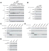
Effects of RagC mutants on mTORC1 signaling. (a) Rag heterodimers containing the RagC mutants co-immunoprecipitate the largest amounts of raptor, an mTORC1 component, similar to the previously characterized RagCS75N mutant. Anti-FLAG immunoprecipitates were collected from HEK-293T cells transiently expressing the indicated cDNAs, and cell lysates and immunoprecipitates were analyzed by immunoblotting. RagBQ99L and RagCQ120L are ‘GTP-locked’ mutants, , while RagCS75N and RagBT54N function as ‘GDP-binding’ mutants, –. (b) Two recurrent mutants from FL, RagCT90N and RagCW115R, and the previously characterized RagCS75N mutant co-immunoprecipitate more endogenous raptor than wild-type RagC in Karpas-422 cells, a B cell lymphoma line of germinal center origin. Anti-FLAG immunoprecipitates from Karpas-422 cells stably expressing the indicated proteins were collected and analyzed as in (a). (c) Stable overexpression of RagCS75N, RagCS75F, RagCT90N, and RagCW115R render cells partially or fully insensitive to leucine deprivation. HEK-293T cells stably expressing the indicated proteins were starved of leucine for 50 minutes and re-stimulated with leucine for 10 minutes. Cell lysates were analyzed by immunoblotting for the indicated proteins. (d) Stable overexpression of the indicated RagC mutants leads to an increase in mTORC1 signaling in the absence of arginine. HEK-293T cells stably expressing the indicated proteins were starved of arginine for 50 minutes, restimulated with arginine for 10 minutes, and analyzed as in (b). (e) Stable overexpression of the indicated RagC mutants, but not wild-type RagC, leads to increased mTORC1 signaling in the absence of leucine in a B cell lymphoma line. Karpas-422 cells stably expressing the indicated proteins were treated and analyzed as in (b).
Figure 4
Figure 4
RagC mutants alter the affinity of nucleotide binding. (a) Three RagC mutants exhibit increased GDP binding, while RagCS75F decreased GDP binding. Nucleotide binding assays were performed with the indicated RagC heterodimer incubated with [3H]GDP and binding assessed using a filter-binding assay. Each value represents the normalized mean +/− SD for n = 3. Statistical differences are assessed comparing each sample to the binding observed with the RagB-RagC wild-type heterodimer. (b) RagCS75N and RagCS75F significantly decrease GTP binding, while RagCT90N does not affect GTP binding with RagCW115R slightly increasing this activity. Nucleotide binding assays were performed as in (B) but incubated with [3H]GTP. Each value represents the normalized mean +/− SD for n = 3. Statistical differences are assessed comparing each sample to the binding observed with the RagB-RagC wild-type heterodimer. (c) RagB and RagC protein levels in the nucleotide binding assays are consistent. Aliquots of the purified Rag heterodimers used in the nucleotide binding assays were resolved on an SDS-PAGE gel and stained with Coomassie. *P<0.05; **P<0.01; ***P<0.005.

Similar articles

Cited by

References

    1. Swenson WT, et al. Improved survival of follicular lymphoma patients in the United States. J Clin Oncol. 2005;23:5019–26. - PubMed
    1. Okosun J, et al. Integrated genomic analysis identifies recurrent mutations and evolution patterns driving the initiation and progression of follicular lymphoma. Nat Genet. 2014;46:176–81. - PMC - PubMed
    1. Pasqualucci L, et al. Genetics of follicular lymphoma transformation. Cell Rep. 2014;6:130–40. - PMC - PubMed
    1. Karube K, et al. Recurrent mutations of NOTCH genes in follicular lymphoma identify a distinctive subset of tumours. J Pathol. 2014;234:423–30. - PubMed
    1. Yildiz M, et al. Activating STAT6 mutations in follicular lymphoma. Blood. 2014 - PMC - PubMed

Publication types

MeSH terms

Substances