. 2016 Feb 19;291(8):3932-46.
doi: 10.1074/jbc.M115.676817. Epub 2015 Dec 17.

Pro-inflammatory Macrophages Sustain Pyruvate Oxidation through Pyruvate Dehydrogenase for the Synthesis of Itaconate and to Enable Cytokine Expression

Affiliations

Pro-inflammatory Macrophages Sustain Pyruvate Oxidation through Pyruvate Dehydrogenase for the Synthesis of Itaconate and to Enable Cytokine Expression

Johannes Meiser et al. J Biol Chem. .

Abstract

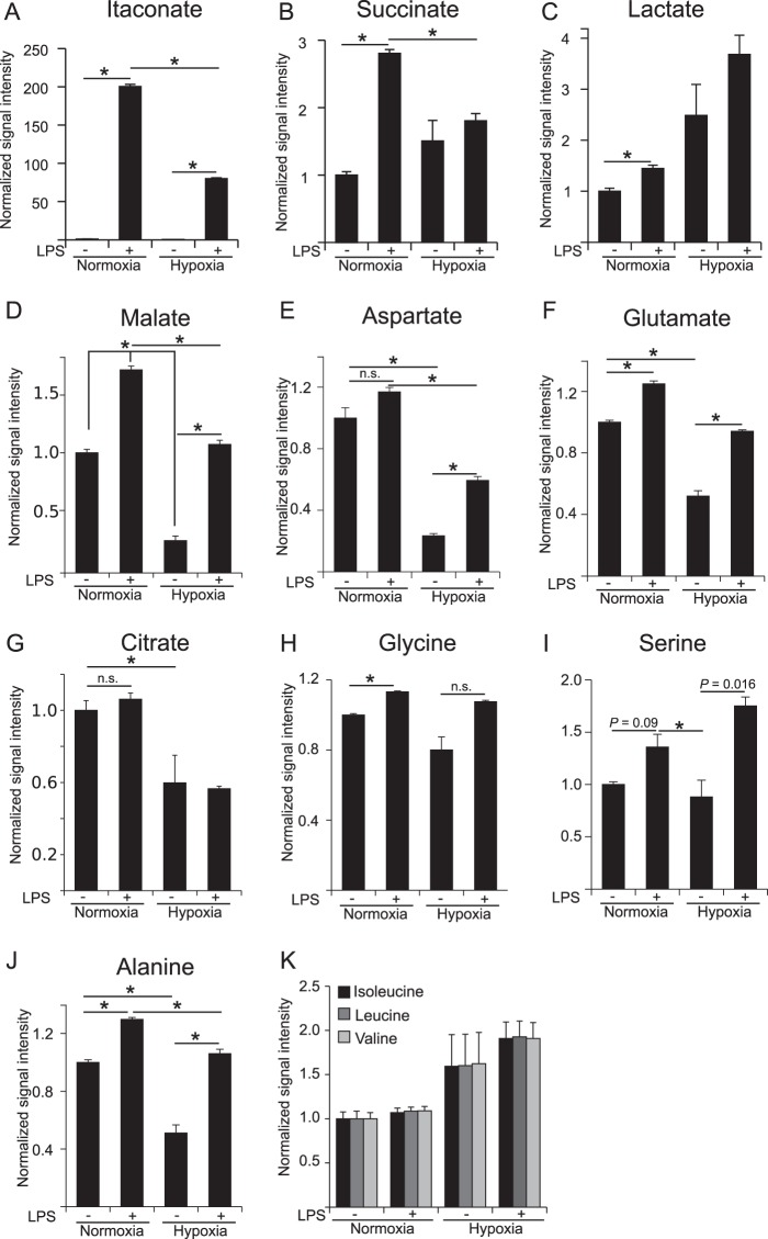
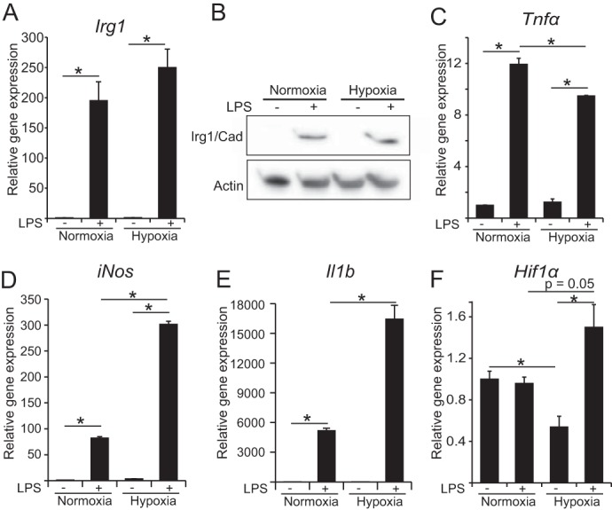
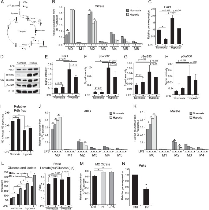
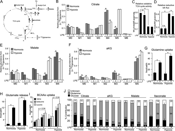
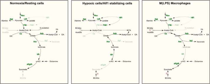
Upon stimulation with Th1 cytokines or bacterial lipopolysaccharides, resting macrophages shift their phenotype toward a pro-inflammatory state as part of the innate immune response. LPS-activated macrophages undergo profound metabolic changes to adapt to these new physiological requirements. One key step to mediate this metabolic adaptation is the stabilization of HIF1α, which leads to increased glycolysis and lactate release, as well as decreased oxygen consumption. HIF1 abundance can result in the induction of the gene encoding pyruvate dehydrogenase kinase 1 (PDK1), which inhibits pyruvate dehydrogenase (PDH) via phosphorylation. Therefore, it has been speculated that pyruvate oxidation through PDH is decreased in pro-inflammatory macrophages. However, to answer this open question, an in-depth analysis of this metabolic branching point was so far lacking. In this work, we applied stable isotope-assisted metabolomics techniques and demonstrate that pyruvate oxidation is maintained in mature pro-inflammatory macrophages. Glucose-derived pyruvate is oxidized via PDH to generate citrate in the mitochondria. Citrate is used for the synthesis of the antimicrobial metabolite itaconate and for lipogenesis. An increased demand for these metabolites decreases citrate oxidation through the tricarboxylic acid cycle, whereas increased glutamine uptake serves to replenish the TCA cycle. Furthermore, we found that the PDH flux is maintained by unchanged PDK1 abundance, despite the presence of HIF1. By pharmacological intervention, we demonstrate that the PDH flux is an important node for M(LPS) macrophage activation. Therefore, PDH represents a metabolic intervention point that might become a research target for translational medicine to treat chronic inflammatory diseases.

Keywords: Itaconate; immunology; inflammation; macrophage; metabolic regulation; metabolism; mitochondrial metabolism; pyruvate;.


Figures

FIGURE 1.
FIGURE 1.
Metabolome analysis of RAW 264 macrophages in the context of LPS activation and hypoxia. Analysis of intracellular metabolite abundance in RAW 264 cells, cultivated under normoxia (21% O2) and hypoxia (2% O2), unstimulated or stimulated with LPS. Metabolites were extracted and analyzed by GC/MS. Signal intensities (peak area) are normalized to unstimulated cells under normoxia. Cells were treated with 10 ng/ml LPS for 6 h. Error bars indicate S.E. (Welsh's t test; *, p < 0.01, n = 3 wells). n.s., not statistically significant. One representative experiment with three individual wells per condition is presented. The experiment was performed three times.
FIGURE 2.
FIGURE 2.
Expression analysis of pro-inflammatory associated genes. A, relative gene expression of Irg1. B, Western blot against IRG1/CAD, the protein that catalyzes the conversion of cis-aconitate to itaconate (no significant difference (data not shown)). C–F, relative gene expression of Tnfα, iNos, Il1β, and Hif1α, normalized to normoxia control (−LPS). Error bars indicate S.E. (Welsh's t test, *, p < 0.01, n = 3). Gene expression data represents the mean over three independent experiments.
FIGURE 3.
FIGURE 3.
PDH flux analysis in M(LPS) macrophages. A, schematic of atom transitions in central metabolism using [U-13C]glucose as a tracer for determination of MIDs to infer relative intracellular fluxes. 13C-carbons are in gray and 12C in black. The dotted line indicates end of one route. Aco, aconitase; Idh, isocitrate dehydrogenase; Akgdh, αKG dehydrogenase; OA, oxaloacetate. B, MID of citrate. M0 to M6 indicates the different mass isotopologues. C, gene expression analysis of Pdk1. D–H, Western blot analysis of PDK1 and PDH phosphorylation on Ser-232, -293, and -300. α-Tubulin serves as loading control. D shows one representative Western blot of three independent experiments. α-Tub, α-tubulin. E–H shows quantification of three independent experiments. I, ratio of M2 citrate/M3 pyruvate indicating relative pyruvate oxidation through PDH. J and K, MID of αKG and malate. L, absolute quantification of glucose uptake and lactate release, and ratio of lactate release/glucose uptake to infer fractional lactate formation per glucose. M, activation of RAW 264 cells with 10 ng/ml LPS or 50 ng/ml interferon-γ (INF) in medium with [U-13C]glucose. Presented are citrate M2 isotopologues as a readout for relative glucose flux through PDH. N, relative gene expression of Pdk1 normalized to normoxia control (Ctrl) (−LPS). Error bars indicate S.E. (Welsh's t test, *, p < 0.01, n = 3). One representative experiment with three individual wells per condition is presented. Each experiment was performed at least three times, except for C. Presented is the mean ± S.E. over three independent experiments. (Welsh's t test, *, p < 0.01, n = 3).
FIGURE 4.
FIGURE 4.
Contribution of glutamine to central metabolism in M(LPS) macrophages. A, schematic of atom transitions in central metabolism using [U-13C]glutamine as a tracer. 13C-carbons are in gray, 12C in black. Citrate molecules derived from reductive carboxylation of αKG are M5 isotopologues, whereas citrate molecules from the oxidative route of the TCA cycle are M4 isotopologues. The dotted line indicates end of one route. Aco, aconitase; Idh, isocitrate dehydrogenase; Akgdh, αKG dehydrogenase; Acly, ATP-dependent citrate lyase; FA, fatty acid. B, MID of citrate. M0 to M6 indicates the different mass isotopologues. C and D, determination of oxidative TCA cycle activity: ratio of M4 citrate/M5 glutamate and ratio of M5 citrate/M5 glutamate indicating relative reductive Idh flux. E and F, MID of malate and αKG. G–I, absolute quantification of glutamine uptake, glutamate release, and uptake of the branched chain amino acids (valine, isoleucine, and leucine). J, carbon contribution (%) of glucose, glutamine, and other carbon sources to citrate, αKG, malate, and itaconate. Carbon contributions are based on MIDs from [U-13C]glucose labeling (Fig. 3) and [U-13C]glutamine labeling. Carbon contribution to itaconate under non-LPS conditions should not be considered because itaconate levels are negligibly low under these non-stimulated conditions (Fig. 1A). Error bars indicate S.E. (Welsh's t test, *, p < 0.01, n = 3). One representative experiment with three individual wells per condition is presented. Each experiment was performed at least three times.
FIGURE 5.
FIGURE 5.
Morphological changes upon LPS activation require sustained lipogenesis in macrophages. A, microscopy (bright field image) of RAW macrophages unstimulated or stimulated with 10 ng/ml LPS for 6 h. White bar indicates 100 μm. B, mean average of cell size (pixels) (left) and analysis of adhesion index (right) of LPS-stimulated and -non-stimulated cells obtained from bright field microscopy. Analysis demonstrates morphological adaption of macrophages during LPS activation. For further details regarding the analysis approach, see under “Experimental Procedures.” C, relative gene expression of carnitine palmitoyl transferase1 (Cpt1) normalized to normoxia control (Ctrl) (−LPS), indicating inhibition of β-oxidation upon LPS stimulation. D and E, MID of palmitate using [U-13C]glucose (D) and [U-13C]glutamine (E) as a tracer. Contribution of each carbon source is depicted in the top right corner of each panel. Error bars indicate S.E. (Welsh's t test, *, p < 0.01, n ≥ 3). One representative experiment with three individual wells per condition is presented. Each experiment was performed at least three times, except for C. Presented is the mean ± S.E. over three independent experiments. (Welsh's t test, *, p < 0.01, n = 3).
FIGURE 6.
FIGURE 6.
Inhibition of pyruvate transport into mitochondria suppresses pro-inflammatory responses in M(LPS) macrophages. A, application of the specific pyruvate transport inhibitor UK5099 to inhibit flux through PDH. Cells were treated with 100 μm UK5099 for 6 h with or without 10 ng/ml LPS at normoxia. Prior to treatment start, cells were cultivated for 24 h in [U-13C]glucose. Presented are citrate M2 isotopologues as a readout for relative glucose flux through PDH. B–D, intracellular metabolite levels of citrate, itaconate, and succinate upon LPS stimulation and after application of 100 μm UK5099. Metabolite levels were determined using GC/MS and normalized to cell number. E–J, relative gene expression of Il1β, iNos, Irg1, Tnfα, Cpt1, and Pdk1 normalized to normoxia control (Ctrl) (−LPS), upon LPS stimulation and after application of 100 μm UK5099 for 6 h. K, viability assay to test a potential effect of UK5099 on cell viability. Assay was performed using trypan blue, dead and live cells were counted. L, to validate the effect of UK5099 a modified experimental setup has additionally been performed. Cells were first activated with 10 ng/ml LPS for 3 h and then UK5099 was added to the cells for additional 6 h. In total, LPS activation was 9 h in this case. Non-UK5099 treated and non-activated cells served as control. M–O, intracellular metabolite levels for citrate, itaconate, and succinate (analysis as in B–D). P–S, relative gene expression analysis of Tnfα, iNos, Irg1, and Il1β. T and U, MID of palmitate using [U-13C]glucose as a tracer. Experimental setup as in A. U, glucose contribution to palmitate, determined from T. Error bars indicate S.E. (Welsh's t test, *, p < 0.01, n = 3). One representative experiment with three individual wells per condition is presented. Each experiment was performed at least three times.
FIGURE 7.
FIGURE 7.
Model summarizing the metabolic adaptations in M(LPS) macrophages. Resting cells use glucose via PDH and TCA oxidation for energy production. Depending on the cell type, anabolic processes run in parallel. Hypoxic cells and cancer cells with stabilized HIF1α increase their glycolytic rate, inhibit PDH flux, and use glutamine-derived carbon for increased reductive carboxylation of αKG for subsequent lipogenesis. Highly proliferating cells promote anabolic processes to support proliferation. M(LPS) macrophages show stabilized HIF1α but do not decrease pyruvate oxidation through PDH. Under this condition, Hif1α does not increase PDK1 abundance. Compared with resting cells, less citrate is oxidized through the TCA cycle and is rerouted to serve as a precursor for itaconate and fatty acid synthesis. Glutamine serves to replenish the TCA cycle by increasing its carbon contribution to αKG and subsequent metabolites. BCAAs might also support carbon supply to the TCA cycle, but this needs additional research. Despite sustained pyruvate oxidation through PDH, oxygen consumption rates are lower than in resting cells.

Similar articles

Cited by

References

    1. Gordon S., and Taylor P. R. (2005) Monocyte and macrophage heterogeneity. Nat. Rev. Immunol. 5, 953–964 - PubMed
    1. Murray P. J., Allen J. E., Biswas S. K., Fisher E. A., Gilroy D. W., Goerdt S., Gordon S., Hamilton J. A., Ivashkiv L. B., Lawrence T., Locati M., Mantovani A., Martinez F. O., Mege J. L., Mosser D. M., et al. (2014) Macrophage activation and polarization: nomenclature and experimental guidelines. Immunity 41, 14. - PMC - PubMed
    1. Kelly B., and O'Neill L. A. (2015) Metabolic reprogramming in macrophages and dendritic cells in innate immunity. Cell Res. 25, 771–784 - PMC - PubMed
    1. Basler T., Jeckstadt S., Valentin-Weigand P., and Goethe R. (2006) Mycobacterium paratuberculosis, Mycobacterium smegmatis, and lipopolysaccharide induce different transcriptional and post-transcriptional regulation of the IRG1 gene in murine macrophages. J. Leukocyte Biol. 79, 628–638 - PubMed
    1. Degrandi D., Hoffmann R., Beuter-Gunia C., and Pfeffer K. (2009) The proinflammatory cytokine-induced IRG1 protein associates with mitochondria. J. Interferon Cytokine Res. 29, 55–67 - PubMed

Publication types

MeSH terms

LinkOut - more resources