. 2015 Dec 16;90(5):2356-71.
doi: 10.1128/JVI.01994-15.

Human Cytomegalovirus Promotes Survival of Infected Monocytes via a Distinct Temporal Regulation of Cellular Bcl-2 Family Proteins

Affiliations

Human Cytomegalovirus Promotes Survival of Infected Monocytes via a Distinct Temporal Regulation of Cellular Bcl-2 Family Proteins

Donna Collins-McMillen et al. J Virol. .

Abstract

Monocytes play a key role in the hematogenous dissemination of human cytomegalovirus (HCMV) to target organ systems. To infect monocytes and reprogram them to deliver infectious virus, HCMV must overcome biological obstacles, including the short life span of monocytes and their antiviral proapoptotic response to infection. We have shown that virally induced upregulation of cellular Mcl-1 promotes early survival of HCMV-infected monocytes, allowing cells to overcome an early apoptotic checkpoint at around 48 h postinfection (hpi). Here, we demonstrate an HCMV-dependent shift from Mcl-1 as the primary antiapoptotic player to the related protein, Bcl-2, later during infection. Bcl-2 was upregulated in HCMV-infected monocytes beginning at 48 hpi. Treatment with the Bcl-2 antagonist ABT-199 only reduced the prosurvival effects of HCMV in target monocytes beginning at 48 hpi, suggesting that Mcl-1 controls survival prior to 48 hpi, while Bcl-2 promotes survival after 48 hpi. Although Bcl-2 was upregulated following viral binding/signaling through cellular integrins (compared to Mcl-1, which is upregulated through binding/activation of epidermal growth factor receptor [EGFR]), it functioned similarly to Mcl-1, adopting the early role of Mcl-1 in preventing caspase-3 cleavage/activation. This distinct, HCMV-induced shift from Mcl-1 to Bcl-2 occurs in response to a cellular upregulation of proapoptotic Bax, as small interfering RNA (siRNA)-mediated knockdown of Bax reduced the upregulation of Bcl-2 in infected monocytes and rescued the cells from the apoptotic effects of Bcl-2 inhibition. Our data demonstrate a distinct survival strategy whereby HCMV induces a biphasic regulation of cellular Bcl-2 proteins to promote host cell survival, leading to viral dissemination and the establishment of persistent HCMV infection.

Importance: Hematogenous dissemination of HCMV via infected monocytes is a crucial component of the viral survival strategy and is required for the establishment of persistent infection and for viral spread to additional hosts. Our system of infected primary human blood monocytes provides us with an opportunity to answer specific questions about viral spread and persistence in in vivo-relevant myeloid cells that cannot be addressed with the more traditionally used replication-permissive cells. Our goal in examining the mechanisms whereby HCMV reprograms infected monocytes to promote viral dissemination is to uncover new targets for therapeutic intervention that would disrupt key viral survival and persistence strategies. Because of this important role in maintaining survival of HCMV-infected monocytes, our new data on the role of Bcl-2 regulation during viral infection represents a promising molecular target for mitigating viral spread and persistence.


Figures

FIG 1
FIG 1
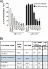
Bcl-2 family proteins are required for monocyte survival beyond the 48-h viability gate. (A) Isolated monocytes were mock infected or HCMV infected and treated with DMSO or with the indicated concentrations of ABT-737 (Bcl-2, Bcl-xL, Bcl-w antagonist). Cells were incubated under adherent conditions on 96-well plates at 37°C, and an MTT viability assay was performed at 0 and 72 h. Results are expressed as percentage of living cells at 72 h compared to that at 0 h. The graph shows the average of results of three replicates from three different blood donors. Paired Student's t tests were performed to determine statistically significant differences in cell survival between drug-treated groups and DMSO controls (indicated by asterisks). (B) Temporal transcriptome analyses of mock-infected versus HCMV-infected monocytes reveal that multiple antiapoptotic mRNAs from the Bcl-2 family are induced following HCMV infection. Data were obtained via reanalysis of Affymetrix gene data from previous studies (GSE19772 [43], GSE11408 [62], and GSE17948 [55]). Significantly upregulated transcripts (≥1.5-fold) are marked with an asterisk.
FIG 2
FIG 2
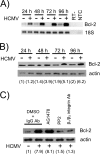
HCMV infection enhances Bcl-2 expression via integrin/Src signaling. (A) Isolated monocytes were mock infected or HCMV infected (MOI = 5) and incubated under adherent conditions at 37°C for 24, 48, 72, and 96 h. RNA was isolated at each time point, and RT-PCR was performed, using primers for Bcl-2 and 18S. Reverse transcriptase-negative (RT-) and nontemplate controls (NTC) were used to show primer specificity for the indicated mRNAs. The image is representative of three repeats from three separate blood donors. The image display has been inverted to show dark bands on a light background for ease of viewing. (B) Isolated monocytes were mock infected or HCMV infected (MOI = 5) and incubated under adherent conditions at 37°C for 24, 48, 72, and 96 h. Whole-cell lysates were collected at each time point, and Western blot analysis was performed using antibodies against Bcl-2 and actin. Densitometric analysis was performed using Quantity One Basic software (Bio-Rad); the numbers below each sample reflect a ratio of Bcl-2 expression normalized to control β-actin expression. The experiment was repeated three times with monocytes from three different donors, with similar results. (C) Isolated monocytes were pretreated for 1 h with DMSO, control IgG antibodies, a 25 μM concentration of the EGFR inhibitor AG1478, a 25 μM concentration of the Src inhibitor PP2, or blocking anti-integrin antibodies, washed in PBS, and then mock infected or HCMV infected (MOI = 5) for 96 h. The medium was supplemented with inhibitors and blocking antibodies every 24 h over the course of infection to ensure continuous blocking of signaling. Total lysates were harvested, and Western blot analysis was performed to examine Bcl-2 expression. Actin was used as a loading control. Densitometric analysis was performed using Quantity One Basic software (Bio-Rad) to obtain a ratio of Bcl-2 expression normalized to actin. The blots and densitometry values are representative of data from three repeats with three different donors.
FIG 3
FIG 3
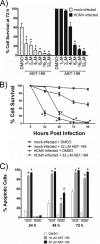
HCMV-induced upregulation of Bcl-2 promotes extended survival of infected monocytes beyond the 48-h viability gate. (A) Monocytes were mock infected or HCMV infected (MOI = 5) and treated with DMSO or with the indicated concentrations of ABT-199 (Bcl-2 antagonist). Cells were incubated under adherent conditions on 96-well plates at 37°C, and an MTT viability assay was performed at 0 and 72 h. Results are expressed as the percentage of living cells at 72 h compared to 0 h. The graph is the result of a single experiment performed in triplicate; similar results were seen in two additional repeats with different blood donors. Paired Student's t tests were performed to determine statistical significance (indicated by an asterisk) between the ABT-199-treated groups and the DMSO controls. (B) Mock-infected or HCMV-infected (MOI = 5) monocytes were treated with 32 μM ABT-199 (Bcl-2 antagonist) and incubated at 37°C on 96-well plates for 24, 48, 72, and 96 h before performance of an MTT viability assay. Results are expressed as the percentage of living cells at each time point compared to the viable cell count at 0 h. The graph is the average of results of three replicates from an experiment repeated with three different blood donors. Asterisks represent statistical significance (as determined by paired Student's t test) between the ABT-199-treated groups and the DMSO-treated controls at each time point. (C) Isolated monocytes were mock infected or HCMV infected and treated with ABT-199 (16 μM or 32 μM) (Bcl-2 antagonist) for 24, 48, and 72 h. The frequency of apoptosis was determined by TUNEL analysis. Cells were visualized by microscopy; the numbers of TUNEL-positive cells and total cells were counted (three fields per well, three wells per experimental arm, and an average of 53 cells per field). Results are expressed as the percentage of apoptotic cells compared to the total number of cells counted at each time point. The graph is the average of results of three replicates from five repeated experiments with different donors (total of 15 data points for each experimental group). Paired Student's t tests were performed; asterisks are used to show statistical significance between the ABT-199-treated groups and the DMSO-treated controls in each time point.
FIG 4
FIG 4
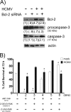
Bcl-2 promotes the extended survival of HCMV-infected monocytes by adopting the early role of Mcl-1 as a regulator of caspase-3 activation. (A) Isolated monocytes were mock infected or HCMV infected (MOI = 5) for 1 h and then nucleofected with nontargeting control siRNA or Bcl-2-specific siRNA and incubated at 37°C for 72 h. Whole-cell lysates were collected, and Western blot analyses were performed using antibodies against Bcl-2, caspase-3, and actin. The experiment was repeated in triplicate using three different donors, with similar results. Densitometric analyses were performed using Quantity One Basic software (Bio-Rad) to quantify the relative expression levels of Bcl-2, procaspase-3, and caspase-3 normalized to the actin control. Quantification is included in parentheses under each corresponding band. (B) Isolated monocytes were mock infected or HCMV infected (MOI = 5) for 1 h at 37°C and then seeded into 96-well plates. Following adhesion, cells were treated with DMSO, 6 μM ABT-199 (Bcl-2 antagonist), 20 μM Z-DEVD-FMK (caspase-3 inhibitor), or 20 μM Z-VAD-FMK (pan-caspase inhibitor). An MTT assay was performed immediately following drug treatment at 0 h and again at 72 h. Results are expressed as percent cell survival at 72 h normalized to 100% survival at 0 h. The graph is representative of results of three replicates from a single experiment; similar results were seen when repeated with two additional donors. Asterisks are used to indicate statistical significance (as determined by paired Student's t tests) between the drug-treated groups and the DMSO-treated controls.
FIG 5
FIG 5
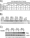
An upregulation of proapoptotic Bax parallels the Mcl-1-to-Bcl-2 shift seen at 48 hpi. (A) The potential binding interactions among various Bcl-2 family proteins are depicted (75). Specifically, the potential binding interactions of the antiapoptotic proteins (Mcl-1, A1, Bcl-2, Bcl-xL, and Bcl-w) with the proapoptotic proteins (Bax and Bak) are shown. (B) Isolated monocytes were mock infected, HCMV infected (MOI = 5), or treated with an alternative activating agent, LPS (20 μM). RNA was isolated at 4, 24, 48, 72, and 96 hpi and used to complete RT-PCR analyses using primers specific for Bax and control 18S RNA. Reverse transcriptase negative (RT-) and nontemplate controls (NTC) were used to ensure that the bands represent proper amplification of the indicated mRNAs. The image display has been inverted to show dark bands on a light background for ease of viewing. The experiment was repeated three times with three different donors; similar results were seen. (C) Monocytes were mock infected or HCMV infected (MOI = 5) for 24, 48, 72, and 96 h. At these time points, whole-cell lysates were collected for a Western blot analysis of cellular Bax expression. Actin was also probed as a loading control. Densitometric analysis was performed using Quantity One Basic software (Bio-Rad). The relative expression of Bax normalized to actin is indicated below each individual sample. Results are representative of data from three repeats with different donors.
FIG 6
FIG 6
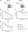
HCMV-induced upregulation of Bax drives the increase in Bcl-2 expression, an event which is required to inactivate Bax and rescue infected cells from apoptosis. (A) Monocytes were mock infected or HCMV infected (MOI = 5) for 1 h and then nucleofected with a nontargeting control siRNA, a Bax-specific siRNA (panel 1), or a Bcl-2-specific siRNA (panel 2). Whole-cell lysates were collected after 72 h of incubation at 37°C. Western blot analysis was performed to analyze expression levels of Bax and Bcl-2. Actin was used as a loading control. Results are representative of three repeats using monocytes from different donors. (B to D) Isolated monocytes were mock infected (B), HCMV infected (MOI = 5) (C), or stimulated with 20 μM LPS for 1 h (D) and then nucleofected with nontargeting control siRNA (circles and triangles) or a Bax-specific siRNA (squares and inverted triangles) and plated in 96-well microtiter plates. Following adhesion, cells were treated with DMSO (circles and squares) or 6 μM ABT-199 (Bcl-2 antagonist) (triangles and inverted triangles). MTT analysis was used to assess cell viability at each of the indicated time points. Data are expressed as percent cell survival normalized to 100% at 0 h (immediately following ABT-199 treatment). Results are the average of three replicates from a single experiment and are representative of data analyzed from two additional repeats with different blood donors.
FIG 7
FIG 7
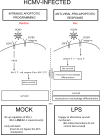
Our model of the role of cellular Bcl-2 family proteins in the survival of HCMV-infected monocytes. Viral binding to EGFR enhances Mcl-1 expression levels in infected monocytes (49), allowing cells to successfully navigate the early cell viability checkpoint by blocking the cleavage of proapoptotic caspase-3 (47). HSP-27 is also upregulated in response to viral infection to ensure complete control over caspase-3 activation at early times postinfection, when cell survival is of primary importance. At later times postinfection, when survival and differentiation become equally important to the viral persistence strategy, Mcl-1 expression is extinguished (49), and integrin-mediated signaling controls Bcl-2 expression in infected monocytes. Bcl-2 is upregulated in HCMV-infected monocytes beginning at 48 hpi to assume the role of Mcl-1 as an inhibitor of caspase-3 activation; however, without additional inhibition by HSP-27, a low-level activation of caspase-3 allows monocyte-to-macrophage differentiation without causing apoptosis. This shift from Mcl-1 to Bcl-2 as the principal antiapoptotic player occurs in response to an upregulation of the proapoptotic protein, Bax, beginning at around 48 hpi. We speculate that this increase in Bax occurs as part of the antiviral proapoptotic response to HCMV infection. In contrast, the majority of mock-infected cells undergo apoptosis at the 48-h checkpoint. For those that survive due to adhesion-dependent signals, it appears that both Mcl-1 and Bcl-2 are required prior to the viability gate for continued survival. LPS-stimulated monocytes appear to engage an unknown alternative survival mechanism, as our data show that Bax/Bcl-2 interactions do not affect survival outcomes in these cells.

Similar articles

Cited by

References

    1. Mocarski E Jr, Shenk T, Griffiths PD, Pass R. 2013. Cytomegaloviruses, p 1960–2014. In Knipe DM, Howley PM (ed), Fields virology, vol 2 Lippincott Williams & Wilkins, Philadelphia, PA.
    1. Nogalski MT, Collins-McMillen D, Yurochko AD. 2014. Overview of human cytomegalovirus pathogenesis, p 15–28. In Yurochko AD, Miller WE (ed), Human cytomegaloviruses: methods & protocols. Humana Press, New York, NY. - PubMed
    1. Cohen JI, Corey GR. 1985. Cytomegalovirus infection in the normal host. Medicine 64:100–114. - PubMed
    1. Wreghitt TG, Teare EL, Sule O, Devi R, Rice P. 2003. Cytomegalovirus infection in immunocompetent patients. Clin Infect Dis 37:1603–1606. doi:10.1086/379711. - DOI - PubMed
    1. Speir E, Modali R, Huang ES, Leon MB, Shawl F, Finkel T, Epstein SE. 1994. Potential role of human cytomegalovirus and p53 interaction in coronary restenosis. Science 265:391–394. doi:10.1126/science.8023160. - DOI - PubMed

Publication types

Substances