. 2016 Jul;36(7):1281-94.
doi: 10.1177/0271678X15606463. Epub 2015 Oct 13.

BACE-1 is expressed in the blood-brain barrier endothelium and is upregulated in a murine model of Alzheimer's disease

Affiliations

BACE-1 is expressed in the blood-brain barrier endothelium and is upregulated in a murine model of Alzheimer's disease

Kavi Devraj et al. J Cereb Blood Flow Metab. 2016 Jul.

Abstract

Endothelial cells of the blood-brain barrier form a structural and functional barrier maintaining brain homeostasis via paracellular tight junctions and specific transporters such as P-glycoprotein. The blood-brain barrier is responsible for negligible bioavailability of many neuroprotective drugs. In Alzheimer's disease, current treatment approaches include inhibitors of BACE-1 (β-site of amyloid precursor protein cleaving enzyme), a proteinase generating neurotoxic β-amyloid. It is known that BACE-1 is highly expressed in endosomes and membranes of neurons and glia. We now provide evidence that BACE-1 is expressed in blood-brain barrier endothelial cells of human, mouse, and bovine origin. We further show its predominant membrane localization by 3D-dSTORM super-resolution microscopy, and by biochemical fractionation that further shows an abluminal distribution of BACE-1 in brain microvessels. We confirm its functionality in processing APP in primary mouse brain endothelial cells. In an Alzheimer's disease mouse model we show that BACE-1 is upregulated at the blood-brain barrier compared to healthy controls. We therefore suggest a critical role for BACE-1 at the blood-brain barrier in β-amyloid generation and in vascular aspects of Alzheimer's disease, particularly in the development of cerebral amyloid angiopathy.

Keywords: Alzheimer’s disease; BACE-1; blood–brain barrier; endothelium; β-amyloid.


Figures

Figure 1.
Figure 1.
BACE-1 mRNA expression analysis in mouse brain microvessels and cultured endothelial cells. In freshly isolated mouse brain microvessels (MBMVs) and primary cultured mouse brain microvascular endothelial cells (MBMECs) mRNA expression analysis by qRT-PCR showed significant expression of BACE-1 using two different primer pairs. Whole brain mRNA served as a positive control for BACE-1 expression as neurons are known to express high levels of BACE-1. BACE-1 expression was present even in pure cultured endothelial cells that have no contamination from neurons thus indicating a specific expression of BACE-1. Cldn-5 served as a marker for endothelium, which was at much higher levels in cultured and freshly isolated brain microvascular endothelial cells. The Ct range for BACE-1 qRT-PCR was in the range 23–26 cycles with non-template control about 35 cycles indicating specificity of expression. Statistical significance was by One-way ANOVA followed by TUKEY-HSD test for multiple groups (n = 4 experiments, *p < 0.05, **p < 0.01, and ***p < 0.001 with MBMV set as 1 with 2–4 mice in each experiment).
Figure 2.
Figure 2.
BACE-1 protein expression analysis in mouse brain microvessels. (a) Western Blots for BACE-1 using a highly specific antibody (B0681, Sigma) made against the N-terminus of BACE-1 (AA 46-62 of BACE, N-terminal) show the staining of the 75 and 50 kDa isoforms of BACE-1 (lane 1) in purified microvessels from wild-type mouse brains; the staining is specific as a blocking peptide (lane 2) completely abolished the staining of mouse brain microvessels. Lanes 3 and 4 show silver-stained 1D gels of recombinant BACE-1 (Invitrogen P2947 and Sigma S4195) as positive controls; lanes 5 and 6 show Western blots of the recombinant BACE-1 proteins with B0681. The blot is representative of three independent preparations of brain microvessels using 8–10 mice each time. The recombinant proteins were made to the extracellular domain of BACE-1 fused to Fc region of human IgG1 (Invitrogen P2947) or with a C-terminal FLAG-tag (Sigma S4195), hence they migrate at a lower molecular weight than the endogenous full-length BACE-1, which runs at 75 kDa. (b) In addition to B0681 (N-terminal epitope, Sigma) ab2077 (C-terminal epitope, Abcam) and 2882-1 (C-terminal, Epitomics) were also used to confirm the expression of BACE-1 in mouse brain microvessels. Competing peptides (for the antibody epitopes) from the vendor were used to obtain the specificity of the bands. For the Epitomics antibody for which the vendor competing peptides were not available only the C-terminal peptide for ab2077 but not the N-terminal one corresponding to B0681 abolished the specific band. The N-terminal antibody from Sigma recognizes two isoforms (full length 75 kDa, soluble 50 kDa) but the C-terminal antibodies (Abcam, Epitomics) recognize only the higher molecular weight full-length isoform. The red arrowheads indicate the specific bands and the red cross marks point the loss of these specific bands.
Figure 3.
Figure 3.
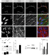
Localization and activity of BACE-1 in brain microvessels and cultured endothelial cells. Immunofluorescence for BACE-1 showing specific staining for BACE-1 at the PM and intracellular vesicles in (a) freshly isolated MBMVs (representative pictures from 3 preparations using 3–4 mice) and in (b) primary cultured MBMECs (representative of three preparations using 4–6 mice each time). Claudin-5 (Cldn5) served as an endothelial marker. (c) Secondary antibody control for BACE-1, showing no staining for BACE-1 when the primary antibody was omitted. (d) Localization analysis in fractionated freshly isolated bovine brain capillary endothelial cells shows the predominant expression of BACE-1 in the abluminal membranes (*p < 0.05, 2-tailed paired t-test). PgP and EAAT-2 served as markers of luminal and abluminal membranes, respectively. GLUT-1 served as a marker expressed equally on both membranes. Quantitation was performed using blots for BACE-1 from three preparations utilizing 10 bovine brains each time. (e) Activity of BACE-1 is shown by inhibition of BACE-1 (Merck IV, 24 h) in MBMECs in medium with 20% bovine serum that is reported to contain full-length APP. The blots show an increase in the mature form of APP (mAPP) in the medium indicating reduced cleavage by BACE-1 and a concomitant increase in α-secretase activity resulting in increased levels of sAPPα. Loading differences accounted for by taking equal volume fraction from cells seeded at the same density along with a Ponceau S protein stain (not shown). Quantitation of the blots was performed from three experiments utilizing two animals in each preparation. Significance was by 2-tailed paired t-tests (*p < 0.05, **p < 0.01).
Figure 4.
Figure 4.
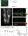
Subcellular localization of BACE-1 in human brain endothelial cells (a) Primary human brain endothelial cells (HBMECs, P4) were stained for BACE-1 (B0681, Sigma), the expression being similar to murine brain endothelial cells showing a punctate pattern. VE-cadherin served as an endothelial marker staining the adherens-junctions. (b) Western blots for BACE-1 in HBMECs (P3) and hCMEC/D3 (P28), a human brain endothelial line confirmed the staining data. Cldn-5 was used as an endothelial marker. (a, b) are representative of two experiments using P3-P4 HBMECs and P28-P30 D3 cells. (c) 3D-dSTORM-imaging of Alexa-Fluor 647 labeled BACE-1. Diffraction-limited widefield fluorescence image shows the spindle shape of differentiated HBMECs (left image). BACE-1 is preferentially located close to the PM as shown by the cross-sections of small insets (XZ). The yellow and white dotted lines mark the apical (top) and basal (bottom) PMs, respectively. Insets are color-coded according to the relative z-position in nm, as indicated by the numbers. Scale bar 10 and 1 µm (overview and insets, respectively). The histograms (on the right) show quantification for the localization. (d) Western blotting on fractionated HBMECs shows enrichment of BACE-1 in PM and TGN fractions with significantly lower levels in cytoplasmic (CYTO) and endosomal vesicle fraction (EN). TUB1A1, lamin B1, and CDH5 served as markers for CYTO, NU, and PM fractions, respectively. (N = 3 sets of fractionation using HBMECs from three different donors, significance was by One-way ANOVA followed by TUKEY-HSD test for multiple groups. **p < 0.01, ***p < 0.001).
Figure 5.
Figure 5.
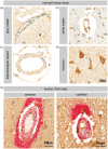
BACE-1 expression in normal and CAA human brain vessels. (a–d) Immunohistochemistry for BACE-1 (C-terminal epitopes, Abcam antibody) shows expression of BACE-1 in the human brain vessels from both the white and grey matter. Leptomeningeal vessels also were stained indicating that even the non-BBB forming vessels express BACE-1. Neurons served as a positive control. Figures (a–d) are representative of five cases. (e) Arteriole and capillary of a single human CAA case. Double staining for amyloid-β (red) and BACE-1 (brown) shows intense staining of amyloid-β around vessels and endothelial as well as neuronal staining for BACE-1.
Figure 6.
Figure 6.
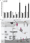
Expression analysis in brain microvessels from an AD mouse model and schematic for APP/Aβ processing at the BBB. (a) MBMVs from transgenic mice (hAPPSL) over-expressing the 751 amino acid form of human amyloid precursor protein (hAPP) with London (V717I) and Swedish (KM670/671NL) mutations under the control of the murine Thy-1 promoter were compared with wild-type mice. We observed a four-fold increase of BACE-1 expression in the BBB microvessels isolated from hAPPSL mice when compared to age-matched wild-type mice suggesting an increase in the APP cleavage activity of BACE-1 at the BBB in the mutant animals. Endothelial marker genes such as VE-cadherin and tight junction molecules ZO-1, claudin-5 were unchanged whereas GLUT-1, the primary glucose transporter at the BBB was downregulated. Interestingly, the luminal Aβ transporter RAGE was upregulated in the AD mice suggesting that circulating Aβ could potentially contribute to the brain amyloidosis. Furthermore the luminally expressed p-glycoprotein (PgP) known to be involved in efflux of Aβ into the circulation was downregulated suggesting a decreased clearance of amyloid peptides from the brain. The abluminally located LRP-1 was upregulated, which is known to endocytose APP, also supporting an increase in the BBB BACE-1 activity in this AD model. Abluminal Aβ antibody transporter FcRN was unchanged. Statistical significance was obtained from three qRT-PCR experiments using six transgenic mice (10 months age) or age-matched wild-type animals (***p < 0.001 using 2-tailed paired t-tests). (b) APP internalized from circulation via LRP2 or from brain parenchyma via LRP-1 or from the ECs is cleaved by BACE-1 to form Aβ. The predominant localization of BACE-1 at the abluminal membrane supports neuronal APP processing within the BBB endothelium. This Aβ is deposited as cerebrovascular plaques and/or is cleared from the brain parenchyma into circulation via LRP-1, FcRn from the abluminal side, and PgP from the luminal side. Additionally Aβ influx via RAGE can potentially regulate the Aβ transport across the BBB.

Similar articles

Cited by

References

    1. Probst G, Xu Y-Z. Small-molecule BACE1 inhibitors: a patent literature review (2006-2011). Expert Opin Ther Pat 2012; 22: 511–540. - PubMed
    1. Revesz T, Holton JL, Lashley T, et al. Genetics and molecular pathogenesis of sporadic and hereditary cerebral amyloid angiopathies. Acta Neuropathol 2009; 118: 115–130. - PMC - PubMed
    1. Bush AI, Martins RN, Rumble B, et al. The amyloid precursor protein of Alzheimer's disease is released by human platelets. J Biol Chem 1990; 265: 15977–15983. - PubMed
    1. Bush AI, Beyreuther K, Masters CL. The beta A4 amyloid protein precursor in human circulation. Ann N Y Acad Sci 1993; 695: 175–182. - PubMed
    1. Ho L, Fukuchi KI, Younkin SG. The alternatively spliced Kunitz protease inhibitor domain alters amyloid beta protein precursor processing and amyloid beta protein production in cultured cells. J Biol Chem 1996; 271: 30929–30934. - PubMed

Publication types

MeSH terms

Substances

LinkOut - more resources