

Nanostructured materials for ocular delivery: nanodesign for enhanced bioadhesion, transepithelial permeability and sustained delivery
- PMID: 26652282
- PMCID: PMC4893795
- DOI: 10.4155/tde.15.75
Nanostructured materials for ocular delivery: nanodesign for enhanced bioadhesion, transepithelial permeability and sustained delivery
Abstract
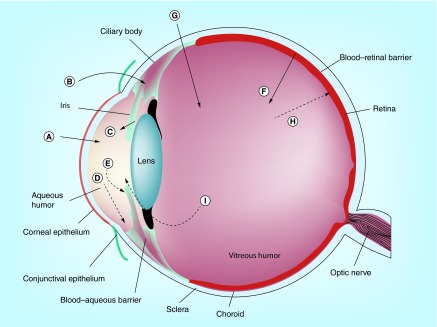
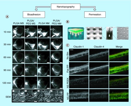
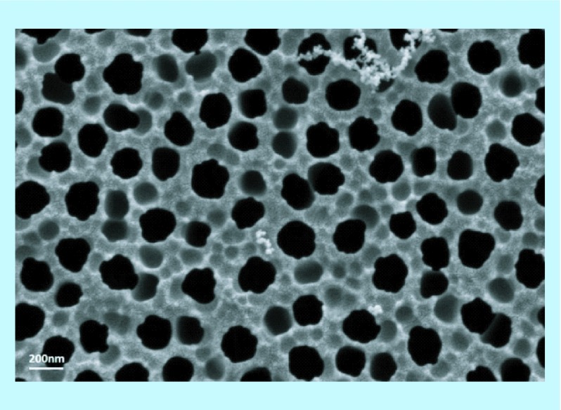
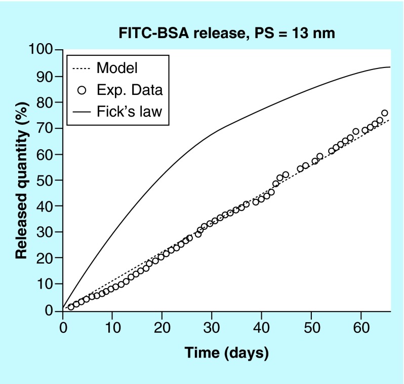
Effective drug delivery to the eye is an ongoing challenge due to poor patient compliance coupled with numerous physiological barriers. Eye drops for the front of the eye and ocular injections for the back of the eye are the most prevalent delivery methods, both of which require relatively frequent administration and are burdensome to the patient. Novel drug delivery techniques stand to drastically improve safety, efficacy and patient compliance for ocular therapeutics. Remarkable advances in nanofabrication technologies make the application of nanostructured materials to ocular drug delivery possible. This article focuses on the use of nanostructured materials with nanoporosity or nanotopography for ocular delivery. Specifically, we discuss nanotopography for enhanced bioadhesion and permeation and nanoporous materials for controlled release drug delivery. As examples, application of polymeric nanostructures for greater transepithelial permeability, nanostructured microparticles for enhanced preocular retention time and nanoporous membranes for tuning drug release profile are covered.
Conflict of interest statement
Financial & competing interests disclosure This work was supported by the NIH R01-EY021574 and R01-EB018842. The authors have no other relevant affiliations or financial involvement with any organization or entity with a financial interest in or financial conflict with the subject matter or materials discussed in the manuscript apart from those disclosed. No writing assistance was utilized in the production of this manuscript.
Figures
Similar articles
-
Nanostructured lipid carriers-based thermosensitive eye drops for enhanced, sustained delivery of dexamethasone.Nanomedicine (Lond). 2018 Jun;13(11):1239-1253. doi: 10.2217/nnm-2017-0318. Nanomedicine (Lond). 2018. PMID: 29949466
-
Nanostructured lipid carriers as novel ophthalmic delivery system for mangiferin: improving in vivo ocular bioavailability.J Pharm Sci. 2012 Oct;101(10):3833-44. doi: 10.1002/jps.23251. Epub 2012 Jul 5. J Pharm Sci. 2012. PMID: 22767401
-
Nanostructured mucoadhesive microparticles for enhanced preocular retention.Acta Biomater. 2014 Jan;10(1):77-86. doi: 10.1016/j.actbio.2013.08.026. Epub 2013 Aug 23. Acta Biomater. 2014. PMID: 23978409
-
Polymeric micelles for ocular drug delivery: From structural frameworks to recent preclinical studies.J Control Release. 2017 Feb 28;248:96-116. doi: 10.1016/j.jconrel.2017.01.012. Epub 2017 Jan 11. J Control Release. 2017. PMID: 28087407 Free PMC article. Review.
-
Emphasis on Nanostructured Lipid Carriers in the Ocular Delivery of Antibiotics.Pharm Nanotechnol. 2024;12(2):126-142. doi: 10.2174/2211738511666230727102213. Pharm Nanotechnol. 2024. PMID: 37519002 Review.
Cited by
-
Ultra-small micelles based on polyoxyl 15 hydroxystearate for ocular delivery of myricetin: optimization, in vitro, and in vivo evaluation.Drug Deliv. 2019 Dec;26(1):158-167. doi: 10.1080/10717544.2019.1568624. Drug Deliv. 2019. PMID: 30822157 Free PMC article.
-
Ocular delivery of proteins and peptides: Challenges and novel formulation approaches.Adv Drug Deliv Rev. 2018 Feb 15;126:67-95. doi: 10.1016/j.addr.2018.01.008. Epub 2018 Jan 13. Adv Drug Deliv Rev. 2018. PMID: 29339145 Free PMC article. Review.
-
Progress in Ocular Drug Delivery: Challenges and Constraints.Handb Exp Pharmacol. 2024;284:267-288. doi: 10.1007/164_2023_693. Handb Exp Pharmacol. 2024. PMID: 37620616
-
Core/shell nanoassembly of amphiphilic naproxen-polyethylene glycol: synthesis, characterisation and evaluation as drug delivery system.IET Nanobiotechnol. 2018 Sep;12(6):814-821. doi: 10.1049/iet-nbt.2017.0219. IET Nanobiotechnol. 2018. PMID: 30104456 Free PMC article.
-
Recent Advancements in Non-Invasive Formulations for Protein Drug Delivery.Comput Struct Biotechnol J. 2019 Sep 11;17:1290-1308. doi: 10.1016/j.csbj.2019.09.004. eCollection 2019. Comput Struct Biotechnol J. 2019. PMID: 31921395 Free PMC article. Review.
References
-
- O'Roruke M. Next generation ocular drug delivery platforms. OIS. 2014;1:7.
-
- Rupenthal I. Sector overview: ocular drug delivery technologies: exciting times ahead. 2015. www.ondrugdelivery.com
-
- Jarvinen K, Jarvinen T, Urtti A. Ocular absorption following topical delivery. Adv. Drug Deliv. Rev. 1995;16:3–19.
-
- Mannermaa E, Vellonen KS, Ryhanen T, et al. Efflux protein expression in human retinal pigment epithelium. Pharm. Res. 2009;26(7):1785–1791. - PubMed
Publication types
MeSH terms
Substances
Grants and funding
LinkOut - more resources
Full Text Sources
Other Literature Sources
Research Materials