. 2016 Jan;126(1):123-36.
doi: 10.1172/JCI81108. Epub 2015 Nov 30.

Endothelial LRP1 transports amyloid-β(1-42) across the blood-brain barrier

Endothelial LRP1 transports amyloid-β(1-42) across the blood-brain barrier

Steffen E Storck et al. J Clin Invest. 2016 Jan.

Abstract

According to the neurovascular hypothesis, impairment of low-density lipoprotein receptor-related protein-1 (LRP1) in brain capillaries of the blood-brain barrier (BBB) contributes to neurotoxic amyloid-β (Aβ) brain accumulation and drives Alzheimer's disease (AD) pathology. However, due to conflicting reports on the involvement of LRP1 in Aβ transport and the expression of LRP1 in brain endothelium, the role of LRP1 at the BBB is uncertain. As global Lrp1 deletion in mice is lethal, appropriate models to study the function of LRP1 are lacking. Moreover, the relevance of systemic Aβ clearance to AD pathology remains unclear, as no BBB-specific knockout models have been available. Here, we developed transgenic mouse strains that allow for tamoxifen-inducible deletion of Lrp1 specifically within brain endothelial cells (Slco1c1-CreER(T2) Lrp1(fl/fl) mice) and used these mice to accurately evaluate LRP1-mediated Aβ BBB clearance in vivo. Selective deletion of Lrp1 in the brain endothelium of C57BL/6 mice strongly reduced brain efflux of injected [125I] Aβ(1-42). Additionally, in the 5xFAD mouse model of AD, brain endothelial-specific Lrp1 deletion reduced plasma Aβ levels and elevated soluble brain Aβ, leading to aggravated spatial learning and memory deficits, thus emphasizing the importance of systemic Aβ elimination via the BBB. Together, our results suggest that receptor-mediated Aβ BBB clearance may be a potential target for treatment and prevention of Aβ brain accumulation in AD.


Figures

Figure 10
Figure 10. Deletion of Lrp1 in brain endothelial cells leads to cognitive deficits in 5xFAD Lrp1BE–/– mice.
7-month-old female 5xFAD Lrp1BE–/– (n = 5), 5xFAD Lrp1BEfl/fl (n = 7), Lrp1BE–/– (n = 5), and WT (n = 6) control mice were tested. (A) Animals underwent acquisition training to learn to use proximal and distal cues to navigate a path to a hidden platform. A significant difference in the escape latency of 5xFAD Lrp1BE–/– mice compared with that of all other groups was seen on days 3 to 5. (B and E) Swimming speed was not affected in all mice tested. (C) Spatial reference memory deficits in 5xFAD Lrp1BE–/– mice were shown in the probe trial, in which 5xFAD Lrp1BE–/– mice spent significantly less time in the target quadrant than all other groups of mice. The probe trial was given after the acquisition training phase to assess spatial reference memory. (D) 5xFAD Lrp1BEfl/fl mice showed no impairment of spatial reference memory, as reflected by the significant greater percentage of time spent in the target quadrant (P < 0.001 target vs. left, right, and opposite quadrant). The probe trial revealed a significant impairment of spatial reference memory in 5xFAD Lrp1BE–/– mice, as they showed no preference for the target quadrant. T, target quadrant; L, left quadrant; R, right quadrant; O, opposite quadrant. Data represent mean ± SEM. For statistical analyses, the following test was used: repeated-measures ANOVA followed by Bonferroni multiple comparisons. *P < 0.05, ***P < 0.001.
Figure 9
Figure 9. Preferential clearance of Aβ1–40 species by brain endothelial LRP1.
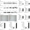
Contrasting the Aβ1–42/Aβ1–40 ratio in 5xFAD Lrp1BE–/– and 5xFAD Lrp1BEfl/fl Aβ pools demonstrates a differential clearance of Aβ species. Densitometry analysis of immunoprecipitated soluble brain Aβ, insoluble brain Aβ, and plasma Aβ with 6E10 antibody from 7-month-old mice (n = 3, n = 5, n = 7, n = 6, n = 3, n = 3 from left to right) showed higher Aβ1–40 and Aβ1–42/Aβ1–40 ratios in plasma and lower and Aβ1–42/Aβ1–40 ratios in brain fractions when LRP1 is present in brain endothelial cells. Data represent mean ± SEM. For statistical analyses, unpaired t test was used. *P < 0.05, **P < 0.01, ***P < 0.001.
Figure 8
Figure 8. No difference in astrogliosis and microgliosis due to brain endothelial knockout of LRP1 in 5xFAD mice.
Immunoreactivity for (A and B) Iba1 (microgliosis) and (C and D) GFAP (astrogliosis) in hippocampi of 5xFAD Lrp1BE–/– (n = 5) and 5xFAD Lrp1BEfl/fl (n = 7) mice (mean ± SEM). Representative results are shown in A and C. For statistical analyses, unpaired t test was used. Scale bar: 200 μm.
Figure 7
Figure 7. BBB clearance of Aβ species in 5xFAD mice is regulated by brain endothelial LRP1.
Representative immunoprecipitations of (A) plasma Aβ, (B) soluble brain Aβ, and (C) insoluble brain Aβ, with 6E10 antibody from 7-month-old female mice show impaired brain-to-blood clearance of Aβ1–40 and Aβ1–42. Quantification of (D) plasma Aβ1–40, (E) plasma Aβ1–42, (F) soluble brain Aβ1–40, (G) soluble brain Aβ1–42, (H) insoluble brain Aβ1–40, and (I) insoluble brain Aβ1–42. n = 3 (D and E); n = 4 (F and G); n = 5 (fl/fl) and n = 3 (–/–) (H and I). (J and K) No effect on plaque deposition in hippocampus. n = 5 (fl/fl) and n = 7 (–/–) (K). Quantification of Aβx–40 and Aβx–42 using ELISA showed (L) insoluble and (M) significantly elevated soluble Aβx–40 and Aβx–42 levels in 7-month-old 5xFAD Lrp1BE–/– mice. n = 12, n = 5, n = 12, n = 5 (L) and n = 10, n = 5, n = 12, n = 5 (M) from left to right. Data represent mean ± SEM of n = 5. All samples except for those shown in lanes 1 and 2 in C were analyzed on the same Western blot but rearranged for clearer presentation. A shorter exposure is shown for Aβ1–40 and Aβ1–42 standards in A. For statistical analyses, unpaired t test was used. *P < 0.05, **P < 0.01, ***P < 0.001. Scale bar: 200 μm.
Figure 6
Figure 6. Brain endothelial LRP1 substantially mediates Aβ clearance in vivo.
(A) 5.14 nM [125I] Aβ1–42 and 40 μCi/ml [14C]-inulin, a paracellular marker, were microinfused into brain ISF of the caudate nucleus. Efflux was studied at designated time points by determining remaining radioactivity in the brain (n = 5, n = 5, n = 3, n = 3, n = 4, n = 5 mice per group from left to right). (B) No alteration was observed in the bulk flow clearance of [14C]-inulin (n = 5 mice per group). (C) Efflux across the BBB of 5.14 nM [125I] Aβ1–42 15 minutes after microinfusion in brain ISF demonstrated the substantial contribution of LRP1 (n = 5 mice per group). (D and E) Scarce presence of tracers in CSF 15 minutes after microinjection into the caudate nucleus (n = 3 mice per group). (F) Contribution of brain endothelial LRP1 to total and BBB clearance of 5.14 nM [125I] Aβ1–42 within 15 minutes (n = 5 mice per group). Data represent mean ± SEM. For statistical analyses, unpaired t test was used. *P < 0.05, **P < 0.01.
Figure 5
Figure 5. LRP1 in brain endothelial cells substantially contributes to [125I] Aβ1–42 transcytosis in vitro.
[125I] Aβ1–42 transport across the primary mouse brain capillary endothelial cell monolayer was studied in the presence of 1 μCi/ml [14C]-inulin to determine the transcytosis quotient (TQ). Transcytosis was analyzed in the brain-to-blood direction (abluminal to luminal) by measuring the dpm for [14C]-inulin and the cpm for the TCA-precipitable [125I] radioactivity. The TQ of Lrp1BE–/– brain endothelial cells was normalized to Lrp1BEfl/fl brain endothelial cells. (A) Transport at a physiological concentration of 0.1 nM Aβ (Lrp1BEfl/fl, n = 18; Lrp1BE–/–, n = 14; 4 independent experiments). (B) Higher contribution of LRP1 in transport of [125I] Aβ1–42 at higher Aβ concentrations (n = 6, n = 4, n = 5, n = 4, n = 3, n = 4, n = 3, n = 3 from left to right). Data represent mean ± SEM. For statistical analyses, unpaired t test was used. *P < 0.05, **P < 0.01, ***P < 0.001.
Figure 4
Figure 4. Deletion of Lrp1 in Lrp1BE–/– mice does not affect BBB integrity.
(A) Brain IgG extravasation and (B) fluorescein permeability studies in Lrp1BEfl/fl (n = 5) and Lrp1BE–/– (n = 8) mice showed no effect of Lrp1 knockout on BBB permeability in vivo. (A) Brain lysates were analyzed by Western blotting and normalized to β-actin. (B) Mice were injected with Na-fluorescein, and fluorescence intensity of brain homogenates (emission at 519 nm, excitation at 488 nm) was normalized to brain weight. Data represent mean ± SEM. For statistical analyses, unpaired t test was used.
Figure 3
Figure 3. Brain endothelial–specific deletion of Lrp1 in Lrp1BE–/– mice.
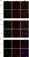
Immunofluorescent staining in cortical brain sections for LRP1 and (A) NeuN-positive neuronal cells, (B) GFAP-positive astrocytes, and (C) CD11b-positive microglia and macrophages to determine potential recombination in macrophages/microglia, neurons, and astrocytes revealed no differences between genotypes. Scale bar: 20 μm. Data show representative results from experiments performed in triplicate.
Figure 2
Figure 2. No difference in Lrp1 expression in capillary-depleted brain fractions of Lrp1BE–/– and Lrp1BEfl/fl mice.
(A) Immunostaining of LRP1 α chain and β chain in brain fractions deprived of capillaries. An anti–β-actin immunoblot is shown as a loading control. MEF, mouse embryonic fibroblast. (B and C) Quantification of relative abundance of (B) LRP1 α chain and (C) LRP1 β chain in brain fractions deprived of capillaries. Data show representative results of 3 to 5 mice per group from experiments performed in triplicate (mean ± SEM). For statistical analyses, unpaired t test was used.
Figure 1
Figure 1. Full deletion of Lrp1 in Lrp1BE–/– mice.
(A) Immunofluorescent staining for endothelial marker CD31 and LRP1 in cortical brain sections demonstrated complete knockout of Lrp1 in brain endothelium of Lrp1BE–/– animals, while Lrp1 expression in surrounding cells remained unaffected (white arrows). DRAQ5 was used to stain cell nuclei. Scale bar: 20 μm. (B) Immunostaining in isolated endothelial cells showed knockout of Lrp1 in Lrp1BE–/– mice. Primary cortical endothelial cell and control lysates of LRP1-expressing CHO cells (K1) and LRP1 knockout (13‑5-1) cells were analyzed on the same Western blot but rearranged for clearer presentation. An anti–β-actin immunoblot is shown as a loading control. (C) PCR analysis revealed complete Cre-mediated excision of the loxP-flanked Lrp1 allele in brain endothelium. Endothelial genomic DNA was used for PCR detecting the WT (WT/WT, 507 bp), the loxP-flanked (fl/fl, 541 bp), and the excised allele (–/–, 325 bp) simultaneously in one reaction. Data show representative results from experiments performed in triplicate.

Similar articles

Cited by

References

    1. Mawuenyega KG, et al. Decreased clearance of CNS β-amyloid in Alzheimer’s disease. Science. 2010;330(6012): doi: 10.1126/science.1197623. - DOI - PMC - PubMed
    1. Naslund J, et al. Relative abundance of Alzheimer Aβ amyloid peptide variants in Alzheimer disease and normal aging. Proc Natl Acad Sci U S A. 1994;91(18):8378–8382. doi: 10.1073/pnas.91.18.8378. - DOI - PMC - PubMed
    1. Zlokovic BV. Neurovascular mechanisms of Alzheimer’s neurodegeneration. Trends Neurosci. 2005;28(4):202–208. doi: 10.1016/j.tins.2005.02.001. - DOI - PubMed
    1. Tarasoff-Conway JM, et al. Clearance systems in the brain-implications for Alzheimer disease. Nat Rev Neurol. 2015;11(8):457–470. doi: 10.1038/nrneurol.2015.119. - DOI - PMC - PubMed
    1. Nazer B, Hong S, Selkoe DJ. LRP promotes endocytosis and degradation, but not transcytosis, of the amyloid-β peptide in a blood-brain barrier in vitro model. Neurobiol Dis. 2008;30(1):94–102. doi: 10.1016/j.nbd.2007.12.005. - DOI - PMC - PubMed

Publication types