Review
. 2016 Apr;98(4):398-416.
doi: 10.1007/s00223-015-0079-1. Epub 2015 Nov 21.

Alkaline Phosphatase and Hypophosphatasia

Affiliations
Review

Alkaline Phosphatase and Hypophosphatasia

José Luis Millán et al. Calcif Tissue Int. 2016 Apr.

Abstract

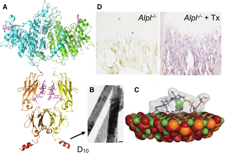
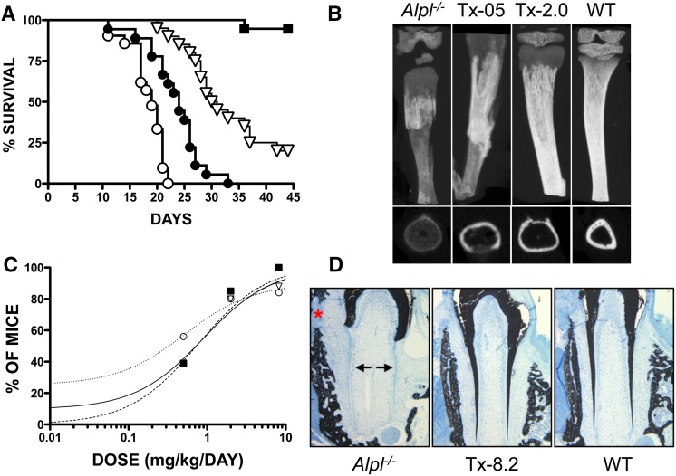
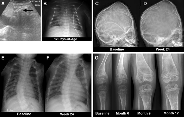
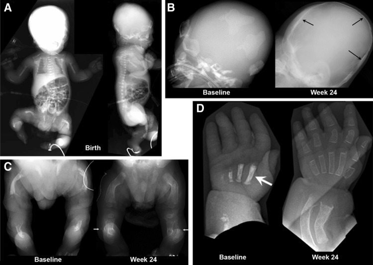
Hypophosphatasia (HPP) results from ALPL mutations leading to deficient activity of the tissue-non-specific alkaline phosphatase isozyme (TNAP) and thereby extracellular accumulation of inorganic pyrophosphate (PPi), a natural substrate of TNAP and potent inhibitor of mineralization. Thus, HPP features rickets or osteomalacia and hypomineralization of teeth. Enzyme replacement using mineral-targeted TNAP from birth prevented severe HPP in TNAP-knockout mice and was then shown to rescue and substantially treat infants and young children with life-threatening HPP. Clinical trials are revealing aspects of HPP pathophysiology not yet fully understood, such as craniosynostosis and muscle weakness when HPP is severe. New treatment approaches are under development to improve patient care.

Keywords: Calcification; Enzyme replacement; Osteomalacia; Rickets; Seizures.


Figures

Fig. 1
Fig. 1
Ribbon representation of the 3D structure of alkaline phosphatase (1EW2) [10]. The active site phosphate (PO4 3−) bound to Ser92 during catalysis is shown in green. The three active site metal ions, two Zn2+ (Zn1 and Zn2) and one Mg2+ (Mg), are shown in white as well as the structural Ca2+ ion (Ca). Also indicated are the flexible exposed sequence known as the “crown domain”; the N-terminal helix of one subunit that reaches close to the active site of the contralateral subunit; and the location where the glycosylphosphatidylinositol (GPI) anchor is attached to the C-terminus of the mature enzyme (Color figure online)
Fig. 2
Fig. 2
Radiographic images of HPP patients. a This middle-aged man who manifested signs and symptoms of hypophosphatasia during childhood shows some healing of a right femoral pseudofracture treated with intramedullary fixation. b Lateral and anteroposterior radiograph of the skull of this 14-week-old baby with infantile hypophosphatasia shows characteristic hypomineralization of areas of the calvarium that give the appearance of widened sutures (arrows). c At 23 weeks-of-age, this radiograph of the chest of a baby with infantile hypophosphatasia shows gracile, deformed, and fractured ribs that contribute to the respiratory compromise that is often lethal for such patients. d This radiograph of the right knee of a 9-year-old girl with childhood hypophosphatasia shows characteristic findings including areas of osteopenia and osteosclerosis especially in the metaphyseal regions, and marked physeal widening and irregularity of the head of the fibula
Fig. 3
Fig. 3
Phenotype of the Alpl −/− mouse model of infantile HPP. a X-ray images of a hind paw of a post-natal day 22 (P22) WT and Alpl −/− knockout (KO) mouse. b X-ray of the femur, tibia, and fibula of a P22 WT and KO mouse. Images from a and b were taken from [126] and reproduced with permission from BONE. c Radiographs of the ribs of a P16 WT and KO mouse. Images taken from [124] and reproduced with permission from the Journal of Bone and Mineral Research. d Micro-CT isosurface images of a P15 WT and KO mouse skull. Multiple cranial vault and facial bones are so severely hypomineralized in P15 that they do not appear on isosurface images calibrated to a bone threshold. The KO skull appears decreased in anterior–posterior length but increased in height when compared to the WT skull, and is more dome-shaped in overall appearance. Images taken from [100] and reproduced with permission from Bone. e Micro-CT demonstrates poorly mineralized molar roots and incisor in 10-day-old KO mice compared with WT, in addition to generalized reduction of bone mineralization in the mandible. Images taken from [47] and reproduced with permission from the Journal of Bone and Mineral Research. f Immunohistochemical localization of osteopontin (red, arrows and inset), as a marker for acellular cementum, shows a distinct line of acellular cementum in the WT sample, but an absence of a discrete immunostained layer in a 16-day-old KO mouse. PDL periodontal ligament, En-S enamel space after decalcification. Magnification bars equal 100 μm. Taken from [46] and reproduced with permission from the Journal of Dental Research. g Scanning electron microscopy (SEM) analysis of incisors (top) and molars (bottom) of WT and KO mice at 20 days-of-age. The SEM images show well-decussated enamel rods and inter-rod in the molar crowns and crown analogs of incisors of WT mice. Note that there is a lack of rod–inter-rod organization in the KO mice. Images taken from [48] and reproduced with permission from the Journal of Bone and Mineral Research (Color figure online)
Fig. 4
Fig. 4
Mechanisms of initiation and propagation of skeletal mineralization. Scanning electron microscopy images of matrix vesicles (MV) displaying mineral confined to the interior of the vesicles (a), with mineral breaking through the MV membranes (b) and with mineral propagating onto the collagenous scaffold (c). d Diagram detailing our current understanding of the biochemical bases for these three steps of MV-mediated initiation of biomineralization. MVs appear to initiate HA mineral deposition by accumulation of Pi generated intravesicularly by the action of PHOSPHO1 on phosphocholine and also via PiT-1-mediated incorporation of Pi generated extravesicularly by TNAP or NPP1. The extravesicular propagation of mineral onto the collagenous matrix is mainly controlled by the pyrophosphatase activity of TNAP that restricts the concentration of this potent mineralization inhibitor and establishes a PPi/Pi ratio conducive for controlled calcification. Additionally, osteopontin (OPN), another potent mineralization inhibitor that binds to HA mineral as soon as it is exposed to the extracellular fluid, also controls the degree of extracellular matrix mineralization. Both elevated levels of PPi and phosphorylated OPN are found in HPP mice. ECM extracellular matrix, HA hydroxyapatite, OPN osteopontin
Fig. 5
Fig. 5
Enzymes involved in the catabolism of ATP to form adenosine, and thus regulate the ATP/adenosine ratio important for purinergic signaling. ENPP1 ectonucleotide pyrophosphatase/phosphodiesterase, ENTPD/CD39 ectonucleotide triphosphate diphosphohydrolase, NT5E/CD73 ecto-5′-nucleotidase, PAP prostatic acid phosphatase, ADA adenosine deaminase, TNAP tissue-non-specific alkaline phosphatase
Fig. 6
Fig. 6
Structure and binding of asfotase alfa to bone mineral. a Three-dimensional modeling of ENB-0040. The model shows rigid ALP and Fc modules connected by a highly flexible linker. The terminal poly-Asp region is exposed on the opposite site of the ALP module. The whole structure is dimeric that conforms to the preferred oligomeric state of the ALP as well as the Fc region of the antibody. The three active site metal ions (two Zn2+ and one Mg2+) are marked with blue spheres. b Transmission electron micrograph showing the binding of sALP-FcD10 to synthetic hydroxyapatite crystals as revealed by immunogold labeling (inset is control incubation without sALP-FcD10 showing an absence of gold-particle labeling). Magnification bar equals 100 nm. c RosettaSurface-simulated model of D10 binding to a calcium-rich plane of the [100] crystallographic face of hydroxyapatite. d Histochemical staining for ALP activity in long bones of an ENB-0040–treated Alpl KO mouse compared with an age-matched untreated Alpl KO mouse. Images shown in b and c were taken from [46] (Color figure online)
Fig. 7
Fig. 7
Preclinical efficacy of asfotase alfa. a Percentage survival of Alpl −/− mice receiving either vehicle (white circle) or escalating doses of asfotase alfa, i.e., Tx-0.5 (black circle), Tx-2.0 (white down-pointing triangle), or Tx-8.2 (black square). b μCT images of tibiae of the 22-day-old Alpl −/− mice treated with vehicle, Tx-0.5, Tx-2.0, and untreated WT mice. The images clearly show improved tissue mineral density and callus formation at the site of fractures in the treated mice. Transaxial views at the bottom. c Percentage of Alpl −/− mice considered normal as a function of asfotase alfa dose for feet (black square), rib cage (white down-pointing triangle), and lower limbs (white circle). d Asfotase alfa treatment maintains complete mineralization of all molar dentin as well as the surrounding alveolar bone such that no mineralization differences are seen between the molar teeth and bone of the treated Alpl −/− mice (8.2 mg/kg/day) compared with WT mice. Images taken from [128] with permission from BONE
Fig. 8
Fig. 8
Efficacy of asfotase alfa treatment in a 36-month-old girl (at therapy baseline) with life-threatening HPP. a She has a short, bowed femur detected in utero by ultrasound. b At 12 days-of-age, her chest radiograph showed thin, osteopenic ribs with lytic areas and fractures. c, d Images of the skull before and after 24 weeks of ENB-0040 treatment. Note the severe pan-suture closure, including a marked increase in “digital” markings (“beaten-copper” appearance). e The ribs at baseline were osteopenic and had fracture deformities with thin cortices. f By week 24 of treatment, the ribs were wider and better mineralized with sharper cortical margins and less deformity. g Improvement of the rickets with therapy is apparent. Images taken for the online supplementary data in [9] and reproduced with permission from The New England Journal of Medicine
Fig. 9
Fig. 9
Efficacy of asfotase alfa in a 20-day-old (at therapy baseline) patient with life-threatening HPP. a This boy had shortened and bowed extremities and “fractures” detected by prenatal sonography at 17–18 weeks gestation. b Before asfotase alfa treatment, little or decreased mineral was present in the frontal, parietal, or occipital bones, skull base, facial bones, and sphenoid. c Before asfotase alfa, the femora were short, sclerotic, bowed, irregular, and lacked defined medullary cavities, cortices, and mineralized metaphyses and epiphyses. The fibulae were not calcified. After therapy, striking mineralization was evident. At week 24 of therapy, all areas showed striking remineralization. d The improvement in the left hand and wrist was remarkable. Images taken for the online supplementary data in [9] and reproduced with permission from The New England Journal of Medicine

Similar articles

Cited by

References

    1. Robison R (1923) The possible significance of hexosephosphoric esters in ossification. Biochem J 17:286 - PMC - PubMed
    1. Robison R (1932) The significance of phosphoric esters in metabolism. New York University Press, New York
    1. Meyer JL. Can biological calcification occur in the presence of pyrophosphate? Arch Biochem Biophys. 1984;231:1–8. doi: 10.1016/0003-9861(84)90356-4. - DOI - PubMed
    1. Weiss MJ, Cole DE, Ray K, Whyte MP, Lafferty MA, Mulivor RA, Harris H. A missense mutation in the human liver/bone/kidney alkaline phosphatase gene causing a lethal form of hypophosphatasia. Proc Natl Acad Sci USA. 1988;85:7666–76669. doi: 10.1073/pnas.85.20.7666. - DOI - PMC - PubMed
    1. Rathbun JC. Hypophosphatasia; a new developmental anomaly. Am J Dis Child. 1948;75:822–831. doi: 10.1001/archpedi.1948.02030020840003. - DOI - PubMed

Publication types