. 2015 Dec 15;85(24):2116-25.
doi: 10.1212/WNL.0000000000002220. Epub 2015 Nov 18.

Loss of TBK1 is a frequent cause of frontotemporal dementia in a Belgian cohort

Collaborators, Affiliations

Loss of TBK1 is a frequent cause of frontotemporal dementia in a Belgian cohort

Ilse Gijselinck et al. Neurology. .

Abstract

Objective: To assess the genetic contribution of TBK1, a gene implicated in amyotrophic lateral sclerosis (ALS), frontotemporal dementia (FTD), and FTD-ALS, in Belgian FTD and ALS patient cohorts containing a significant part of genetically unresolved patients.

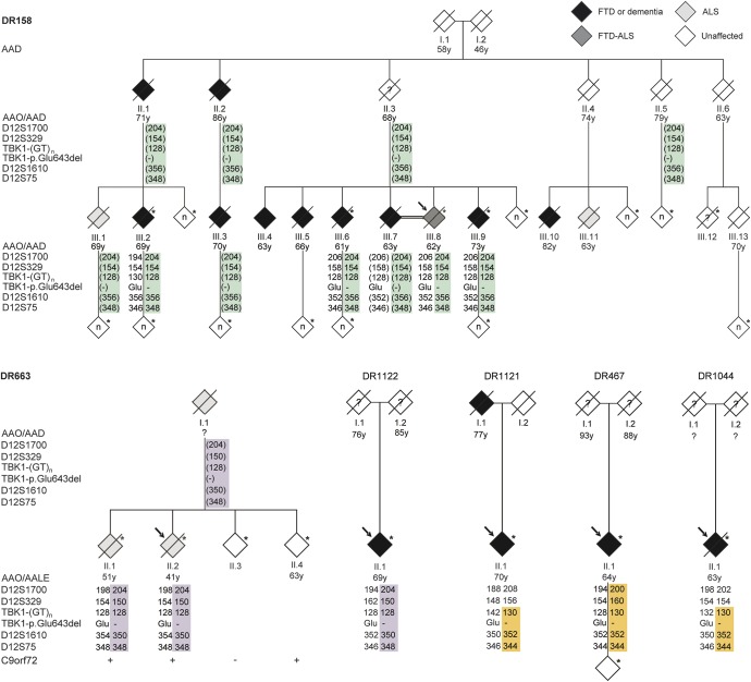
Methods: We sequenced TBK1 in a hospital-based cohort of 482 unrelated patients with FTD and FTD-ALS and 147 patients with ALS and an extended Belgian FTD-ALS family DR158. We followed up mutation carriers by segregation studies, transcript and protein expression analysis, and immunohistochemistry.

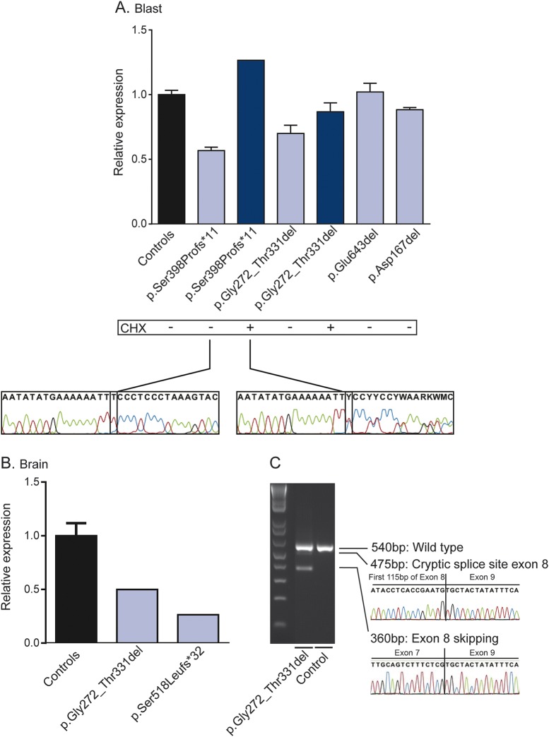
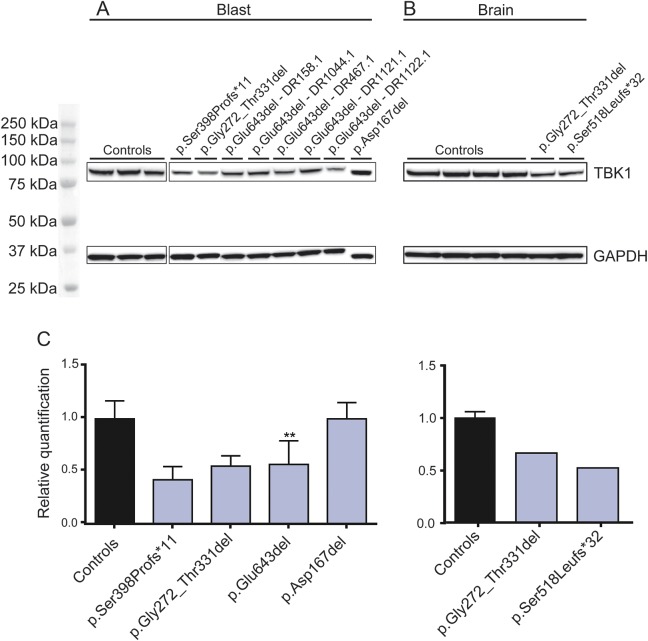
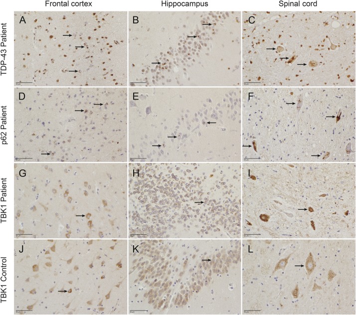
Results: We identified 11 patients carrying a loss-of-function (LOF) mutation resulting in an overall mutation frequency of 1.7% (11/629), 1.1% in patients with FTD (5/460), 3.4% in patients with ALS (5/147), and 4.5% in patients with FTD-ALS (1/22). We found 1 LOF mutation, p.Glu643del, in 6 unrelated patients segregating with disease in family DR158. Of 2 mutation carriers, brain and spinal cord was characterized by TDP-43-positive pathology. The LOF mutations including the p.Glu643del mutation led to loss of transcript or protein in blood and brain.

Conclusions: TBK1 LOF mutations are the third most frequent cause of clinical FTD in the Belgian clinically based patient cohort, after C9orf72 and GRN, and the second most common cause of clinical ALS after C9orf72. These findings reinforce that FTD and ALS belong to the same disease continuum.


Figures

Figure 1
Figure 1. Pedigrees show segregation and disease haplotype sharing in patients and families with the TBK1 mutation p.Glu643del
To protect the privacy of the participants, we masked the sex of each person, scrambled the pedigree of family DR158, and did not specify the number of at-risk individuals tested shown in the white diamonds. The index patient is indicated with an arrow. Filled symbols represent patients, with their age at onset (AAO) in years below their symbol. Age at death (AAD) is shown for individuals who died in old age without symptoms. In family DR663, 3 of the 4 children also carry a repeat expansion mutation in C9orf72, 2 affected (I-1 and II-2) and 1 asymptomatic (II-4), for whom the age at last examination (AALE) is given. An asterisk identifies the family members for whom genomic DNA was available. The Glu643del mutation is shown as “–.” The 5 polymorphic markers, including the TBK1-(GT)n repeat polymorphism located 400 kb upstream of TBK1, are indicated at the left and flank the TBK1 mutation p.Glu643del at both sites. Genotypes of additional polymorphic markers can be found in table e-3. The 3 different disease haplotypes are shown with colored bars: green haplotype of at least 7.5 Mb, purple haplotype of at least 8.7 Mb, and orange haplotype of at least 3.0 Mb. Haplotypes based on segregation data are indicated in regular font, those based on allele sharing are in italics, and those that were inferred are between brackets. ALS = amyotrophic lateral sclerosis; FTD = frontotemporal dementia.
Figure 2
Figure 2. Transcript analysis of TBK1 loss-of-function mutations
Quantitative PCR quantification of TBK1 transcripts from lymphoblast cells (A) and frontal cortex (B) of LOF mutation carriers (blue bars) compared to control persons without mutation (black bars) normalized to different housekeeping genes. Five unrelated p.Glu643del mutation carriers were included. Error bars represent the SD. Below the chart is indicated if cells were treated (+, dark blue bars) or not treated (−, light blue bars) with cycloheximide (CHX), a protein synthesis inhibitor. Sequence traces are obtained by real-time PCR from the lymphoblast cells of the p.Ser398Profs*11 mutation carrier treated or not treated with CHX using primers in exon 6 and exon 21. (C) Real-time PCR with primers in exon 6 and 10 of cDNA of brain of the p.Gly272_Thr331del mutation carriers is shown on agarose gel, with the respective sequence traces from the aberrant transcripts.
Figure 3
Figure 3. Protein analysis of TBK1 loss-of-function mutations
Western blot analysis of protein extracts from lymphoblast cells (A) and frontal cortex (B) of loss-of-function (LOF) mutation carriers compared to control persons without mutation. Of the p.Glu643del mutation, 5 unrelated index patients were included. The upper band represents TBK1 (84 kDa) and the lower band the housekeeping protein GAPDH (37 kDa). The graphs below (C) show the quantification in control samples (black) and patient samples (blue) of the TBK1 signal normalized to the signal of GAPDH. Error bars represent the SD; ** indicates a significant p value of <0.01 compared to controls.
Figure 4
Figure 4. TDP-43, p62, and TBK1 immunohistochemistry
Immunohistochemistry of frontal cortex (A, D, G) and hippocampus (B, E, H) of 1 patient with frontotemporal lobar degeneration with the p.Gly272_Thr331del mutation (DR189) and spinal cord (C, F, I) of 1 patient with amyotrophic lateral sclerosis (ALS) with the p.Ser518Leufs*32 mutation (DR1124) compared to a control individual without mutation (J–L). In frontotemporal dementia patient DR189, a mild neuronal loss of the frontal cortices and to a lesser extent of the temporal neocortex and the hippocampus was shown. A moderate amount of TDP-43 and p62 neuronal cytoplasmic inclusions (NCI) (arrows) and short dystrophic neurites, but no intraneuronal intranuclear inclusions, were found throughout all layers of the frontal (A, D) and temporal neocortices, in the dentate gyrus of the hippocampus (B, E), the parahippocampal transentorhinal cortex, the neostriatum, and the pallidum, but not in the cerebellum. In ALS patient DR1124, no explicit cortical abnormalities were present, but pathologic signs of lower and upper motor neuron disease were apparent. Severe demyelination of the pyramidal tract in mesencephalon, pons, medulla oblongata, and the entire spinal cord and severe neuronal loss in the hypoglossal nucleus, as well as in the ventral horns of the cervical and thoracic spinal cord, were observed. The TDP-43-positive (C) and p62-positive (F) NCI were mainly restricted to the hypoglossal nucleus and the ventral horn neurons of cervical and thoraco-lumbar spinal cord (arrows). Cytoplasmic TBK1 immunoreactivity (arrows) in cortical neurons of the frontal cortex (G, J), in the dentate gyrus of the hippocampus (H, K), and in the cervical ventral horn neurons (I, L) showed no differences between patients and controls. The TDP-43-positive inclusions were not stained with TBK1.

Similar articles

  • Clinical features of TBK1 carriers compared with C9orf72, GRN and non-mutation carriers in a Belgian cohort.
    Van Mossevelde S, van der Zee J, Gijselinck I, Engelborghs S, Sieben A, Van Langenhove T, De Bleecker J, Baets J, Vandenbulcke M, Van Laere K, Ceyssens S, Van den Broeck M, Peeters K, Mattheijssens M, Cras P, Vandenberghe R, De Jonghe P, Martin JJ, De Deyn PP, Cruts M, Van Broeckhoven C; Belgian Neurology consortium. Van Mossevelde S, et al. Brain. 2016 Feb;139(Pt 2):452-67. doi: 10.1093/brain/awv358. Epub 2015 Dec 15. Brain. 2016. PMID: 26674655 Free PMC article.
  • TBK1 mutation frequencies in French frontotemporal dementia and amyotrophic lateral sclerosis cohorts.
    Le Ber I, De Septenville A, Millecamps S, Camuzat A, Caroppo P, Couratier P, Blanc F, Lacomblez L, Sellal F, Fleury MC, Meininger V, Cazeneuve C, Clot F, Flabeau O, LeGuern E, Brice A; French Clinical and Genetic Research Network on FTLD/FTLD-ALS. Le Ber I, et al. Neurobiol Aging. 2015 Nov;36(11):3116.e5-3116.e8. doi: 10.1016/j.neurobiolaging.2015.08.009. Epub 2015 Aug 14. Neurobiol Aging. 2015. PMID: 26476236
  • Association between TBK1 mutations and risk of amyotrophic lateral sclerosis/frontotemporal dementia spectrum: a meta-analysis.
    Cui R, Tuo M, Li P, Zhou C. Cui R, et al. Neurol Sci. 2018 May;39(5):811-820. doi: 10.1007/s10072-018-3246-0. Epub 2018 Jan 18. Neurol Sci. 2018. PMID: 29349657 Review.
  • TBK1 Mutation Spectrum in an Extended European Patient Cohort with Frontotemporal Dementia and Amyotrophic Lateral Sclerosis.
    van der Zee J, Gijselinck I, Van Mossevelde S, Perrone F, Dillen L, Heeman B, Bäumer V, Engelborghs S, De Bleecker J, Baets J, Gelpi E, Rojas-García R, Clarimón J, Lleó A, Diehl-Schmid J, Alexopoulos P, Perneczky R, Synofzik M, Just J, Schöls L, Graff C, Thonberg H, Borroni B, Padovani A, Jordanova A, Sarafov S, Tournev I, de Mendonça A, Miltenberger-Miltényi G, Simões do Couto F, Ramirez A, Jessen F, Heneka MT, Gómez-Tortosa E, Danek A, Cras P, Vandenberghe R, De Jonghe P, De Deyn PP, Sleegers K, Cruts M, Van Broeckhoven C, Goeman J, Nuytten D, Smets K, Robberecht W, Damme PV, Bleecker J, Santens P, Dermaut B, Versijpt J, Michotte A, Ivanoiu A, Deryck O, Bergmans B, Delbeck J, Bruyland M, Willems C, Salmon E, Pastor P, Ortega-Cubero S, Benussi L, Ghidoni R, Binetti G, Hernández I, Boada M, Ruiz A, Sorbi S, Nacmias B, Bagnoli S, Sorbi S, Sanchez-Valle R, Llado A, Santana I, Rosário Almeida M, Frisoni GB, Maetzler W, Matej R, Fraidakis MJ, Kovacs GG, Fabrizi GM, Testi S. van der Zee J, et al. Hum Mutat. 2017 Mar;38(3):297-309. doi: 10.1002/humu.23161. Epub 2017 Jan 19. Hum Mutat. 2017. PMID: 28008748 Free PMC article.
  • Sex differences in the prevalence of genetic mutations in FTD and ALS: A meta-analysis.
    Curtis AF, Masellis M, Hsiung GR, Moineddin R, Zhang K, Au B, Millett G, Mackenzie I, Rogaeva E, Tierney MC. Curtis AF, et al. Neurology. 2017 Oct 10;89(15):1633-1642. doi: 10.1212/WNL.0000000000004494. Epub 2017 Sep 15. Neurology. 2017. PMID: 28916533 Free PMC article. Review.

Cited by

References

    1. Lomen-Hoerth C, Anderson T, Miller B. The overlap of amyotrophic lateral sclerosis and frontotemporal dementia. Neurology 2002;59:1077–1079. - PubMed
    1. Tsuji H, Arai T, Kametani F, et al. Molecular analysis and biochemical classification of TDP-43 proteinopathy. Brain 2012;135:3380–3391. - PubMed
    1. Dejesus-Hernandez M, Mackenzie IR, Boeve BF, et al. Expanded GGGGCC hexanucleotide repeat in noncoding region of C9ORF72 causes chromosome 9p-linked FTD and ALS. Neuron 2011;72:245–256. - PMC - PubMed
    1. Renton AE, Majounie E, Waite A, et al. A hexanucleotide repeat expansion in C9ORF72 is the cause of chromosome 9p21-linked ALS-FTD. Neuron 2011;72:257–268. - PMC - PubMed
    1. Gijselinck I, Van Langenhove T, van der Zee J, et al. A C9orf72 promoter repeat expansion in a Flanders-Belgian cohort with disorders of the frontotemporal lobar degeneration-amyotrophic lateral sclerosis spectrum: a gene identification study. Lancet Neurol 2012;11:54–65. - PubMed

Publication types

Substances