

doi: 10.1038/nmeth.3658.
Epub 2015 Nov 16.
Parmbsc1: a refined force field for DNA simulations
Affiliations
- PMID: 26569599
- PMCID: PMC4700514
- DOI: 10.1038/nmeth.3658
Item in Clipboard
Parmbsc1: a refined force field for DNA simulations
Nat Methods.
2016 Jan.
Abstract
We present parmbsc1, a force field for DNA atomistic simulation, which has been parameterized from high-level quantum mechanical data and tested for nearly 100 systems (representing a total simulation time of ∼ 140 μs) covering most of DNA structural space. Parmbsc1 provides high-quality results in diverse systems. Parameters and trajectories are available at http://mmb.irbbarcelona.org/ParmBSC1/.
Figures
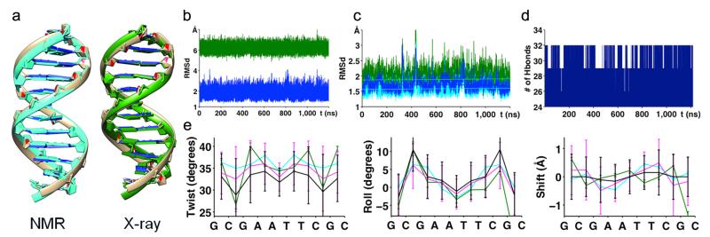
(a) Visual comparison of MD average structure (brown) and NMR structure (PDB id: 1NAJ) (light blue) and X-ray structure (PDB id: 1BNA) (green). (b) RMSd of 1.2 μs trajectory of DDD compared with BDNA (blue) and A-DNA (green) form (coming from Fiber). (c) RMSd compared to experimental structures (with (dark) and without (light) ending base-pairs): X-ray (green) and NMR (blue). Linear fits of all RMSd curves are plotted on top. (d) Evolution of total number of hydrogen bonds formed between base pairs in the whole duplex. (e) Helical rotational parameters (twist, roll, and tilt) comparison of average values per base-pair step (standard deviations are shown by error bars) coming from NMR (cyan), X-ray (dark green), 1 μs parmbsc0 trajectory (black) and 1.2 μs parmbsc1 trajectory (violet).
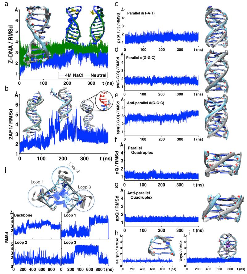
(a) Comparison of Z-DNA (PDB id: 1I0T) simulations in neutralized conditions (green) and in 4 M solution of Na+Cl− (blue). Structural comparisons at given time points are shown above the RMSd curves. (b) Simulation of anti-parallel H-DNA (PDB id: 2AF1) showing deviation of the structure over time (highlighted in red). RMSd of (c) parallel d(T-A•T)10, (d) parallel d(G-G•C)10, and (e) antiparallel d(G-G•C)10 triplexes. (f) Parallel (PDB id: 352D) and (g) anti-parallel (PDB id: 156D) quadruplex showing stable structures over time. (h) Structural stability of d(GCGAAGC) hairpin (PDB id: 1PQT) and (i) OxyQ quadruplex (PDB id: 1JRN) with ions, over time. (j) Human Telomeric Quadruplex (PDB id: 1KF1) with highlighted loops. RMSd of HTQ backbone, loop 1, loop 2 and loop 3 regions are shown below. In all panels, parmbsc1 (final, averaged or at a given trajectory point) structures (light blue; also green for Z-DNA) are overlapped over experimental structure (grey) for comparison. See Supplementary Table 1 for information on the PDB structures.
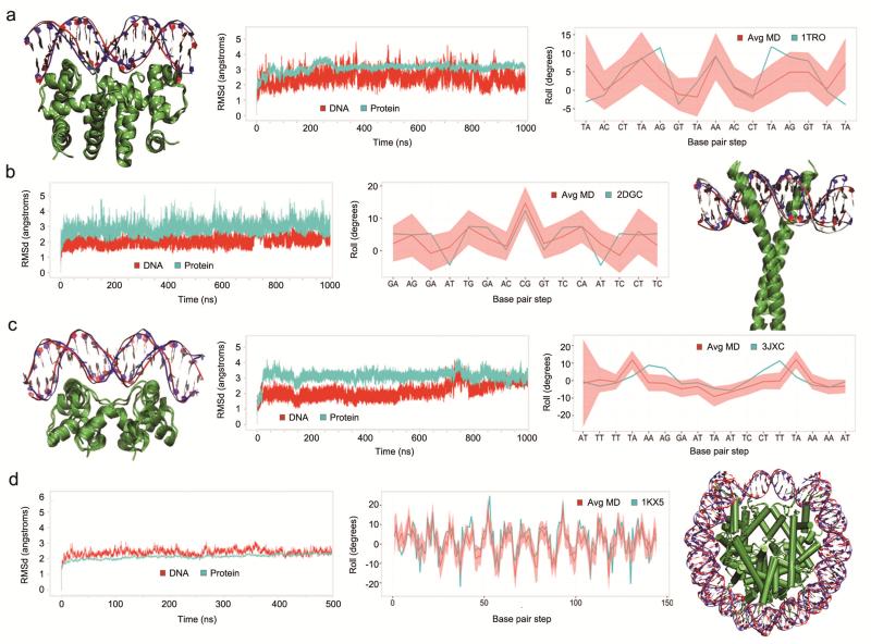
Structural details of microsecond trajectories of four complexes with PDB id: 1TRO (a), 2DGC (b), 3JXC (c) and 1KX5 (d) (500 ns trajectory). Each plot shows overlap of the MD starting (red) and final (blue) structures, time dependent mass-weighted root mean square deviation (RMSD in Å) of all DNA (red) and protein (cyan) heavy atoms, and comparison of the values of rotational helical parameter roll (in degrees) at each base pair step calculated from the X-ray crystal structure (cyan) and averaged along the MD simulation (red line with the standard deviation envelope in light red). For clarity, in the 1KX5 plot of the roll value, the base pair steps are defined by the number of the position along the DNA strand and not by the base pair step name.
Similar articles
-
BIGNASim: a NoSQL database structure and analysis portal for nucleic acids simulation data.Nucleic Acids Res. 2016 Jan 4;44(D1):D272-8. doi: 10.1093/nar/gkv1301. Epub 2015 Nov 26. Nucleic Acids Res. 2016. PMID: 26612862 Free PMC article.
-
Modeling DNA Flexibility: Comparison of Force Fields from Atomistic to Multiscale Levels.J Phys Chem B. 2020 Jan 9;124(1):38-49. doi: 10.1021/acs.jpcb.9b09106. Epub 2019 Dec 28. J Phys Chem B. 2020. PMID: 31805230
-
All-atom molecular dynamics simulations of spin labelled double and single-strand DNA for EPR studies.Phys Chem Chem Phys. 2018 May 16;20(19):13461-13472. doi: 10.1039/c7cp08625c. Phys Chem Chem Phys. 2018. PMID: 29725672
-
Molecular dynamic simulations of environment and sequence dependent DNA conformations: the development of the BMS nucleic acid force field and comparison with experimental results.J Biomol Struct Dyn. 1998 Dec;16(3):487-509. doi: 10.1080/07391102.1998.10508265. J Biomol Struct Dyn. 1998. PMID: 10052609 Review.
-
Mixed Quantum Mechanical/Molecular Mechanical Molecular Dynamics Simulations of Biological Systems in Ground and Electronically Excited States.Chem Rev. 2015 Jun 24;115(12):6217-63. doi: 10.1021/cr500628b. Epub 2015 Apr 16. Chem Rev. 2015. PMID: 25880693 Review. No abstract available.
Cited by
-
Site-specific investigation of DNA Holliday Junction dynamics and structure with 6-Methylisoxanthopterin, a fluorescent guanine analog.Trends Photochem Photobiol. 2023;22:85-102. Trends Photochem Photobiol. 2023. PMID: 39371247 Free PMC article.
-
A new paradigm for molecular dynamics databases: the COVID-19 database, the legacy of a titanic community effort.Nucleic Acids Res. 2024 Jan 5;52(D1):D393-D403. doi: 10.1093/nar/gkad991. Nucleic Acids Res. 2024. PMID: 37953362 Free PMC article.
-
Novel inhibitors of the main protease enzyme of SARS-CoV-2 identified via molecular dynamics simulation-guided in vitro assay.Bioorg Chem. 2021 Jun;111:104862. doi: 10.1016/j.bioorg.2021.104862. Epub 2021 Mar 29. Bioorg Chem. 2021. PMID: 33862474 Free PMC article.
-
OCT4 interprets and enhances nucleosome flexibility.Nucleic Acids Res. 2022 Oct 14;50(18):10311-10327. doi: 10.1093/nar/gkac755. Nucleic Acids Res. 2022. PMID: 36130732 Free PMC article.
-
Unfolding rates of 1:1 and 2:1 complex of CX-5461 and c-MYC promoter G-quadruplexes revealed by single-molecule force spectroscopy.Biophys Rep. 2024 Jun 30;10(3):180-189. doi: 10.52601/bpr.2024.240018. Biophys Rep. 2024. PMID: 39027314 Free PMC article.
References
Online Methods references
-
- Krishnan R, Binkley JS, Seeger R, Pople JA. J. Chem. Phys. 1980;72:650–654.
-
- Woon DE, Dunning TH., Jr J. Chem. Phys. 1993;98:1358–1371.
-
- Becke AD. J. Chem. Phys. 1993;98:5648–5652.
-
- Head–Gordon M, Pople JA, Frisch MJ. Chemical Physics Letters. 1988;153:503–506.
-
- Halkier A, Helgaker T, Jørgensen P, Klopper W, Olsen J. Chem. Phys. Lett. 1999;302:437–446.
Publication types
MeSH terms
Substances
Grants and funding
LinkOut - more resources
Full Text Sources
Other Literature Sources
