. 2015 Dec;33(12):1280-1286.
doi: 10.1038/nbt.3415. Epub 2015 Nov 9.

Soft, stretchable, fully implantable miniaturized optoelectronic systems for wireless optogenetics

Affiliations

Soft, stretchable, fully implantable miniaturized optoelectronic systems for wireless optogenetics

Sung Il Park et al. Nat Biotechnol. 2015 Dec.

Abstract

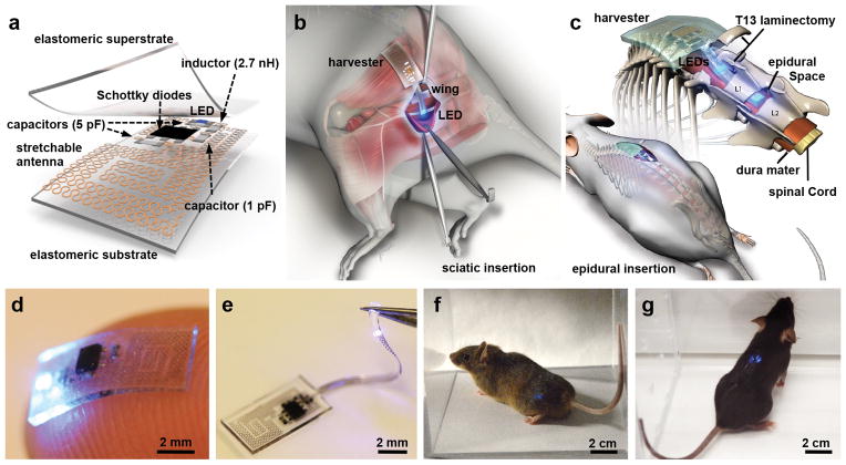
Optogenetics allows rapid, temporally specific control of neuronal activity by targeted expression and activation of light-sensitive proteins. Implementation typically requires remote light sources and fiber-optic delivery schemes that impose considerable physical constraints on natural behaviors. In this report we bypass these limitations using technologies that combine thin, mechanically soft neural interfaces with fully implantable, stretchable wireless radio power and control systems. The resulting devices achieve optogenetic modulation of the spinal cord and peripheral nervous system. This is demonstrated with two form factors; stretchable film appliqués that interface directly with peripheral nerves, and flexible filaments that insert into the narrow confines of the spinal epidural space. These soft, thin devices are minimally invasive, and histological tests suggest they can be used in chronic studies. We demonstrate the power of this technology by modulating peripheral and spinal pain circuitry, providing evidence for the potential widespread use of these devices in research and future clinical applications of optogenetics outside the brain.


Conflict of interest statement

Competing Financial Interests Statement

The authors declare no competing interests as defined by Nature Publishing Group, or other interests that might be perceived to influence the results and discussion reported in this paper.

Figures

Fig. 1
Fig. 1
Miniaturized, fully implantable, soft optoelectronic systems for wireless optogenetics. (a) Exploded view schematic illustration of the energy harvester component of the system, with an integrated LED to illustrate operation. (b) and (c) Illustration of the anatomy and location of the peripheral and epidural devices relative to the sciatic nerve and spinal cord, respectively. (d) Picture of an active device resting on the tip of the index finger. The device is 0.7 mm thick, 3.8 mm wide, and 6 mm long; its weight is 16 mg. (e) Picture of the epidural device, highlighting the soft, stretchable connection to an LED. The diameter of the epidural implant component is 380 μm, with cross sectional dimensions comparable to the epidural space. (f) and (g) Images of mice with wireless devices implanted near the sciatic nerve and the spinal cord, respectively.
Fig. 2
Fig. 2
Electrical and mechanical characteristics of the stretchable optoelectronics systems. (a) and (b) Strain distributions in the stretchable antenna (left), its scattering coefficient S11 (middle), and corresponding optical output power (right) for strain applied in the horizontal (28 %) and vertical directions (30 %) (blue solid), respectively, and for the undeformed (0 %) configuration (red dashed). (c) In vivo monitoring of the temperature of a mouse at the location of an implanted device using infrared imaging, during device operation. (d) Measurements of optical output power from devices operating in saline of immersion at temperatures of 37 °C, 60 °C and 90 °C as a function of time. (e) Measurements of optical output power from devices subjected to cyclical application of strain with magnitudes between 5 % and 20 %. (f) Schematic illustration of the TX system and an experimental assay with computed SAR distributions on a mouse mesh body. Multiple antennas lie in the XY plane, placed below the assay. (g) Simultaneous operation of devices implanted into multiple animals in the same cage (30 × 30 cm). (h) Image of a mouse while running on a wheel with a device interfaced to the sciatic nerve. (i) and (j) Long-exposure pictures of continuous activation of LED devices manually moved through the enclosure.
Fig. 3
Fig. 3
Electrophysiological and anatomical characterization of ChR2 expression in Advillin-ChR2 mice. (a) Schematic of the Ai32 locus and Advillin-Cre mouse locus where stop codons are inserted in all three reading frames and flanked by loxP sites upstream of the coding region for ChR2. The Advillin-Cre mouse locus shows Cre-recombinase driven by the sensory neuron specific Avil promoter. Cre recombinase expression results in recombination between loxP sites and excision of the stop codons, leading to expression of ChR2. Electrophysiological recordings from DRG neurons cultured from Advillin-ChR2 mice. For all traces, 470 nm illumination is delivered at 10 mW/mm2. (b) 1 second-long illumination induces inward currents (lower trace) in voltage clamp recordings, and in some cells produces sustained firing in current clamp recordings (upper trace). (c) Pulsed illumination at 20 Hz induces action potential firing with high fidelity (upper trace) resulting from the inward currents that are generated in voltage clamp (lower trace). Note that the first pulse produces larger amplitude inward currents relative to the 2nd and all subsequent light pulses, consistent with the rapid desensitization to a steady-state current seen with prolonged illumination (b, lower). (d) Immunohistochemical analysis of tissue from adult Advillin-ChR2 mice demonstrates that ChR2 is expressed along the peripheral neuraxis, including termination in lamina IIo and lamina IIi of the spinal cord dorsal horn as evidenced by overlap with CGRP (purple) and IB4 (red), respectively. (e) Staining of DRG demonstrates significant overlap of ChR2 expression with the neuronal marker βIII tubulin (purple) and IB4 (red) within the soma. Longitudinal (f) and cross sections (g) of sciatic nerve demonstrate robust staining along the plasma membrane of the axons of both myelinated (marked with NF200, purple) and unmyelinated neurons. We also note some expression of ChR2 in the circumferential non-excitable epineurial tissue. See Supplementary Fig. 13–14 for comparison to the ChR2 expression pattern seen in TrpV1-ChR2 and SNS-ChR2 mice which, as expected, are more restricted than in these Advillin-ChR2 mice. Scale bars = 100 μm for panels d, f, g, and 50 μm for panel e.
Fig. 4
Fig. 4
Wireless activation of ChR2 expressed in nociceptive pathways results in spontaneous pain behaviors and place aversion. (a) Representation of nociceptive pathways and illumination of nociceptive fibers with a sciatic LED stimulator. (b) Implantation of the sciatic LED stimulator has no effect on motor behavior vs. sham animals in the rotarod test (p = 0.894, n = 5 sham, n = 8 device). (c) Wireless activation of the sciatic LED stimulator causes increased nocifensive behaviors (flinching, hind paw licking, jumping) in Advillin-ChR2 mice but not in controls (17.5 vs. 1.2 flinches, p < 0.0001 vs. without illumination n = 3 per group). No other statistical comparisons reach significance. (d) Mice are placed in a modified Y-maze and one arm is targeted with the RF antenna to operate the LED device (LED ON) while the other is not (LED OFF). Time spent in the center area (dashed lines) is not scored (e) Heat maps from individual mice representing the time spent in each zone. Red indicates a higher amount of time, whereas blue areas indicate regions that animals occupied for less time. In animals implanted with the sciatic LED device, aversion to the LED-ON zone is observed in TrpV1-ChR2 and Advillin-ChR2 mice, but not in controls. (f) Quantification of time spent in each zone of the Y-maze. TrpV1-ChR2 (420.5 vs. 644.5 seconds; p = 0.011, n = 5) and Advillin-ChR2 (491.2 vs. 656 seconds; p = 0.001, n = 8) mice display aversion to the LED-ON zone vs. the LED-OFF zone. No difference is observed in control mice (547.0 vs. 512.1 seconds; p = 0.551, n = 10). (g) Representation of ascending nociceptive pathways and illumination of primary afferent terminals innervating the spinal cord with a wireless epidural implant. (h) Wireless activation of the epidural LED implant increased nocifensive behaviors in SNS-ChR2 mice (64.2 % vs. 0 % of time; p < 0.001, n = 3). (i) Heat maps representing the time spent in each zone of the Y-maze. Red indicates areas where the animals spend a higher proportion of their time. Aversion to the LED-ON zone is observed in SNS-ChR2 mice but not in controls. (j) Quantification of the time spent in each zone of the Y-maze. SNS-ChR2 mice display aversion to the LED-ON zone (73 vs. 251 seconds; p = 0.006, n = 3). No difference is observed in control mice (n = 3). Group data are presented as mean ± s.e.m. Statistical comparisons were made using two-tailed t-tests, except for panel B, which was a two-way ANOVA. * p < 0.05, ** p < 0.01.

Similar articles

Cited by

References

    1. Iyer SM, et al. Virally mediated optogenetic excitation and inhibition of pain in freely moving nontransgenic mice. Nature biotechnology. 2014 - PMC - PubMed
    1. Towne C, Montgomery KL, Iyer SM, Deisseroth K, Delp SL. Optogenetic control of targeted peripheral axons in freely moving animals. PloS one. 2013;8:e72691. - PMC - PubMed
    1. Daou I, et al. Remote optogenetic activation and sensitization of pain pathways in freely moving mice. The Journal of neuroscience : the official journal of the Society for Neuroscience. 2013;33:18631–18640. - PMC - PubMed
    1. Kozai TD, et al. Ultrasmall implantable composite microelectrodes with bioactive surfaces for chronic neural interfaces. Nature materials. 2012;11:1065–1073. - PMC - PubMed
    1. Sparta DR, et al. Construction of implantable optical fibers for long-term optogenetic manipulation of neural circuits. Nature protocols. 2012;7:12–23. - PMC - PubMed