. 2016 Jan 1;27(1):75-89.
doi: 10.1091/mbc.E15-05-0319. Epub 2015 Nov 4.

A complex of Rab13 with MICAL-L2 and α-actinin-4 is essential for insulin-dependent GLUT4 exocytosis

Affiliations

A complex of Rab13 with MICAL-L2 and α-actinin-4 is essential for insulin-dependent GLUT4 exocytosis

Yi Sun et al. Mol Biol Cell. .

Abstract

Insulin promotes glucose uptake into skeletal muscle through recruitment of glucose transporter 4 (GLUT4) to the plasma membrane. Rab GTPases are molecular switches mobilizing intracellular vesicles, and Rab13 is necessary for insulin-regulated GLUT4-vesicle exocytic translocation in muscle cells. We show that Rab13 engages the scaffold protein MICAL-L2 in this process. RNA interference-mediated knockdown of MICAL-L2 or truncated MICAL-L2 (MICAL-L2-CT) impaired insulin-stimulated GLUT4 translocation. Insulin increased Rab13 binding to MICAL-L2, assessed by pull down and colocalization under confocal fluorescence and structured illumination microscopies. Association was also visualized at the cell periphery using TIRF microscopy. Insulin further increased binding of MICAL-L2 to α-actinin-4 (ACTN4), a protein involved in GLUT4 translocation. Rab13, MICAL-L2, and ACTN4 formed an insulin-dependent complex assessed by pull down and confocal fluorescence imaging. Of note, GLUT4 associated with the complex in response to insulin, requiring the ACTN4-binding domain in MICAL-L2. This was demonstrated by pull down with distinct fragments of MICAL-L2 and confocal and structured illumination microscopies. Finally, expression of MICAL-L2-CT abrogated the insulin-dependent colocalization of Rab13 with ACTN4 or Rab13 with GLUT4. Our findings suggest that MICAL-L2 is an effector of insulin-activated Rab13, which links to GLUT4 through ACTN4, localizing GLUT4 vesicles at the muscle cell periphery to enable their fusion with the membrane.


Figures

FIGURE 1:
FIGURE 1:
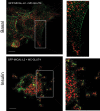
Insulin increases colocalization of Rab13 with MICAL-L2 in L6 myoblasts. L6 cells were cotransfected with GFP-MICAL-L2 and MC-Rab13 for 24 h, serum starved, stimulated with insulin or not, and then processed for spinning-disk confocal microscopy. Representative collapsed optical z-stack images of GFP-MICAL-L2 (green) and MC-Rab13 (red) (>25 cells/condition per experiment). Excerpts are magnifications of the outlined region of interest. (A) Basal, (B) insulin stimulated. (C) Pearson coefficient of colocalization of MC-Rab13 to GFP-MICAL-L2 (mean ± SE, **p < 0.01).
FIGURE 2:
FIGURE 2:
SIM reveals detailed colocalization of MICAL-L2 with Rab13 in response to insulin. L6 cells were cotransfected with GFP-MICAL-L2 and MC-Rab13 overnight and then replated onto coverslips for 24 h, serum starved, stimulated with insulin or not, and processed for SIM. Representative single confocal planes of GFP-MICAL-L2 (green) and MC-Rab13 (red) (>10 cells/condition). (A) Basal, (B) insulin stimulated. Magnified excerpts are also shown. Scale bars, 10 μm.
FIGURE 3:
FIGURE 3:
Insulin promotes colocalization of MICAL-L2 and Rab13 at the TIRF zone. L6 cells cotransfected with GFP-MICAL-L2 and MC-Rab13 were replated onto coverslips for 24 h, serum deprived, stimulated with insulin or not, and examined with an Olympus 1XR1 TIRF microscope. TIRF images of GFP-MICAL-L2 (green) and MC-Rab13 (red; >25 cells/condition). Scale bars, 10 μm. Excerpts show magnified regions. (A) Basal, (B) insulin stimulated. (C) Pearson coefficient of colocalization of MC-Rab13 to GFP-MICAL-L2 at the TIRF zone (mean ± SE, **p < 0.01).
FIGURE 4:
FIGURE 4:
Insulin-dependent GLUT4 translocation is prevented by silencing MICAL-L2 or expressing MICAL-L2-CT. (A) L6-GLUT4myc cells were transfected with GFP-coexpressing vectors containing shRNA interference to rat MICAL-L2 (sh-MICAL-L2) or unrelated shRNA. Cells were replated on coverslips for 48 h, serum starved, and stimulated with insulin (15 min) or not. Surface GLUT4myc was detected with anti-myc and Alexa 555–secondary antibodies (red), imaged by confocal microscopy, and quantified as in Materials and Methods. Fold changes relative to shRNA control from three independent experiments, >25 cells/experiment (mean ± SE, ***p < 0.001 for n = 3). (B) L6-GLUT4myc cells transfected with GFP-MICAL-L2-CT or GFP were stimulated with insulin, and surface GLUT4myc was detected as in A. Results from four experiments, >25 cells/experiment (mean ± SE, **p < 0.01).
FIGURE 5:
FIGURE 5:
Insulin promotes association of Rab13 with ACTN4. (A) L6 GLUT4myc-IR myoblasts were transfected with GFP-Rab13 for 48 h, cross-linked with DSP (1.5 mM, 30 min), serum deprived, and stimulated with insulin (15 min) or not. Lysates were immunoprecipitated with anti-ACTN4 or immunoglobulin G and immunoblotted with mouse anti-GFP, along with lysate aliquots. Phospho-Akt S473 confirmed insulin effectiveness. Graphs show the results of four independent experiments (mean ± SE, **p < 0.01). (B) L6 myoblasts transfected with GFP-Rab13 and stimulated with insulin or not were fixed and permeabilized, and endogenous ACTN4 was labeled with anti-ACTN4 and Alexa 546–secondary antibodies. Collapsed optical z-stack images of basal and insulin-stimulated cells, representative of three experiments. (C) L6 myoblasts grown on coverslips were transfected with GFP-MICAL-L2 and processed as in B. Representative collapsed optical z-stack images of GFP-MICAL-L2 (green) and ACTN4 (red) in basal and insulin-stimulated cells from three experiments. Scale bars, 10 μm. The Pearson correlation coefficients for the colocalization of Rab13 with ACTN4 at the cell periphery are indicated below the images of basal and insulin conditions.
FIGURE 6:
FIGURE 6:
GST-MICAL-L2-ACT binds Rab13, ACTN4, and GLUT4. (A) Lysates from L6 GLUT4myc cells expressing GFP-Rab13 or GFP, stimulated with insulin or not, were pulled down with GST-MICAL-L2-ACT (containing Rab- and ACTN4-binding domains) and immunoblotted with anti-GFP and anti-ACTN4. Phospho-Akt illustrates insulin effectiveness. Pull down by GST-MICAL-L2-ACT of (B) GFP-Rab13, (C) ACTN4, and (D) GLUT4myc (n = 6, mean ± SE, *p < 0.05; ns, not significant).
FIGURE 7:
FIGURE 7:
Insulin-elicited three-way colocalization of MICAL-L2, ACTN4, Rab13, and GLUT4 at cellular ruffles. (A) L6 cells cotransfected with GFP-MICAL-L2 and MC-Rab13 were stimulated with insulin or not and analyzed by spinning-disk confocal fluorescence microscopy. Collapsed optical z-stack images of GFP-MICAL-L2 (green), MC-Rab13 (red), and ACTN4 (blue). (B) L6 cells cotransfected with GFP-Rab13 and MC-GLUT4 and labeled for ACTN4. Collapsed optical z-stack images of GFP-Rab13 (green), MC-GLUT4myc (red), and ACTN4 (blue). All results are representative of three experiments (>25 cells/condition). Scale bars, 10 μm.
FIGURE 8:
FIGURE 8:
SIM reveals the insulin-induced colocalization of Rab13 with GLUT4 at the cell periphery. L6 cells cotransfected with GFP-Rab13 and MC-GLUT4myc were stimulated with insulin or not, fixed, and processed for SIM. Images of single focal planes of GFP-Rab13 (green) and MC-GLUT4myc (red). Scale bars, 10 μm. Excerpts show magnified regions of interest. In those peripheral regions, the Pearson coefficients of colocalization in the experiment shown were 0.05 (basal) and 0.39 (insulin).
FIGURE 9:
FIGURE 9:
SIM reveals the insulin-induced colocalization of MICAL-L2 with GLUT4 at the cell periphery. L6 cells cotransfected with GFP-MICAL-L2 and MC-GLUT4myc were stimulated or not with insulin as indicated, fixed, and processed for SIM. Images of single focal planes of GFP-MICAL-L2 (green) and MC-GLUT4myc (red). Scale bar, 10 μm. Excerpts show magnified regions of interest. The Pearson coefficients of MICAL-L2 and GLUT4 colocalization at the cell periphery in the experiment shown were 0.10 (basal) and 0.29 (insulin).
FIGURE 10:
FIGURE 10:
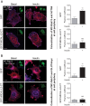
The insulin-stimulated colocalization of MC-Rab13 with both ACTN4 and GLUT4myc at cell periphery is reduced in cells expressing MICAL-L2-CT. (A) L6-GLUT4myc cells cotransfected with either GFP vector and MC-Rab13 or GFP-MICAL-L2-CT and MC-Rab13 were stimulated with insulin or not, fixed, labeled for ACTN4, and analyzed by spinning-disk confocal fluorescence microscopy. Top, collapsed optical z-stack images of GFP (green, inset), MC-Rab13 (red), and ACTN4 (blue). Bottom, images of GFP-MICAL-L2-CT (green, inset), MC-Rab13 (red), and ACTN4 (blue). Graphs show Pearson coefficients of colocalization of MC-Rab13 and ACTN4 at the cell periphery (mean ± SE, **p < 0.01). (B) L6-GLUT4myc cells treated as described were labeled for GLUT4myc and otherwise analyzed as before. Top, images of GFP (green, inset), MC-Rab13 (red), and GLUT4myc (blue). Bottom, images of GFP-MICAL-L2-CT (green, inset), MC-Rab13 (red), and GLUT4myc (blue). Graphs show Pearson coefficient of colocalization of MC-Rab13 and GLUT4myc at the cell periphery (mean ± SE, **p < 0.01, n = 3). Results are representative of three independent experiments (>25 cells/condition per experiment). Scale bars, 10 μm.

Similar articles

Cited by

References

    1. Bogan JS. Regulation of glucose transporter translocation in health and diabetes. Annu Rev Biochem. 2012;81:507–532. - PubMed
    1. Boguslavsky S, Chiu T, Foley KP, Osorio-Fuentealba C, Antonescu CN, Bayer KU, Bilan PJ, Klip A. Myo1c binding to submembrane actin mediates insulin-induced tethering of GLUT4 vesicles. Mol Biol Cell. 2012;23:4065–4078. - PMC - PubMed
    1. Chen Y, Wang Y, Zhang J, Deng Y, Jiang L, Song E, Wu XS, Hammer JA, Xu T, Lippincott-Schwartz J. Rab10 and myosin-Va mediate insulin-stimulated GLUT4 storage vesicle translocation in adipocytes. J Cell Biol. 2012;198:545–560. - PMC - PubMed
    1. Chiu TT, Jensen TE, Sylow L, Richter EA, Klip A. Rac1 signalling towards GLUT4/glucose uptake in skeletal muscle. Cell Signal. 2011;23:1546–1554. - PubMed
    1. Chiu TT, Patel N, Shaw AE, Bamburg JR, Klip A. Arp2/3- and cofilin-coordinated actin dynamics is required for insulin-mediated GLUT4 translocation to the surface of muscle cells. Mol Biol Cell. 2010;21:3529–3539. - PMC - PubMed

Publication types

LinkOut - more resources