. 2015 Dec;5(12):1282-95.
doi: 10.1158/2159-8290.CD-15-1020. Epub 2015 Oct 29.

Convergence of Acquired Mutations and Alternative Splicing of CD19 Enables Resistance to CART-19 Immunotherapy

Affiliations

Convergence of Acquired Mutations and Alternative Splicing of CD19 Enables Resistance to CART-19 Immunotherapy

Elena Sotillo et al. Cancer Discov. 2015 Dec.

Abstract

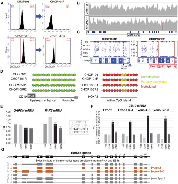
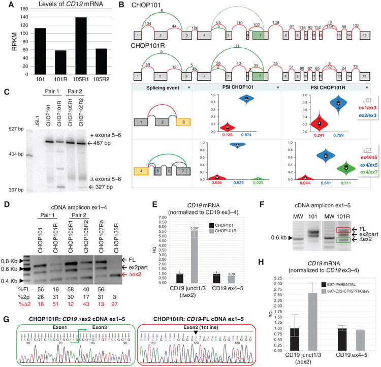
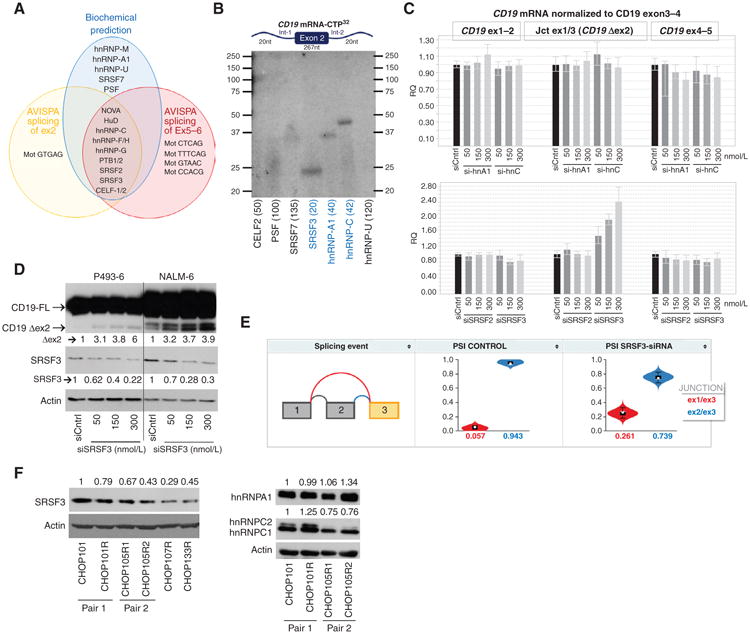
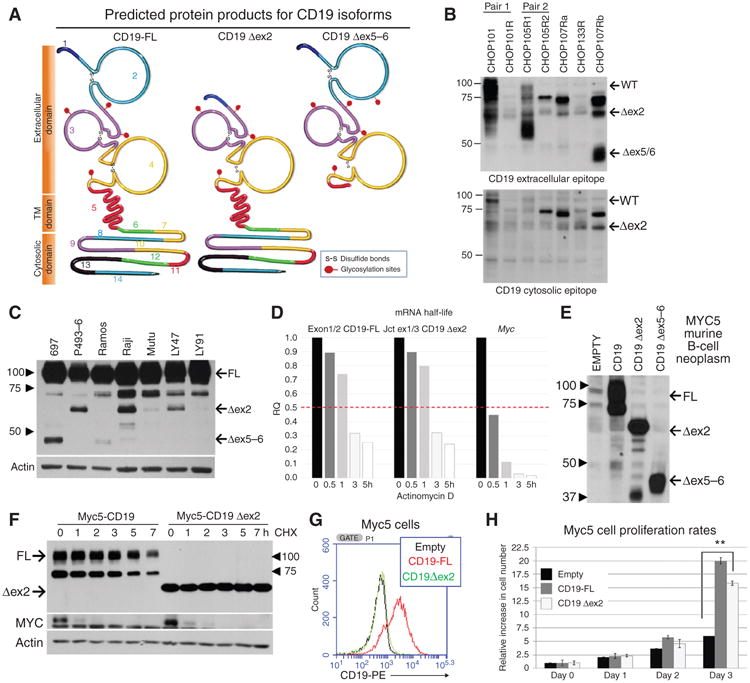
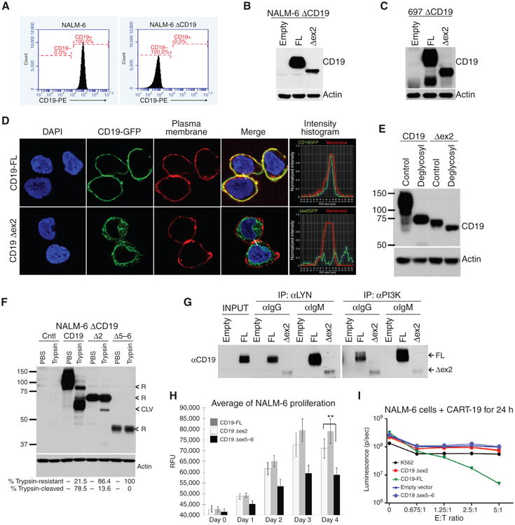
The CD19 antigen, expressed on most B-cell acute lymphoblastic leukemias (B-ALL), can be targeted with chimeric antigen receptor-armed T cells (CART-19), but relapses with epitope loss occur in 10% to 20% of pediatric responders. We detected hemizygous deletions spanning the CD19 locus and de novo frameshift and missense mutations in exon 2 of CD19 in some relapse samples. However, we also discovered alternatively spliced CD19 mRNA species, including one lacking exon 2. Pull-down/siRNA experiments identified SRSF3 as a splicing factor involved in exon 2 retention, and its levels were lower in relapsed B-ALL. Using genome editing, we demonstrated that exon 2 skipping bypasses exon 2 mutations in B-ALL cells and allows expression of the N-terminally truncated CD19 variant, which fails to trigger killing by CART-19 but partly rescues defects associated with CD19 loss. Thus, this mechanism of resistance is based on a combination of deleterious mutations and ensuing selection for alternatively spliced RNA isoforms.

Significance: CART-19 yield 70% response rates in patients with B-ALL, but also produce escape variants. We discovered that the underlying mechanism is the selection for preexisting alternatively spliced CD19 isoforms with the compromised CART-19 epitope. This mechanism suggests a possibility of targeting alternative CD19 ectodomains, which could improve survival of patients with B-cell neoplasms.


Conflict of interest statement

Disclosure of Potential Conflicts of Interest: S.L. Maude is a consultant/advisory board member for Novartis Pharmaceuticals. S. Gill and S.F. Lacey report receiving commercial research grants from Novartis. J.J. Melenhorst reports receiving commercial research support from Novartis. S.A. Grupp reports receiving a commercial research grant and other commercial research support from Novartis; has ownership interest (including patents) in University of Pennsylvania; and is a consultant/advisory board member for Novartis. No potential conflicts of interest were disclosed by the other authors.

Figures

Figure 1
Figure 1
Retention of CD19 genetic material in relapsed leukemias. A, flow cytometric profiles of CD19 surface expression in paired B-ALL samples included in subsequent analyses. B, CD19 gene coverage obtained through whole-genome sequencing of CHOP101 and CHOP101R samples. C, SNP array analysis of Chr16p performed on DNA from 105R1 and 105R2 showing the large hemizygous deletion (red brackets) found in the CHOP105R2 sample. D, direct bisulfite sequencing of the enhancer and promoter regions of CD19 (downstream of the PAX5-binding site) in the paired samples. A CpG island within the HOXA3 locus was analyzed as a positive control. E, qRT-PCR analysis of PAX5 mRNA expression in xenografted patient samples. ACTB and GAPDH were used as reference genes. F, qRT-PCR analysis of different regions of the CD19 mature mRNA in all qPCR panels; graphs show relative quantifications of expression ± 1 SD. G, Genome browser SIB track predicted isoforms of CD19 mRNA, including those skipping exon 2 (Δex2) and exons 5 and 6 (Δex5–6), and the partial deletion of exon 2 (ex2part) that shifts the reading frame.
Figure 2
Figure 2
Alternatively spliced CD19 mRNA species in post–CART-19 relapses. A, levels of CD19 mRNA in xenografts of paired pre– and post–CART-19 B-ALL samples. Values represent reads per kilobase per million mapped reads (RPKM). B, top, splicegraphs of CD19 mRNA species from primary (CHOP101) and relapsed (CHOP101R) tumors. Shown above arcs are raw numbers of RNA-seq reads spanning annotated (red) and novel (green) splice junctions. Bottom, violin plots showing the distribution of PSI values (y-axis) quantified by MAJIQ for primary (101, left) and relapsed (101R, right) samples. Colors correspond to the junctions displayed in the thumbnail (far left) with the expected PSI value for each junction displayed on the x-axis. C, analysis by low-cycle semiquantitative RT-PCR of the region spanning exons 4 to 8. cDNAs were obtained from paired primary and relapsed samples. CD19-negative JSL1 T-cell line was used as negative control. Arrows indicate inclusion of exons 5 to 6 (+) and the Δex5–6 isoform. D, semiquantitative RT-PCR of cDNA from xenografted samples corresponding to exons 1 to 4 of CD19. Arrows indicate full-length (FL), partial deletion (ex2part), and the Δex2 isoform. Quantification of relative isoform abundance in each sample (numbers below) was performed using Image J software (NIH). E, qRT-PCR analysis of CD19 splicing variants using oligos that span conserved and alternative exon/exon junctions. Graph shows relative quantification of expression ± 1 SD. Oligos expanding exon3/4 of CD19 were used as reference. F, semiquantitative RT-PCR of cDNA from xenografted samples corresponding to exons 1 to 5 of CD19. G, direct Sanger sequencing performed from gel-purified bands color-coded in panel F. Exon1/3 junction (left) and single-nucleotide insertion in exon2 (right) are indicated. H, qRT-PCR analysis of CD19 splicing variants was performed on cDNA from 697 cells using oligos as in E, CD19 exon2 was targeted and mutated using CRISPR/Cas9.
Figure 3
Figure 3
The splicing factor SRSF3 binds to and promotes inclusion of exon2 of CD19. A, Venn diagrams of splicing factors predicted by CD19 mRNA pull-down (biochemical predictions) or by the sequence-based algorithm AVISPA to bind to CD19 exon1–exon3 (splicing of exon2) or exon4–exon7 (splicing of exons 5–6) CD19 mRNA. B, RNA immunoprecipitation with antibodies against indicated proteins for detection of splicing factors that bind to mRNA CD19 exon2 and its flanking introns (not drawn to scale). Numbers in parentheses indicated expected molecular weights for each protein C, qRT-PCR analysis of CD19 Δex2 splicing variant in RNA from P493–6 transfected with increasing concentrations of si-hnRNPA1 or si-hRNPC (top graph) and siSRSF2 or siSRSF3 (bottom graph). D, immunoblotting for CD19 and SRSF3 in protein lysates from indicated cell lines transfected with increasing concentrations of siSRSF3 for 24 hours. Arrows indicate full-length (FL) and exon 2 skipping (Δex2) CD19 variants. Quantification of SRSF3 and Δex2 abundance relative to siRNA controls is shown. E, violin plots showing the distribution of PSI values (y-axis) quantified by MAJIQ for control (left) and SRSF3 knockdown (right) GM19238 B cells. Colors correspond to the junctions displayed in the thumbnail (far left) with the expected PSI value for each junction displayed on the x-axis. F, immunoblotting of SRSF3 (top) and hnRNPA1 and hnRNPC1/C2 (right) in xenografted tumor samples. Quantification of relative SRSF3, hnRNPA1, and hnRNPC protein abundance (numbers on left) was performed using Image J software (NIH).
Figure 4
Figure 4
Truncated protein isoforms of CD19 provide proliferative advantage. A, CD19 proteins encoded by the full-length (FL) and the Δex2 and Δex5–6 isoforms of CD19 mRNA. The epitope recognized by CART-19 is encoded by a sequence contained within exons 1–4. The transmembrane domain is encoded by exons 5 and 6. B, immunoblotting for CD19 in protein lysates from xenografted tumor samples using antibodies recognizing the extracellular domain (clone 3F5 from Origene; top) or the cytosolic domain (Santa Cruz Biotechnologies; sc-69735; bottom) C, immunoblotting for CD19 in protein lysates from a panel of cell lines representing human B-cell malignancies. Arrows indicate full-length and the Δex2 isoforms. The antibody used (Santa Cruz Biotechnologies; sc-69735) recognizes the cytosolic domains. D, qRT-PCR analysis of CD19 mRNA splicing variants in NALM-6 that were treated with actinomycin D for indicated periods of time. MYC mRNA was used as internal control for effective inhibition of transcription. E, immunoblotting for CD19 in lysates from CD19-negative Myc5 murine B-lymphoid cells transduced with CD19 retroviral constructs. Arrows indicate full-length, Δex2, and Δex5–6 isoforms. F, immunoblotting analysis of CD19 protein stability in cells from E. Cultures were treated with cycloheximide for indicated periods of time. Labile MYC protein was used as control for effective inhibition of protein synthesis. G, flow cytometry performed on CD19-negative murine Myc5 cells infected with empty (black), full-length CD19 (red), or CD19 Δex2 (green) expressing retrovirus. H, growth rates of first three cultures from D. Average fold increase in cell numbers from triplicate plates is shown. Statistical significance per Student t test, with *, P ≤ 0.05 and **, P < 0.01.
Figure 5
Figure 5
Truncated protein isoforms of CD19 provide proliferative advantage while evading CART-19. A, flow cytometry analysis of CD19 expression on the surface of parental and CD19-negative NALM-6 cells. B, immunoblotting for CD19 in lysates from CD19-negative NALM-6 cells transduced with retroviral constructs from Fig 4D. C, immunoblotting of CD19 in protein lysates from CD19-negative 697 cells with reconstituted expression of full length of CD19 Δex2. D, confocal microscopy of 697 ΔCD19 cells expressing CD19-GFP and CD19 Δex2–GFP fusion proteins. Plasma membranes (red) and DNA (blue) were stained for colocalization studies. Histograms represent the intensity of the CD19-GFP (green line) and membrane (red line) along the cell-to-cell junction highlighted (white line) in the “merge” picture. E, immunoblotting detection of the shift in CD19 protein size in lysates from CD19-negative 697 cells transduced with full length of Δex2 retroviral constructs and treated with a mix of glycosylases. F, immunoblotting for CD19 in protein lysates from NALM-6 ΔCD19 cells with reconstituted expression of full-length, Δex2, or Δex5–6 CD19 variants that were incubated with trypsin. “<R” indicates bands that correspond to CD19 resistant to trypsin (intracellular), “<CLV” indicates CD19 cleaved by trypsin (plasma membrane). Quantification of CD19 resistant or sensitive to trypsin is shown. G, immunoblotting of CD19 present in complexes with PI3K or LYN. These complexes were first coimmunoprecipitated from NALM-6 ΔCD19 cells transduced with the indicated CD19 retroviral constructs. Prior to the experiment, cells were stimulated with α-IgM or control IgG for 10 minutes. H, growth rates of NALM-6 ΔCD19 with reconstituted expression of CD19 as in D. Average fold increase in cell numbers from triplicate plates is shown. Statistical significance per Student t test, with *, P ≤ 0.05 and **, P < 0.01. I, NALM-6 ΔCD19-luciferase+ cells were infected with CD19 retroviral constructs, then incubated with CART-19 cells at indicated ratios of effector T cells (E) to target NALM-6 cells (T), and cell death was assayed by measurement of luminescence. Erythroleukemic K562 cells were used as a negative control.

Comment in

Similar articles

Cited by

References

    1. Roberts KG, Mullighan CG. Genomics in acute lymphoblastic leukaemia: insights and treatment implications. Nat Rev Clin Oncol. 2015;12:344–57. - PubMed
    1. Porter DL, Levine BL, Kalos M, Bagg A, June CH. Chimeric antigen receptor-modified T cells in chronic lymphoid leukemia. N Engl J Med. 2011;365:725–33. - PMC - PubMed
    1. Kalos M, Levine BL, Porter DL, Katz S, Grupp SA, Bagg A, et al. T cells with chimeric antigen receptors have potent antitumor effects and can establish memory in patients with advanced leukemia. Sci Transl Med. 2011;3:95ra73. - PMC - PubMed
    1. Maude SL, Frey N, Shaw PA, Aplenc R, Barrett DM, Bunin NJ, et al. Chimeric antigen receptor T cells for sustained remissions in leukemia. N Engl J Med. 2014;371:1507–17. - PMC - PubMed
    1. Topp MS, Gokbuget N, Zugmaier G, Klappers P, Stelljes M, Neumann S, et al. Phase II trial of the anti-CD19 bispecific T cell-engager blinatumomab shows hematologic and molecular remissions in patients with relapsed or refractory B-precursor acute lymphoblastic leukemia. J Clin Oncol. 2014;32:4134–40. - PubMed

Publication types

MeSH terms