. 2015 Dec 4;290(49):29389-401.
doi: 10.1074/jbc.M115.695171. Epub 2015 Oct 26.

Mapping the Binding Site of the Inhibitor Tariquidar That Stabilizes the First Transmembrane Domain of P-glycoprotein

Affiliations

Mapping the Binding Site of the Inhibitor Tariquidar That Stabilizes the First Transmembrane Domain of P-glycoprotein

Tip W Loo et al. J Biol Chem. .

Abstract

ABC (ATP-binding cassette) transporters are clinically important because drug pumps like P-glycoprotein (P-gp, ABCB1) confer multidrug resistance and mutant ABC proteins are responsible for many protein-folding diseases such as cystic fibrosis. Identification of the tariquidar-binding site has been the subject of intensive molecular modeling studies because it is the most potent inhibitor and corrector of P-gp. Tariquidar is a unique P-gp inhibitor because it locks the pump in a conformation that blocks drug efflux but activates ATPase activity. In silico docking studies have identified several potential tariquidar-binding sites. Here, we show through cross-linking studies that tariquidar most likely binds to sites within the transmembrane (TM) segments located in one wing or at the interface between the two wings (12 TM segments form 2 divergent wings). We then introduced arginine residues at all positions in the 12 TM segments (223 mutants) of P-gp. The rationale was that a charged residue in the drug-binding pocket would disrupt hydrophobic interaction with tariquidar and inhibit its ability to rescue processing mutants or stimulate ATPase activity. Arginines introduced at 30 positions significantly inhibited tariquidar rescue of a processing mutant and activation of ATPase activity. The results suggest that tariquidar binds to a site within the drug-binding pocket at the interface between the TM segments of both structural wings. Tariquidar differed from other drug substrates, however, as it stabilized the first TM domain. Stabilization of the first TM domain appears to be a key mechanism for high efficiency rescue of ABC processing mutants that cause disease.

Keywords: ABC transporter; membrane enzyme; membrane protein; protein cross-linking; protein folding.


Figures

FIGURE 1.
FIGURE 1.
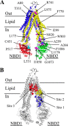
The three predicted tariquidar binding sites. A, predicted structure of human P-gp in an open conformation (NBDs apart and drug-binding pocket closed at the extracellular surface) based on the crystal structure of mouse P-gp (16). The model was viewed using the PyMol system (42). TMD1 is shown in blue and TMD2 is shown in yellow. The branched lines between TM segments 1 and 2 represent glycosylated sites. The locations where cross-linkable cysteines were introduced into the TM segments (A80C(TM1)/R741C(TM7), I299C(TM5)/F770C(TM8), and T333C(TM6/L975C(TM12)), ICL2/ICL3 (L175C(ICL1)/N820C(ICL3), A259C(ICL2)/W803C(ICL3), and A266C(ICL2/F1086C(NBD2)) or the interface between NBD1 and NBD2 (C431(NBD1)/L1176C(NBD2), L531C(NBD1)/C1074(NBD2), and P517C(NBD1)/I1050C(NBD2)) are indicated. B, the colored spheres represent the residues in predicted (9) tariquidar-binding sites 1 (blue), 2 (yellow), and 3 (red).
FIGURE 2.
FIGURE 2.
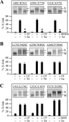
Tariquidar inhibits cross-linking between cysteines in the TM segments. Mutants with cysteines in the TM segments (A), ICLs (B), or NBDs (C) were treated in the absence (−) or presence (+) of copper phenanthroline (CP), BMOE or M4M (BMTS) cross-linker in the absence (−) or presence (+) of tariquidar (Tar). Samples were subjected to immunoblot analysis. The locations of cross-linked (X) and mature (M) P-gps are indicated (insets). The amount of cross-linked P-gp (% X-link) relative to total P-gp was determined. Each value is the mean ± S.D. (n = 3). An asterisk indicates significant difference (p < 0.001) relative to that cross-linked in the absence of tariquidar.
FIGURE 3.
FIGURE 3.
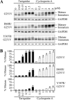
Arginine residues inhibit rescue of a G251V processing mutant with tariquidar and cyclosporine A. A, whole cell SDS extracts of cells expressing P-gp processing mutant G251V, G251V/I868R, or G251V/Y307R in the presence of various concentrations of tariquidar or cyclosporine A were subjected to immunoblot analysis. The positions of mature and immature P-gps are indicated. B, the level of mature P-gp relative to total was determined. Each value is the mean ± S.D. (n = 3–5). An asterisk indicates significant difference (p < 0.001) relative to samples incubated in the absence of drugs.
FIGURE 4.
FIGURE 4.
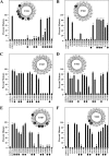
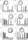
Rescue of G251V arginine mutants in TMs 1–6 with tariquidar. Whole cell SDS extracts of cells expressing the G251V mutants containing arginines at various positions in predicted TM1 (A), TM2 (B), TM3 (C), TM4 (D), TM5 (E), or TM6 (F) in the presence of 0.5 μm tariquidar or the G251V parent in the absence (−) or presence (+) of tariquidar were subjected to immunoblot analysis. The amount of mature P-gp (170-kDa protein) relative to total (mature 170-kDa plus immature 150-kDa protein) (Percent Mature) was quantified. Each value is the mean ± S.D. (n = 3–5). The black dots identify residues predicted to line the drug-binding pocket shown in Fig. 5. An asterisk indicates a significant decrease (p < 0.001) relative to the G251V parent grown in the presence of tariquidar. The insets show the positions of the residues in the TM segments when arranged as α-helical wheels. Arginine mutations predicted to line the drug-binding pocket and show significant difference relative to the G251V parent are shown as black-filled circles.
FIGURE 5.
FIGURE 5.
Location of residues predicted to line the drug-binding pocket. The two halves of the P-gp model shown in Fig. 1A are shown in separate panels. The C-terminal half (C-half) has been rotated to view the residues facing the drug-binding pocket (side chains shown as magenta spheres). The TM segments are numbered.
FIGURE 6.
FIGURE 6.
Rescue of G251V arginine mutants in TMs 7–12 with tariquidar. Whole cell SDS extracts of cells expressing the G251V mutants containing arginines at various positions in predicted TM7 (A), TM8 (B), TM9 (C), TM10 (D), TM11 (E), or TM12 (F) in the presence of 0.5 μm tariquidar or the G251V parent in the absence (−) or presence (+) of tariquidar were subjected to immunoblot analysis. The amount of mature P-gp (170-kDa protein) relative to total (mature 170-kDa plus immature 150-kDa protein) (Percent Mature) was quantified. Each value is the mean ± S.D. (n = 3–5). The black dots identify residues predicted to line the drug-binding pocket shown in Fig. 5. An asterisk indicates a significant decrease (p < 0.001) relative to the G251V parent grown with tariquidar. The positions of the residues in the TM segments when arranged as α-helical wheels are shown in the insets. The black-filled circles represent arginine mutations predicted to line the drug-binding pocket and show significant difference relative to the G251V parent.
FIGURE 7.
FIGURE 7.
Rescue of G251V arginine mutants with cyclosporine A. Cells expressing the G251V parent or G251V/arginine mutants that could not be rescued with tariquidar (identified as black circles in the α-helical wheels shown in Figs. 4 and 6) were expressed in the presence of 5 μm cyclosporine A. Whole cell SDS extracts were subjected to immunoblot analysis. The amount of mature P-gp (170-kDa protein) relative to total (mature 170-kDa plus immature 150-kDa protein) (Percent Mature) was quantified. Each value is the mean ± S.D. (n = 3–5). An asterisk indicates a significant difference (p < 0.001) relative to the G251V parent grown in the presence of cyclosporine A.
FIGURE 8.
FIGURE 8.
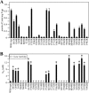
Mutants defective in tariquidar rescue show reduced tariquidar-stimulated ATPase activity. Histidine-tagged wild-type or mutant P-gps (in wild-type background) containing arginines defective in tariquidar rescue (identified as black circles in the α-helical wheels shown in Figs. 4 and 6) were expressed in HEK 293 cells and isolated by nickel-chelate chromatography. The isolated P-gps were mixed with lipid and ATPase activities were measured in the presence of a saturating concentration of tariquidar (60 μm) to determine maximal activity (A) or in the presence of various concentrations of tariquidar to determine the concentration required to stimulate ATPase activity by 50% (S50) (B) are shown. Each value is the mean ± S.D. (n = 3). An asterisk in B indicates a significant difference (p < 0.001) relative to wild-type P-gp.
FIGURE 9.
FIGURE 9.
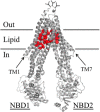
Location of the arginine mutations that inhibit tariquidar rescue of misprocessed mutants and tariquidar-stimulated ATPase activity. Predicted structure of human P-gp in the open conformation as described in the legend to Fig. 1. The red balls show the positions of residues in the TM segments (His61, Gly64, Leu65, Met68, Met69, Phe72, Ala129, Phe303, Ile306, Tyr307, Ser309, Tyr310, Phe336, Phe343, Gln725, Phe728, Phe732, Val865, Ile868, Gly872, Phe942, Thr945, Gln946, Met949, Tyr950, Ser952, Tyr953, Leu975, Phe978, and Val982) that when changed to arginine significantly reduced both arginine rescue (Figs. 4 and 6) and tariquidar-stimulated ATPase activity (Fig. 8).
FIGURE 10.
FIGURE 10.
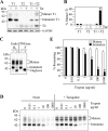
Tariquidar stabilizes TMD1. A, A52-tagged TMD1 (T1) (residues 1–379), A52-tagged TMD2 (T2) (residues 681–1025), or both A52-tagged TMD1 plus A52-tagged TMD2 (T1 + T2) were expressed in the absence (−) or presence (+) of 1 μm tariquidar (Tar). Whole cell SDS extracts were subjected to immunoblot analysis. The locations of TMD2 (T2) and mature and immature forms of TMD1 (T1) are indicated. B, the amount of mature TMD1 (T1) relative to total (mature and immature TMD1) (% Mature T1) was quantified. Each value is the mean ± S.D. (n = 3–5). An asterisk indicates a significant difference (p < 0.001) relative to samples grown in the absence of tariquidar. C, HEK 293 cells expressing A52-tagged TMD1 plus untagged TMD2 and grown in the presence of tariquidar were treated without (−) or with (+) endoglycosidase H (Endo Hf) or PNGase F. The reactions were stopped by addition of SDS sample buffer and subjected to immunoblot analysis. The positions of mature, immature, and unglycosylated (Unglycos) forms of TMD1 are indicated. D, membranes prepared from cells expressing A52-tagged TMD1 plus untagged TMD2 and grown in the absence (None) or presence (+Tariquidar) of tariquidar were treated with various concentrations of trypsin. The reactions were stopped by addition of trypsin inhibitor and samples were subjected to immunoblot analysis. The positions of mature and immature forms of TMD1 are indicated. E, the levels of mature (filled bars) or immature (unfilled bars) forms of TMD1 were determined and the amount remaining (% Remaining) after treatment with various levels of trypsin was compared with the untreated control. An asterisk indicates a significant difference (p < 0.001; n = 3) relative to that without trypsin.
FIGURE 11.
FIGURE 11.
Residues predicted to lie close to the tariquidar-binding site by arginine mutagenesis and molecular docking studies. Residues predicted to lie close to the tariquidar-binding site by arginine mutagenesis (A, this study) or by in silico molecular docking (9) to site 1 (D), site 2 (C), and site 3 (B) are shown. The number in the bracket indicates the TM segment where the residue is located.

Similar articles

Cited by

References

    1. Borst P., and Elferink R. O. (2002) Mammalian abc transporters in health and disease. Annu. Rev. Biochem. 71, 537–592 - PubMed
    1. Loo T. W., and Clarke D. M. (2014) Tariquidar inhibits P-glycoprotein drug efflux but activates ATPase activity by blocking transition to an open conformation. Biochem. Pharmacol. 92, 558–566 - PubMed
    1. Wagner C. C., Bauer M., Karch R., Feurstein T., Kopp S., Chiba P., Kletter K., Löscher W., Müller M., Zeitlinger M., and Langer O. (2009) A pilot study to assess the efficacy of tariquidar to inhibit P-glycoprotein at the human blood-brain barrier with (R)-11C-verapamil and PET. J. Nucl. Med. 50, 1954–1961 - PMC - PubMed
    1. Kannan P., Telu S., Shukla S., Ambudkar S.V., Pike V.W., Halldin C., Gottesman M.M., Innis R.B., and Hall M.D. (2011) The “specific” P-glycoprotein inhibitor tariquidar is also a substrate and an inhibitor for breast cancer resistance protein (BCRP/ABCG2). ACS Chem. Neurosci. 2, 82–89 - PMC - PubMed
    1. Loo T. W., and Clarke D. M. (1997) Correction of defective protein kinesis of human P-glycoprotein mutants by substrates and modulators. J. Biol. Chem. 272, 709–712 - PubMed

Publication types

LinkOut - more resources