. 2015 Oct 27;10(10):e0141267.
doi: 10.1371/journal.pone.0141267. eCollection 2015.

Comparing the Assembly and Handedness Dynamics of (H3.3-H4)2 Tetrasomes to Canonical Tetrasomes

Affiliations

Comparing the Assembly and Handedness Dynamics of (H3.3-H4)2 Tetrasomes to Canonical Tetrasomes

Rifka Vlijm et al. PLoS One. .

Abstract

Eukaryotic nucleosomes consists of an (H3-H4)2 tetramer and two H2A-H2B dimers, around which 147 bp of DNA are wrapped in 1.7 left-handed helical turns. During chromatin assembly, the (H3-H4)2 tetramer binds first, forming a tetrasome that likely constitutes an important intermediate during ongoing transcription. We recently showed that (H3-H4)2 tetrasomes spontaneously switch between a left- and right-handed wrapped state of the DNA, a phenomenon that may serve to buffer changes in DNA torque induced by RNA polymerase in transcription. Within nucleosomes of actively transcribed genes, however, canonical H3 is progressively replaced by its variant H3.3. Consequently, one may ask if and how the DNA chirality dynamics of tetrasomes is altered by H3.3. Recent findings that H3.3-containing nucleosomes result in less stable and less condensed chromatin further underline the need to study the microscopic underpinnings of H3.3-containing tetrasomes and nucleosomes. Here we report real-time single-molecule studies of (H3.3-H4)2 tetrasome dynamics using Freely Orbiting Magnetic Tweezers and Electromagnetic Torque Tweezers. We find that the assembly of H3.3-containing tetrasomes and nucleosomes by the histone chaperone Nucleosome Assembly Protein 1 (NAP1) occurs in an identical manner to that of H3-containing tetrasomes and nucleosomes. Likewise, the flipping behavior of DNA handedness in tetrasomes is not impacted by the presence of H3.3. We also examine the effect of free NAP1, H3.3, and H4 in solution on flipping behavior and conclude that the probability for a tetrasome to occupy the left-handed state is only slightly enhanced by the presence of free protein. These data demonstrate that the incorporation of H3.3 does not alter the structural dynamics of tetrasomes, and hence that the preferred incorporation of this histone variant in transcriptionally active regions does not result from its enhanced ability to accommodate torsional stress, but rather may be linked to specific chaperone or remodeler requirements or communication with the nuclear environment.


Conflict of interest statement

Competing Interests: The authors have declared that no competing interests exist.

Figures

Fig 1
Fig 1. Domains and illustration of the (H3.3-H4)2 tetrasome.
(A) The Drosophila H3 and H3.3 vary by only four amino acids that are marked in red. One is located in the N-terminal tail domain and three are located in the α-helix 2 domain, at locations 87, 89 and 90 (after [3]). (B) The crystal structures for H3-H4 and H3.3-H4 tetrasomes are not known, but to illustrate the most likely configuration, we here show a visual rendering of the structure of the human nucleosome containing the histone variant H3.3 (3AV2 from PDB). Only histones H3.3 (blue) and H4 (grey) are shown, as well as part of the nucleosomal DNA (grey). The amino acid locations 87, 89 and 90 are marked in red to indicate the region that deviates from the canonical histone H3 in the histone fold domain. The amino acid variant in the N-terminal tail is not marked, since it is outside of this region. This image was created using the software described in Ref. [43].
Fig 2
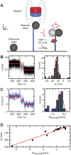
Fig 2. NAP1-assisted (H3.3-H4)2 tetrasome assembly.
(A) Schematic of the in vitro assay showing a single DNA molecule (blue) tethered between a glass surface and a paramagnetic bead. The circular magnet above the bead applies a stretching force to the bead (and hence to the DNA), but leaves it free to rotate about the DNA-tether axis. A nonmagnetic reference bead is fixed to the surface to allow for drift correction. After flushing in NAP1 preincubated with histones H3.3-H4, tetrasomes are loaded onto the DNA. (B) Time-dependence of the end-to-end length z (μm) (left) of a single DNA tether during the assembly of two (H3.3-H4)2 tetrasomes. The step sizes are -25 and -27 ± 5 nm. The green arrow at t = 420 s indicates the flushing in of the proteins. Data was acquired at 100 Hz, and red lines indicate the mean values of each assembly step. The histogram on the right derives from 19 independent assembly experiments (69 steps). A Gaussian fit shows that the average step in z during tetramer assembly is -25 ± 6 nm. (C) Time-dependence of bead rotations θ (turns) (left) of the same DNA tether as in B). Compaction of the DNA (shown in B)) occurs concurrently with a change in linking number (changes in θ). The step sizes in θ are -1.17 ± 0.24 and -0.97 ± 0.24 turns. The green arrow at t = 420 s indicates the flushing in of the proteins. Data was acquired at 100 Hz, and red lines indicate the mean values of each assembly step. The histogram on the right derives from 15 independent assembly experiments (23 steps). It can be fitted to Gaussian peaks. The most likely step in θ during tetramer assembly is -0.8 ± 0.1 turns. A small number of steps appears to result from the simultaneous assembly of two tetramers, with a mean step size in θ of -1.9 ± 0.1 turns. (D) The total degree of compaction (Δz) plotted versus the total change in linking number (Δθ assembly) on 25 individual DNA molecules following the assembly of tetrasomes (black squares). Fits to a linear relationship yield Δz/Δθ assembly = 32 ± 2 nm (solid red line).
Fig 3
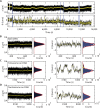
Fig 3. (H3.3-H4)2 tetrasomes undergo dynamic changes in linking number and form a viable intermediate for nucleosomes.
(A) Assembly of a single complete nucleosome from a single assembled tetrasome. By flushing in H3.3-H4 preincubated with NAP1 at t = 708 s, we assembled one tetrasome (Δz = 23 ± 5 nm, Δθ assembly = -0.81 ± 0.25 turns). Dynamic changes in the linking number were observed immediately following assembly and continued for ~8000 s. When we then flushed in histones H2A and H2B preincubated with NAP1 at 8536 and 8935 s, we observed an additional assembly step (Δz = 31 ± 5 and Δθ = -0.55 ± 0.25 turns). Subsequently, the linking number remained stable (i.e. the flipping behavior of the handedness ceased). Blue lines mark flushing in of NAP and core histones H3.3-H4 at t = 708 s and of H2A-H2B at t = 8536 s and t = 8935 s. All proteins are flushed out at t = 11400 s. Parts of the data shown in A) are highlighted in panels B)—D). The left panels show a typical segment (350 s) of the end-to-end length z (left) and the angular coordinate θ (right). Side panels show histograms with fits to Gaussian functions (red lines) that are derived from the full portion of the trace acquired under the indicated conditions. (B) Bare DNA, before the proteins are flushed in. (C) DNA loaded with a single tetrasome. The centers of the Gaussian fits are at -0.80 and 0.86 turns. (D) DNA loaded with a single nucleosome. In B-D), the mean extension, z, remains constant in time, with fluctuations merely arising from Brownian motion (standard deviations of σbare DNA, σtetrasome, and σnucleosome are 23 nm). Both bare DNA and DNA loaded with nucleosomes exhibit a fixed mean linking number in time, with comparable fluctuations about the mean (σ = 0.66 and 0.77 turns, respectively). However, tetrasomes exhibit clear fluctuations in the linking number over time.
Fig 4
Fig 4. Analysis of θ flipping of (H3.3-H4)2 tetrasomes.
(A) A single (H3.3-H4)2 tetrasome (for a different molecule than that of Fig 3, to emphasize repeatability). The histogram of θ flipping, the difference in angle between the left- and right-handed states from a single (H3.3-H4)2 tetrasome, shows two peaks. The peak has a maximum at θ = -1.011 ± 0.003 turns. The positively wrapped state has a peak at θ = 0.63 (± 0.03) turns. (B) Histogram of θ flipping of a DNA molecule loaded with four tetrasomes. Data are collected after flushing out free proteins. The most pronounced peaks are for 1 (θ = -2.1 turns), 2 (θ = -0.44 turns) and 3 (θ = 1.0 turns) tetrasomes in the right-handed state (values extracted from Gaussian fitting to the histogram). When any one tetrasome flipped to the right-handed state, the linking number increased on average by 1.7 ± 0.2 turns. (C) Histogram of dynamical linking number steps observed following assembly of tetrasomes on distinct DNA molecules (N = 10) before (black) and after (grey) flushing out free proteins (N = 33), which yields a mean value of <Δθ flipping> = 1.7 ± 0.1 turns both before and after flushing out of free proteins. (D) Determination of the probability p of finding a tetrasome in the left-handed state in the presence (N = 12, dark blue crosses, <p> = 0.91 ± 0.03) and absence (N = 7, green plusses, <p> = 0.84 ± 0.09) of free proteins. Using the formula ΔG = -kBT ln((1/p)-1), the difference in the free energy between the two states can be computed (red datapoints). We deduce ΔG = 2.3 ± 0.4 kBT prior to flushing out free proteins (red open squares) and ΔG = 1.6 ± 0.8 kBT following the flushing out of free proteins (red filled circles).
Fig 5
Fig 5. Torque response of DNA loaded with (H3.3-H4)2 tetrasomes.
(A) Diagram of the eMTT configuration used in these experiments. The eMTT resembles the FOMT configuration, but additionally has two pairs of Helmholtz coils placed around the flow cell to permit the application of torque in the horizontal plane. (B) The torque stored in DNA loaded with 5 tetrasomes plotted as a function of the number of applied rotations, θ. The black squares represent the data for a bare DNA molecule, prior to assembly. Following assembly, the torque response of DNA loaded with tetrasomes is measured by decreasing the number of applied turns from +17 to -17 (red triangles, labeled by ‘1’). Consecutively, the torque response of DNA loaded with tetrasomes is measured in the opposite direction by increasing the number of applied turns -17 to +17 (green diamonds, labeled by ‘2’). The solid lines are segmented fits to the plateau regions (with slope 0) and to the sloped regions in which torque is built up. The widths of the plateaus for positive (red) and negative (green) rotation directions are 7.5 ± 1 and 5.9 ± 1 turns, respectively, as determined from the intersections between the segmented fits. The applied force is ~0.8 pN. (C) The DNA end-to-end length plotted as a function of the number of rotations, θ. The applied stretching force is 0.3 pN. The black squares show the data for a bare DNA molecule, prior to any tetrasome assembly. Following tetrasome assembly, a broad plateau (of ~8 ± 1 turns) is observed surrounding 0 turns (red circles). The solid lines are linear fits to the data (5 per trace).

Similar articles

Cited by

References

    1. Luger K, Mader AW, Richmond RK, Sargent DF, Richmond TJ. Crystal structure of the nucleosome core particle at 2.8 A resolution. Nature. 1997;389(6648):251–60. 10.1038/38444 . - DOI - PubMed
    1. Luger K. Structure and dynamic behavior of nucleosomes. Current opinion in genetics & development. 2003;13(2):127–35. . - PubMed
    1. Kamakaka RT, Biggins S. Histone variants: deviants? Genes & development. 2005;19(3):295–310. 10.1101/gad.1272805 . - DOI - PubMed
    1. Daban JR, Cantor CR. Structural and kinetic study of the self-assembly of nucleosome core particles. Journal of molecular biology. 1982;156(4):749–69. . - PubMed
    1. Hamiche A, Carot V, Alilat M, De Lucia F, O'Donohue MF, Revet B, et al. Interaction of the histone (H3-H4)2 tetramer of the nucleosome with positively supercoiled DNA minicircles: Potential flipping of the protein from a left- to a right-handed superhelical form. Proceedings of the National Academy of Sciences of the United States of America. 1996;93(15):7588–93. - PMC - PubMed

Publication types

LinkOut - more resources