. 2015 Dec 4;290(49):29375-88.
doi: 10.1074/jbc.M115.681908. Epub 2015 Oct 21.

Retinoblastoma-binding Protein 4-regulated Classical Nuclear Transport Is Involved in Cellular Senescence

Affiliations

Retinoblastoma-binding Protein 4-regulated Classical Nuclear Transport Is Involved in Cellular Senescence

Akira Tsujii et al. J Biol Chem. .

Abstract

Nucleocytoplasmic trafficking is a fundamental cellular process in eukaryotic cells. Here, we demonstrated that retinoblastoma-binding protein 4 (RBBP4) functions as a novel regulatory factor to increase the efficiency of importin α/β-mediated nuclear import. RBBP4 accelerates the release of importin β1 from importin α via competitive binding to the importin β-binding domain of importin α in the presence of RanGTP. Therefore, it facilitates importin α/β-mediated nuclear import. We showed that the importin α/β pathway is down-regulated in replicative senescent cells, concomitant with a decrease in RBBP4 level. Knockdown of RBBP4 caused both suppression of nuclear transport and induction of cellular senescence. This is the first report to identify a factor that competes with importin β1 to bind to importin α, and it demonstrates that the loss of this factor can trigger cellular senescence.

Keywords: IBB; cellular regulation; cellular senescence; importin α; importin β1; nuclear transport; protein-protein interaction; structural model.


Figures

FIGURE 1.
FIGURE 1.
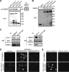
Importin α binds to RBBP4 but does not mediate its nuclear transport. A and B, GST pulldown assays using three GST-fused importin α subtypes (α1, α3, and α5) with HEK-293F cell lysates expressing HA-tagged RBBP4 (A) or with recombinant RBBP4 (B). Bound proteins were analyzed by Western blotting using the antibodies indicated. C, HeLa cell lysates were immunoprecipitated (IP) with RBBP4 or importin α1 antibody, followed by Western blotting (WB) using importin α1 or RBBP4 antibody, respectively. D and E, in vitro transport assays. Nuclear import of either GFP-RBBP4 or GST-SV40T-NLS-GFP was examined in digitonin-permeabilized HeLa cells, in HeLa cell cytosols with or without Q69LRanGTP (D), or in a reconstitution system that included importin α1, importin β1, RanGDP, and an ATP-regenerating system (E). After 30 min at 30 °C, GFP-tagged proteins were visualized by fluorescence microscopy.
FIGURE 2.
FIGURE 2.
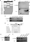
Identification of the importin α1-binding site in RBBP4. A, schematic representation of RBBP4 wild type (WT) and its deletion mutants. A, 1–141 aa; B, 142–283 aa; C, 284–425 aa; AB, 1–283 aa; BC, 142–425 aa; AC, 1–141 and 284–425 aa; ΔN5, 6–425 aa; ΔN10, 11–425 aa; ΔN20, 21–425 aa; ΔN30, 31–425 aa; ΔC5, 1–420 aa; ΔC10, 1–415 aa; ΔC20, 1–405 aa; ΔC30, 1–395 aa. B and C, GST pulldown assays for importin α1 and RBBP4 deletion mutants. HEK-293F cells were transfected with the HA-tagged RBBP4 mutants described in A, and proteins bound to GST-importin α1 were detected using an anti-HA antibody (B: WT, A, B, C, AB, BC, and AC; C: WT, ΔN5, ΔN10, ΔN20, ΔN30, ΔC5, ΔC10, ΔC20, and ΔC30). D, amino acid mutations or deletions in the region of 301–315 aa in RBBP4. E and F, GST pulldown assays for importin α1 and RBBP4 mutants (E, WT and Δ305–310; F, WT, K307/309A, K307/309E, L306/308/310A, and L306/308/310E). All binding assays were analyzed using the same procedure described in (B).
FIGURE 3.
FIGURE 3.
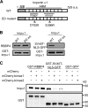
Importin α1 binds to RBBP4 through a site other than the major or minor NLS-binding sites. A, schematic representation of importin α1 wild type (WT) and ED mutant. B, binding assay between GST-importin α1 (Impα1) and RBBP4. Either WT or the ED mutant (D192K, E396R) of GST-importin α1 was incubated with recombinant RBBP4 or SV40T-NLS-GFP. The SV40T-NLS-GFP was used as a positive control for binding with the WT protein and detected by an anti-GFP antibody. C, examination of binding inhibition by cNLS peptides, bimax1 and bimax2. GST-RBBP4, GST-SV40T-NLS-GFP, or GST-GFP immobilized on GSH beads was incubated with importin α1 in the presence of mCherry-bimax1 or bimax2 (100 pmol each). The unconjugated mCherry protein was used as a negative control.
FIGURE 4.
FIGURE 4.
IBB domain of importin α1 is an RBBP4-binding site. A, schematic representation of wild type (WT) and deletion mutants of importin α1 recombinant protein. B and C, GST pulldown assays were performed using GST-fused importin α1 WT, the ΔIBB mutant, or the ΔC mutant incubated with HEK-293 cell lysates expressing HA-tagged RBBP4 (B) or with recombinant RBBP4 (C) and analyzed by Western blotting using the indicated antibodies. Endogenous importin β1 was detected to confirm the lack of interaction with the ΔIBB mutant. Signal intensities (HA/GST or RBBP4/GST) were normalized to the control (GST-importin α1 WT). Values represent averages ± S.D. from three experiments. Significant differences from control are indicated (*, p < 0.01; **, p < 0.05, Student's t test). D, GST pulldown assays using GST-IBB-GFP with RBBP4. CBB, Coomassie Brilliant Blue. E, GST pulldown assays using GST-importin α1 with SV40T-NLS-GFP in the presence of either importin β1 or RBBP4. Signal intensities (NLS/GST) were normalized to the control (impβ-/RBBP4-) and statistical analyses were conducted as described in (B and C).
FIGURE 5.
FIGURE 5.
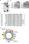
Biochemical and structural analysis of RBBP4-IBB domain interaction. A, GST pulldown assay using GST, GST-importin β1, and GST-importin α1 with RBBP4. The assays were analyzed by Western blotting. B, competition assays for GST-IBB-GFP with RBBP4 (50 pmol) in the presence of increasing amounts of importin β1 or GFP (50, 100, and 200 pmol, respectively). The assays were analyzed by Western blotting. GST-IBB and applied importin β1 or GFP are shown by Coomassie staining (CBB, Coomassie Brilliant Blue). C, multiple sequence alignment of all subtypes of human and mouse importin α (IBB domains) was constructed using ClustalW (28). The underlined residues in hIMPα1 are involved in importin β1 binding based on the structure of the importin β1·IBB domain complex (PDB code 1qgk), and residues boxed in thin lines are conserved among all the subtypes. D, helical wheel projection of the helix in the IBB domain was generated using a script on the web site. Conserved residues and importin β1-binding residues are circled in red and cyan, respectively. Importin β1 (PDB code 1qgk) binds to three distinct regions in the IBB domain as follows: the N-terminal short segment and two regions on the helix.
FIGURE 6.
FIGURE 6.
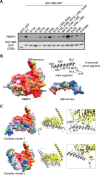
Structural models of the complex between RBBP4 and the IBB domain. A, GST pulldown assays for GST-IBB-GFP with RBBP4. Either WT or mutants of GST-IBB-GFP were incubated with RBBP4 and then detected by Western blotting. GST-IBB-GFP proteins are shown by Coomassie staining (CBB, Coomassie Brilliant Blue). B, RBBP4-binding residues in the IBB domain. Left, electrostatic potential and hydrophobicity on the molecular surface of the RBBP4 structure (PDB code 4pby), which was obtained from the eF-site database (60). The figure is colored to identify negatively charged (red), positively charged (blue), or neutral (white) non-hydrophobic residues and negatively charged (orange), positively charged (green), or neutral (yellow) hydrophobic residues. Upper right, IBB domain structure (PDB code 1qgk), with the putative RBBP4-binding residues shown as ball and stick models. Lower right, electrostatic potential and hydrophobicity on the molecular surface of the IBB domain. We predict that the hydrophobic and positively charged surface in the IBB domain binds to the hydrophobic and negatively charged surface spanning from Leu-306/-308/-310 to the C terminus of RBBP4. C, two structural models of the RBBP4·IBB domain complex. We used the interactive molecular viewer, jV (60), and superimposed manually the IBB domain structure in the importin β1·IBB domain complex (PDB code 1qgk) onto the RBBP4 structure in the RBBP4·MTA1 complex (PDB code 4pby), so that Leu-306/-308/-310 of RBBP4 would interact with Met-27/Val-34/Leu-38 of the IBB domain, and either the N or C terminus of RBBP4 would also interact with the IBB domain. Finally, two possible models were obtained showing the C terminus of RBBP4 mediating IBB domain binding. Note that in these models, we allowed some collisions between the PP loop (Ser-347–Glu-364) of RBBP4 and the IBB domain, and loose interactions between the N-terminal short segment and RBBP4, because of possible conformational changes upon RBBP4-IBB domain binding in the PP loop and the relative position of the N-terminal short segment to the helix in the IBB domain.
FIGURE 7.
FIGURE 7.
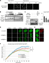
Classical nuclear import of cNLS is impaired in HeLa cells in which RBBP4 is knocked down. A, in vitro transport assays using digitonin-permeabilized HeLa cells. GST-SV40T-NLS-GFP was incubated with or without importin α1 and β1 in the presence of increasing amounts of RBBP4 as indicated. After 30 min at 30 °C, the GFP-tagged proteins were visualized by fluorescence microscopy. B, GST pulldown assays to examine the effect of RBBP4 on the IBB domain·importin β1 complex. GST-IBB-GFP was incubated first with importin β1 (200 pmol) for 30 min to form a complex (pre-incubation). Then increasing amounts of RBBP4 (25, 50, and 100 pmol) were added (B, left panel). GST-IBB-GFP, importin β1, and the same increasing amounts of RBBP4 were added simultaneously (B, right panel). Importin β1 and RBBP4 were detected by Western blotting. GST-IBB is shown by Coomassie staining. CBB, Coomassie Brilliant Blue. C, GST pulldown assays to examine the cooperative effect of Q69LRanGTP and RBBP4 against the IBB domain·importin β1 complex. GST-IBB-GFP and importin β1 were pre-incubated, and then Q69LRanGTP (400 pmol) was added either with or without RBBP4 (400 pmol). Importin β1, RBBP4, and GST-IBB were detected by Western blotting. The graph indicates the signal intensities of importin β1 relatively to GST-IBB (values represent averages ± S.D.). D, in vitro transport assays to measure the nuclear import efficiency of GST-SV40T-NLS-GFP. HeLa cells were transfected with two siRNAs against RBBP4: si-RBBP4-1 or si-RBBP4-2. At 48 h after transfection, the cells were permeabilized with digitonin, and an in vitro transport assay was performed using importin α1 and β1 recombinant proteins. The fluorescence intensity of the cNLS substrate in the nucleus was analyzed in time-lapse imaging and is displayed for each indicated time. E, immunofluorescence staining of RBBP4 in the nucleus of HeLa cells fixed after performing transport assays. At 20 min after the incubation described in D, the cells were stained with anti-RBBP4 antibody. Green fluorescence corresponds to GST-SV40T-NLS-GFP, and red fluorescence corresponds to RBBP4. F, averaged nuclear fluorescence signal was collected for 20 min and is presented as nuclear import curves. Relative signal intensities of 10 different nuclei per experiment were measured in three independent experiments (error bars represent standard error of the mean) and statistically analyzed using Bonferroni's post hoc test after a two-way analysis of variance (*, p < 0.05; **, p < 0.001; NS, not significant).
FIGURE 8.
FIGURE 8.
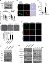
Silencing RBBP4 induces cellular senescence. A, endogenous RBBP4 in different PDLs of TIG-1 cells. p21 was used to show that cells were in senescent states, and actin was used as an internal control in Western blotting (WB). The graphs indicate the protein level or mRNA level of RBBP4 normalized to actin (Western blotting) or HPRT (quantitative RT-PCR), respectively (values represent averages ± S.D.). B and C, comparisons of nuclear import ability in different stages of TIG-1 cells. Senescent cells (PDL of >65) are compared with young cells (PDL of <30). GST-SV40T-NLS-GFP and Alexa-Fluor-594-conjugated antibody as an injection marker were microinjected into the cytoplasm of both stages of TIG-1 cells. The cells were incubated at room temperature and fixed at 20 min. After the fluorescent examination, cells were stained by SA β-gal (B). The nuclear and cytoplasmic fluorescence intensities were determined for at least 10 different cells to enable the calculation of the nuclear to cytoplasmic ratio (Fn/c). Statistical analysis was performed using the Student's t test (C, *, p < 0.001). D–F, TIG-1 cells were transfected with two independent siRNAs (si-RBBP4-1 and si-RBBP4-2) or with control (si-control) for 48 h. Scale bars, 20 μm. D, cellular senescence was detected by SA β-gal staining. The graph indicates the percentage of SA β-gal-positive cells. Values represent averages ± S.D. from three experiments; n > 200. Statistical analysis was performed using the Student's t test (*, p < 0.001). E, detection of endogenous Ki-67 in the cells using a specific antibody. F, Western blotting with indicated antibodies using whole cell lysates prepared from RBBP4 knockdown TIG-1 cells. G, Western blotting for histone modifications in whole cell lysates from si-RBBP4 treated TIG-1 cells or from three different PDLs of TIG-1 cells.

Similar articles

Cited by

References

    1. Terry L. J., Shows E. B., and Wente S. R. (2007) Crossing the nuclear envelope: hierarchical regulation of nucleocytoplasmic transport. Science 318, 1412–1416 - PubMed
    1. Imamoto N., Shimamoto T., Takao T., Tachibana T., Kose S., Matsubae M., Sekimoto T., Shimonishi Y., and Yoneda Y. (1995) In vivo evidence for involvement of a 58-kDa component of nuclear pore-targeting complex in nuclear protein import. EMBO J. 14, 3617–3626 - PMC - PubMed
    1. Goldfarb D. S., Corbett A. H., Mason D. A., Harreman M. T., and Adam S. A. (2004) Importin α: a multipurpose nuclear-transport receptor. Trends Cell Biol. 14, 505–514 - PubMed
    1. Miyamoto Y., Boag P. R., Hime G. R., and Loveland K. L. (2012) Regulated nucleocytoplasmic transport during gametogenesis. Biochim. Biophys. Acta 1819, 616–630 - PubMed
    1. Lee S. J., Matsuura Y., Liu S. M., and Stewart M. (2005) Structural basis for nuclear import complex dissociation by RanGTP. Nature 435, 693–696 - PubMed

Publication types

MeSH terms

LinkOut - more resources