. 2015 Oct 14:6:8516.
doi: 10.1038/ncomms9516.

Sleep disruption impairs haematopoietic stem cell transplantation in mice

Affiliations

Sleep disruption impairs haematopoietic stem cell transplantation in mice

Asya Rolls et al. Nat Commun. .

Abstract

Many of the factors affecting the success of haematopoietic cell transplantation are still unknown. Here we show in mice that donor sleep deprivation reduces the ability of its haematopoietic stem cells (HSCs) to engraft and reconstitute the blood and bone marrow of an irradiated recipient by more than 50%. We demonstrate that sleep deprivation downregulates the expression of microRNA (miR)-19b, a negative regulator of the suppressor of cytokine signalling (SOCS) genes, which inhibit HSC migration and homing. Accordingly, HSCs from sleep-deprived mice have higher levels of SOCS genes expression, lower migration capacity in vitro and reduced homing to the bone marrow in vivo. Recovery of sleep after sleep deprivation restored the reconstitution potential of the HSCs. Taken together, this study provides insights into cellular and molecular mechanisms underlying the effects of sleep deprivation on HSCs, emphasizing the potentially critical role of donor sleep in the success of bone marrow transplantation.


Figures

Figure 1
Figure 1. HSCs isolated from sleep-deprived mice have reduced mid-term and long-term reconstitution potential in lethally irradiated hosts
(a) Mice were sleep-deprived, by gentle handling, for four hours, immediately after light onset (ZT0-ZT4; n=8 per group). Rapid eye movement (REM) and non-REM (NREM) duration were determined using electroencephalography (EEG) and electromyography (EMG), individually plotted for each hour (sleep deprived points in red; mean±s.e.m). (b) Plasma of the sleep and sleep-deprived mice was analyzed by ELISA for corticosterone levels (n=8 per group; mean±s.e.m). (c) We isolated HSCs from mice that were allowed to sleep or sleep-deprived mice. The flow cytometry gating scheme for HSCs isolation is shown on a representative mouse. (d) To test the mid-term and long-term transplantation potential of the isolated HSCs we intravenously injected 300 HSCs mice into lethally irradiated congenic recipients (to distinguish between the donor and recipient cells we used CD45.1 mice as donors and CD45.2 as recipients). Peripheral blood (PB) samples were collected from the recipient mice at (e) eight weeks (peripheral blood), (f) 16 weeks and (g) bone marrow (BM) post-transplantation. Cells were analyzed for myeloid chimerism, as determined by the percentage of myeloid cells derived from the donor compared to the total number of myeloid cells in the indicated tissue (mean±s.e.m; Student’s t-test; ***p<0.0001; **p<0.001). In all experiments, HSCs were transplanted along with 1×10⁶ bone marrow mononuclear cells to support mouse survival (d–g; n=12 mice per group). (h) Primary recipients received HSCs from sleep or sleep deprived donors as indicated in d–g. Then, at 16 weeks, 2.5 × 10⁶ BM cells from primary recipients were transplanted into a new group of lethally irradiated CD45.2+ (secondary recipients) and donor-derived reconstitution was assessed in the secondary recipients (mean±s.e.m; Student’s t-test; p=0.83).
Figure 2
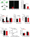
Figure 2. HSCs from sleep-deprived mice have reduced homing capacity in vivo and in vitro
(a) HSCs (2000 FACS-purified cells) derived from GFP-expressing, sleep-deprived mice were transplanted into lethally irradiated congenic mice that did not express GFP (control mice were allowed to sleep for the same duration). The donor HSCs were visualized 12 hours later in the recipient’s bone, under a fluorescence microscope. GFP expression was validated using anti-GFP staining and a representative image is shown in the insert. Scale 10µm (n=6–11 mice per group; mean±s.e.m). (b) The number of GFP-labeled cells in the recipient’s bone marrow was determined. Results are presented as the percentage of homing, assuming that two tibias and two femurs represent 20% of total mouse bone marrow (student’s t-test; p=0.021; t=2.56; df=15). (c) Migration towards SDF-1α was determined in vitro using a transwell migration assay. KLS cells were placed in the upper chamber and the chemoattractant (SDF-1α, 50, 100, or 200ng/mL) was immersed in the medium of the lower chamber. Migration across the membrane in response to the chemoattractant was determined using flow cytometry, after four hours of incubation, and compared to baseline migration in the absence of the chemoattractant. Results are presented as a migration index (50ng/mL SDF-1α: 9.6 ± 1.7% in the sleep group compared to 2.06 ± 0.4% in the sleep-deprived group; 100ng/mL SDF-1α: 14.5 ± 3.4% in the sleep compared to 3.6 ± 1.4% in the sleep-deprived mice; Repeated measures ANOVA- sleep: F(2,15)=10.18, p<0.0061; n=6 mice per group; mean ±s.e.m. (d) Plasma was collected and analyzed by ELISA for SDF-1α levels, from sleep or sleep-deprived mice. (e) CXCR4 levels on HSCs derived from sleep and sleep-deprived mice (mean fluorescent intensity (MFI)±s.e.m). (f) VLA4 (α4) levels on HSCs derived from sleep and sleep-deprived mice. (g) Cells were cultured with 0.5 or 10 nM LDV-FITC to evaluate VLA4 binding affinity. Binding was evaluated as FITC MFI on HSCs (mean±s.e.m). Statistical significance was determined by Student’s t-test. (h) Migration towards S1P was determined in vitro using a transwell migration assay as described in (c). S1P concentration is indicated. Repeated measure ANOVA (n=4 mice per group).
Figure 3
Figure 3. Sleep deprivation decreases the levels of miR19b and increases the levels of SOSC3 in HSCs
(a) miRNA profiles of HSCs from sleep and sleep-deprived mice were analyzed by multiplex quantitative real-time PCR after HSC sorting by FACS. The ΔΔCt method was used, with U6, sno-202 and sno-135 as normal controls. MicroRNA levels underwent > 4-fold changes and potentially targeted SOCS genes are highlighted. (b) qPCR analysis of SOCS1, SOCS3 and SOCS5 levels. Results are presented as fold change normalized to GAPDH (ND=not detected; 3 independent repeats, n=4 mice per group; SOCS3: 4.13± 0.5-fold and SOCS1: 1.9± 0.8-fold). (c) A schematic presentation of the luciferase reporter system in 293T cells that were transfected with a microRNA mimic or scramble control and pMir Target construct containing the 3'UTR of SOCS3 and an RFP reporter. Luciferase activity was measured 48 hours after transfection and normalized to RFP and then to the values measured for vector alone (student’s t-test; t(4)=8.313; p=0.0011; mean±s.e.m). (d) Migration of HSCs, transfected with 50nM miR-19b mimic or scramble control, towards 50ng/mL SDF-1α. Migration was compared to baseline migration in the absence of chemoattractant (migration index). (e) Mx-1 cre mice were crossed with SOCS3-floxed mice. Mx-1 cre mice express Cre recombinase under the control of an inducible Mx1 promoter, active after induction with polyinosinic-polycytidylic acid (pI:pC). When bred with SOCS3-floxed mice (carrying SOCS3 gene flanked by loxP recognition sites), the expression of Cre recombinase causes SOCS3 gene deletion. Upper panel: Genomic DNA was extracted from FACS-sorted KLS cells and analyzed using PCR. The PCR product obtained from the SOCS fl+ locus is 250 bp band indicates cre-mediated deletion of SOCS3 (SOCS3del). Lower panel: Migration of KLS cells isolated from sleep mice and from mice that were deprived of sleep for four hours. We compared two sets of mice, mice that express mx-1-cre and SOCS3- floxed gene and their littermates that do not express mx-1-cre. This allows controlling for potential effect of pI:pC injection required to induce cre expression. Migration towards 50 ng/mL SDF-1 α was determined for cells from sleep or sleep-deprived mice. The migration index was determined for each group and the ratio between the migration index of the control and the SOCS-3 deleted mice was calculated (mean±s.e.m).
Figure 4
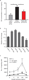
Figure 4. Growth hormone alters the expression of miR-19b and cell migration
(a) Mice were either sacrificed with light onset, after 1.5h or 3h of sleep deprivation or after 1.5h or 3h of sleep, and plasma GH levels were determined by ELISA (one-way ANOVA followed by Tukey’s multiple comparisons test; mean±s.e.m). (b) KLS cells were treated with GH (100ng/mL- dose was determined based on previous studies,) for 30 min and RNA was extracted using Trizol for analysis of miRs levels. All values were statistically significant within each group (technical replicas) and are reported as fold of change relative to control that was used as a reference (mean±s.e.m). (c) Cells from control mice were pre-incubated for 30 min with GH (0, 5, 25, 50 or 100ng/mL), washed and the migration of KLS to increasing concentrations of SDF-1α was determined on transwell migration plates. Statistical analyses was analyzed using one-way ANOVA followed by Tukey’s multiple comparisons test; ***p<0.01; n=4 mice per group.
Figure 5
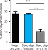
Figure 5. Sleep rebound restores the reconstitution potential in lethally irradiated hosts
Chimerism in the bone marrow of mice transplanted with HSC derived from mice that were allowed to recover their sleep for 2h, mice deprived of sleep for 6h and mice allowed to sleep for 6h. Analysis of was performed at 16 weeks after transplantation (F(2,15)=192.9; p<0.0001). *p<0.05; **p<0.01; ***p<0.001. Mean ± s.e.m; n=6 mice per group.

Similar articles

Cited by

References

    1. Besedovsky L, Lange T, Born J. Sleep and immune function. Pflugers Arch. 2012;463:121–137. - PMC - PubMed
    1. Krueger JM, Majde JA, Rector DM. Cytokines in immune function and sleep regulation. Handb. Clin. Neurol. 2011;98:229–240. - PMC - PubMed
    1. Bryant PA, Trinder J, Curtis N. Sick and tired: Does sleep have a vital role in the immune system? Nat. Rev. Immunol. 2004;4:457–467. - PubMed
    1. Guariniello LD, Vicari P, Lee KS, de Oliveira AC, Tufik S. Bone marrow and peripheral white blood cells number is affected by sleep deprivation in a murine experimental model. J. Cell. Physiol. 2012;227:361–366. - PubMed
    1. Tsinkalovsky O, et al. Circadian expression of clock genes in purified hematopoietic stem cells is developmentally regulated in mouse bone marrow. Exp. Hematol. 2006;34:1249–1261. - PubMed

Publication types

MeSH terms

Substances

LinkOut - more resources