

Diagnostic role of DOG1 and p63 immunohistochemistry in salivary gland carcinomas
- PMID: 26464669
- PMCID: PMC4583901
Diagnostic role of DOG1 and p63 immunohistochemistry in salivary gland carcinomas
Abstract
Background: The differential diagnosis of salivary carcinomas is always difficult and challenging. Salivary neoplasms often shows more than one growth pattern and significant morphologic variability may exist within a single tumor and between different tumors. The aim of this study was to examine the role of DOG1 (discovered on gastrointestinal tumor-1) and p63 immunohistochemistry in the diagnosis and differential diagnosis of salivary carcinomas.
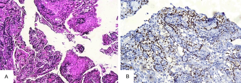
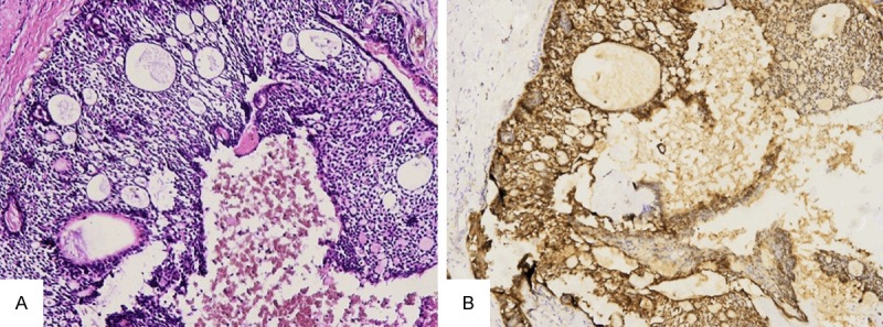
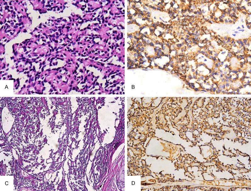
Methods: we examined the expression of DOG1 and p63 immunohistochemistry in 33 mucoepidermoid carcinomas (MEC), 9 acinic cell carcinomas (ACC), 10 adenoid cystic carcinomas (AdCC) and 4 myoepithelial carcinomas.
Results: All ACC showed strong to moderate positivity for DOG1 (P=0.001) and all were totally negative for p63. All MEC expressed strong to moderate positivity for p63 (P=0.001) while only (9.1%) were weak to moderately positive for DOG1. (80%) AdCC were moderately positive for DOG1 in ductal and myoepithelial components and (100%) showed moderate positivity for p63 in myoepithelial cells only (P=0.001). All myoepithelial carcinomas were DOG1 negative, 2 (50%) were weakly positive for p63 while the other 2 were moderately positive (P=0.5).
Conclusion: DOG1 is a sensitive marker in the diagnosis of acinic cell carcinoma, p63 is sensitive in the diagnosis of mucoepidermoid carcinoma, the combined use of both markers is helpful and statistically significant in the differential diagnosis of acinic cell carcinoma versus mucoepidermoid carcinoma, both markers can help in the diagnosis of adenoid cystic carcinoma but they have no role in the diagnosis of myoepithelial carcinoma.
Keywords: DOG1; Salivary carcinomas; immunohistochemistry; p63.
Figures
Similar articles
-
DOG1 as an Immunohistochemical Marker of Acinic Cell Carcinoma: A Systematic Review and Meta-Analysis.Int J Mol Sci. 2022 Aug 26;23(17):9711. doi: 10.3390/ijms23179711. Int J Mol Sci. 2022. PMID: 36077107 Free PMC article. Review.
-
Lysozyme Expression Can be Useful to Distinguish Mammary Analog Secretory Carcinoma from Acinic Cell Carcinoma of Salivary Glands.Head Neck Pathol. 2016 Dec;10(4):429-436. doi: 10.1007/s12105-016-0718-5. Epub 2016 May 13. Head Neck Pathol. 2016. PMID: 27177644 Free PMC article.
-
P63 expression can be used in differential diagnosis of salivary gland acinic cell and mucoepidermoid carcinomas.Head Neck Pathol. 2013 Mar;7(1):64-8. doi: 10.1007/s12105-012-0403-2. Epub 2012 Oct 10. Head Neck Pathol. 2013. PMID: 23054955 Free PMC article.
-
Characterisation of DOG-1 Expression in Salivary Gland Tumours and Comparison with Myoepithelial Markers.Head Neck Pathol. 2019 Jun;13(2):140-148. doi: 10.1007/s12105-018-0917-3. Epub 2018 Apr 18. Head Neck Pathol. 2019. PMID: 29671211 Free PMC article.
-
Salivary gland-like tumors of the breast express basal-type immunohistochemical markers.Appl Immunohistochem Mol Morphol. 2013 Jul;21(4):283-6. doi: 10.1097/PAI.0b013e31826a277e. Appl Immunohistochem Mol Morphol. 2013. PMID: 22935826 Review.
Cited by
-
Diagnostic role of DOG-1, GFAP and B-catenin in Basal cell Adenoma and Cellular Pleomorphic Adenoma of the Salivary Gland.Head Neck Pathol. 2023 Jun;17(2):339-346. doi: 10.1007/s12105-022-01498-7. Epub 2022 Oct 28. Head Neck Pathol. 2023. PMID: 36307634 Free PMC article.
-
Increased p63 Expression in Canine Perianal Gland Tumours.J Vet Res. 2018 Oct 24;62(2):229-235. doi: 10.2478/jvetres-2018-0020. eCollection 2018 Jun. J Vet Res. 2018. PMID: 30364817 Free PMC article.
-
DOG1 as an Immunohistochemical Marker of Acinic Cell Carcinoma: A Systematic Review and Meta-Analysis.Int J Mol Sci. 2022 Aug 26;23(17):9711. doi: 10.3390/ijms23179711. Int J Mol Sci. 2022. PMID: 36077107 Free PMC article. Review.
-
Lysozyme Expression Can be Useful to Distinguish Mammary Analog Secretory Carcinoma from Acinic Cell Carcinoma of Salivary Glands.Head Neck Pathol. 2016 Dec;10(4):429-436. doi: 10.1007/s12105-016-0718-5. Epub 2016 May 13. Head Neck Pathol. 2016. PMID: 27177644 Free PMC article.
-
Distinct TP63 Isoform-Driven Transcriptional Signatures Predict Tumor Progression and Clinical Outcomes.Cancer Res. 2018 Jan 15;78(2):451-462. doi: 10.1158/0008-5472.CAN-17-1803. Epub 2017 Nov 27. Cancer Res. 2018. PMID: 29180475 Free PMC article.
References
-
- Cheuk W, Chan JK. Advances in salivary gland pathology. Histopathology. 2007;51:1–20. - PubMed
-
- Simpson RH. Classification of tumors of the salivary glands. Histopathology. 1994;24:187–191. - PubMed
-
- Speight PM, Barrett AW. Prognostic factors in malignant tumours of the salivary glands. Br J Oral Maxillofac Surg. 2009;47:587–593. - PubMed
-
- Barnes L, Eveson JW, Reichart P, Sudansky D, editors. World healh organization classification of tumors: Pathology and genetics of the head and neck tumors. Lyon, France: IARC Press; 2005.
-
- Luukkaa H, Klemi P, Leivo I, Koivunen P, Laranne J, Makitie A, Virtaniemi J, Hinkka S, Grenman R. Salivary gland cancer in Finland 1991-96: An evaluation of 237 cases. Acta Otolaryngol. 2005;125:207–14. - PubMed
MeSH terms
Substances
LinkOut - more resources
Full Text Sources
Medical