. 2015 Oct 27;112(43):13225-30.
doi: 10.1073/pnas.1518361112. Epub 2015 Oct 12.

Inhibition of ErbB3 by a monoclonal antibody that locks the extracellular domain in an inactive configuration

Affiliations

Inhibition of ErbB3 by a monoclonal antibody that locks the extracellular domain in an inactive configuration

Sangwon Lee et al. Proc Natl Acad Sci U S A. .

Abstract

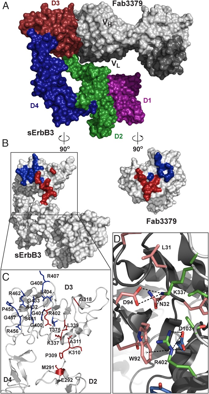
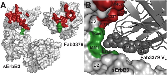
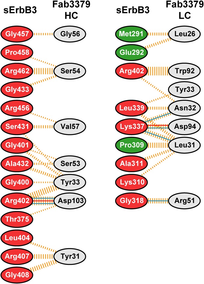
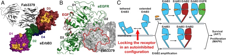
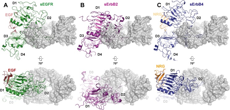
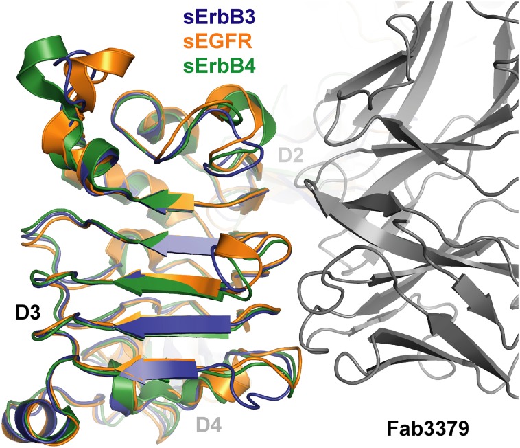
ErbB3 (HER3) is a member of the EGF receptor (EGFR) family of receptor tyrosine kinases, which, unlike the other three family members, contains a pseudo kinase in place of a tyrosine kinase domain. In cancer, ErbB3 activation is driven by a ligand-dependent mechanism through the formation of heterodimers with EGFR, ErbB2, or ErbB4 or via a ligand-independent process through heterodimerization with ErbB2 overexpressed in breast tumors or other cancers. Here we describe the crystal structure of the Fab fragment of an antagonistic monoclonal antibody KTN3379, currently in clinical development in human cancer patients, in complex with the ErbB3 extracellular domain. The structure reveals a unique allosteric mechanism for inhibition of ligand-dependent or ligand-independent ErbB3-driven cancers by binding to an epitope that locks ErbB3 in an inactive conformation. Given the similarities in the mechanism of ErbB receptor family activation, these findings could facilitate structure-based design of antibodies that inhibit EGFR and ErbB4 by an allosteric mechanism.

Keywords: cancer; cell signaling; crystal structure; surface receptor; therapeutic antibodies.


Conflict of interest statement

Conflict of interest statement: J.S. is a founder and consultant of Kolltan. G.F.L., J.S.L., E.J.N., D.A., and Y.H. are Kolltan employees.

Figures

Fig. 1.
Fig. 1.
KTN3379 binds to ErbB3 with high affinity and prevents NRG binding to ErbB3. (A) Binding of fluorescently conjugated KTN3379 (KTN3379-647) to ErbB3-expressing T47D breast cancer cells. The fluorescent intensity of cells labeled with KTN3379-647 was quantitated by flow cytometry analysis. The experiment was performed three times with similar results. (B) SPR sensorgram of sErbB3 to Fab3379 immobilized on the BIAcore sensor chip surface. (C) KTN3379 prevents binding of NRG to Ba/F3 cells expressing ErbB3. ErbB3-expressing Ba/F3 cells were incubated with (open squares) and without (filled circles) 10 nM KTN3379, followed by titration with fluorescently conjugated NRG (NRG-647). The fluorescent intensity of cells labeled with NRG-647 was quantitated by flow cytometry analysis. The experiment was performed three times with similar results. (D) KTN3379 binds to sErbB3 (filled circles) and to isolated domain 3 (open triangles). KTN3379 was titrated on ELISA plates coated with sErbB3 or each purified domain 1, 2, 3, or 4 and subsequently was incubated with an HRP-conjugated anti-human antibody. (E) SPR sensorgram of isolated domain 3 of ErbB3 to Fab3379 immobilized on a BIAcore sensor chip surface. Isolated domain 3 binds Fab3379 with an approximately twofold weaker affinity and accelerated kinetics relative to sErbB3.
Fig. S1.
Fig. S1.
KTN3379 inhibits ligand-dependent and ligand-independent ErbB3 activities in cancer cells. (A) KTN3379 inhibits ErbB3 (open circles) and AKT phosphorylation (filled triangles) in the ligand-dependent cell line MDA-MB-175 with low-nanomolar IC50 values. (B) Similarly, KTN3379 demonstrates antiproliferative activity on MDA-MB-175 cells with similar IC50 values. In this cell line, ErbB3 is activated by a NRG autocrine loop, and cell growth is largely dependent on ErbB3 activity. (C) KTN3379 inhibits ErbB3 (open circles) and AKT phosphorylation (filled triangles) in the ligand-independent cell line BT-474 with mid-picomolar IC50 values. (D) KTN3379 also demonstrates antiproliferative activity on BT-474 cells with a similar potency. This cell line expresses very high levels of ErbB2 and no detectable NRG. (E and F) KTN3379 exhibits significant single-agent antitumor activity in mouse xenograft models of human cancer. Twice weekly dosing of KTN3379 at 20 mg/kg significantly delays tumor growth of the ligand-dependent model of head-and-neck cancer Cal27 (E) and the ligand-independent model of gastric cancer NCI-N87 (F).
Fig. 2.
Fig. 2.
Crystal structure of the sErbB3–Fab3379 complex. (A) Surface representation model of the sErbB3–Fab3379 complex shows that Fab3379 (gray) binds primarily to the side of domain 3 (red) in sErbB3, to domain 2 (green), and to the hinge region between domain 2 and domain 3 and stabilizes the inactive tethered state of sErbB3. The heavy chain region is shown in light gray, and the light chain region is shown in dark gray. (B) “Open-book” representation of the sErbB3–Fab3379 complex shown in A. The Fab3379-binding epitope in sErbB3 is highlighted in blue (heavy chain epitope) and red (light chain epitope). Likewise, the sErbB3-binding paratope in Fab3379 is highlighted in blue (heavy chain) and red (light chain). (C) Detailed view of region in sErbB3 that interacts with Fab3379. The heavy chain binds exclusively to domain 3, and the light chain contacts part of domain 3, the C terminus of domain 2, and the hinge region between both domains. (D) Representative interactions between Fab3379 and domain 3 of sErbB3 in the region centered on Arg402 in sErbB3. Sidechain atoms in stick representation from the epitope in sErbB3, the heavy chain paratope in Fab3379, and the light chain paratope in Fab3379 were colored in light green, light blue, and bright red, respectively. Both Arg402 and Lys337 in sErbB3 make multiple contacts with Fab3379, contributing significantly to the overall stability of receptor antibody complex.
Fig. S2.
Fig. S2.
Overlay of Fab3379-bound sErbB3 with a tethered conformation of sErbB3. Structure-based comparison between Fab3379-bound sErbB3 (dark blue) and a previously determined sErbB3 structure (orange, PDB ID code: 1M6B) in its native tethered conformation. The overall Cα rmsd is 1.41 Å. The Fab3379 molecule is omitted for clarity.
Fig. 3.
Fig. 3.
Contacts between Fab3379 and the hinge region connecting domains 2 and 3 of ErbB3 are critical for the mechanism of inhibition. (A) Contact regions between Fab3379 and domain 3 and domain 2 of sErbB3 are colored in red and green, respectively. Also highlighted are the contacts by domain 3 (red) and domain 2 (green) on the surface of Fab3379. sErbB3 and Fab3379 are shown in same orientation as in Fig. 2B. (B) Detailed view focusing on the interaction between the Fab3379 VL (gray) and the hinge region between domain 2 (red) and domain 3 (green). CDR1 residues in Fab3379 VL wedge the antibody into the hinge region, preventing the conformational rearrangement required for transition from the autoinhibited configuration of sErbB3 to its extended activated form. Listed residues in Fab3379 VL were modified to strengthen the interaction with domain 2 of ErbB3 further, lowering the off-rate of the reaction (Fig. S5).
Fig. S3.
Fig. S3.
Summary of interactions between sErbB3 and Fab3379 identified from the crystal structure. The diagram showing the amino acid residues that make intermolecular contact was generated by PDBsum (36). The residues in sErbB3 are colored according to the domains: green for domain 2, and red for domain 3. The interactions are represented by lines connecting the residues: solid blue lines for hydrogen bonds; solid red lines for salt bridges; and dashed orange lines for nonbonded contacts. The thickness of the lines indicates the number of contacts.
Fig. 4.
Fig. 4.
KTN3379 blocks NRG binding to ErbB3 through an allosteric mechanism. (A) The sErbB3–Fab3379 complex in which NRG-binding sites in domain 1 and 3 of sErbB3 are highlighted in yellow. The NRG-binding sites were identified by superposition of the NRG-bound form of ErbB4 onto the structure of the sErbB3–Fab3379 complex. The model reveals that KTN3379 and NRG bind to mutually exclusive sites in ErbB3. (B) KTN3379 binding is incompatible with the extended form of ErbB3 and other family members. Superimposing the extended conformation of sEGFR (PDB ID code: 1IVO) on the sErbB3–Fab3379 complex centered on domain 3 reveals that KTN3379 would clash significantly with domain 2 in the extended form; the area in the extended conformation of sEGFR that would clash with Fab3379 is marked by the red dotted line. Superimposing the extended conformations of other ErbB receptors, sErbB2 and NRG-bound sErbB4, also demonstrates that KTN3379 would clash with the extended forms of all ErbB receptors (Fig. S4). (C) Mechanism of ErbB3 inhibition by KTN3379. KTN3379 binds and blocks ErbB3 in the first step of ErbB3 activation to lock the ErbB3 extracellular domain in the tethered configuration (marked with a red arrow). Similar to EGFR and ErbB4, ErbB3 exists in equilibrium between a predominantly autoinhibited form and an extended form. The tethered form of ErbB3 (light blue) is stabilized primarily by contacts between domains 2 and 4. During normal physiological stimulation and in several cancers (Upper), NRG binding induces and stabilizes the formation of a dimerization-competent form of the receptor by binding to domains 1 and 3, enabling heterodimerization with ErbB2 (red), ligand-bound EGFR (light green), or ErbB4. In tumors in which ErbB2 is highly overexpressed (Lower), ErbB3 can be activated in the absence of NRG through forced dimerization with ErbB2. In either scenario, ErbB3 activation strongly elicits proliferative and antiapoptotic signals. The dual mechanism of action of KTN3379 therefore hinges on its ability to prevent ErbB3 from sampling a dimerization-competent conformation.
Fig. S4.
Fig. S4.
KTN3379 binding is incompatible with the extended and active form of ErbB receptors. Superimposing available structures of extended ErbB receptors with the sErbB3–Fab3379 complex centered on domain 3 reveals that KTN3379 would clash significantly with domain 2 of all receptors. Ligand-bound sEGFR (PDB ID code: 1IVO) (A), sErbB2 (PDB ID code: 2A91) (B), and NRG-bound sErbB4 (PDB ID code: 3U7U) (C) demonstrate that Fab3379 (gray) clashes with the extended forms of ErbB receptors.
Fig. S5.
Fig. S5.
Improvement of KTN3379 binding kinetics by structure-guided modifications. SPR sensorgrams of sErbB3 to Fab3379 and its VL variants, S23R and S25H, immobilized on a BIAcore sensor chip. A single-cycle kinetics method was used with an extended dissociation time (4,800 s) to compare the off-rates. Measurements were done in triplicate.
Fig. S6.
Fig. S6.
EGFR and ErbB4 share a similar topology with ErbB3 in the KTN3379-binding epitope. An overlay of the autoinhibited structures of EGFR (orange; PDB ID code: 1YY9), ErbB4 (green; PDB ID code: 2AHX), and the sErbB3 (dark blue)–Fab3379 (gray) complex centered on domain 3 highlights the overall architectural conservation of the KTN3379-binding epitope among autoinhibited ErbB receptors. Minor clashes arise from differences in a flexible loop within domain 3. By contrast, this epitope is not present in ErbB2 because it exists in a constitutively extended state. The same mechanism of inhibition therefore could be exploited in EGFR and ErbB4 with KTN3379-like antibodies.

Similar articles

Cited by

References

    1. Lemmon MA, Schlessinger J. Cell signaling by receptor tyrosine kinases. Cell. 2010;141(7):1117–1134. - PMC - PubMed
    1. Arteaga CL, Engelman JA. ERBB receptors: From oncogene discovery to basic science to mechanism-based cancer therapeutics. Cancer Cell. 2014;25(3):282–303. - PMC - PubMed
    1. Arteaga CL. ErbB-targeted therapeutic approaches in human cancer. Exp Cell Res. 2003;284(1):122–130. - PubMed
    1. Falls DL. Neuregulins: Functions, forms, and signaling strategies. Exp Cell Res. 2003;284(1):14–30. - PubMed
    1. Shi F, Telesco SE, Liu Y, Radhakrishnan R, Lemmon MA. ErbB3/HER3 intracellular domain is competent to bind ATP and catalyze autophosphorylation. Proc Natl Acad Sci USA. 2010;107(17):7692–7697. - PMC - PubMed

Publication types

Associated data