Replacement of brain-resident myeloid cells does not alter cerebral amyloid-β deposition in mouse models of Alzheimer's disease
- PMID: 26458770
- PMCID: PMC4612086
- DOI: 10.1084/jem.20150478
Replacement of brain-resident myeloid cells does not alter cerebral amyloid-β deposition in mouse models of Alzheimer's disease
Abstract
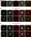
Immune cells of myeloid lineage are encountered in the Alzheimer's disease (AD) brain, where they cluster around amyloid-β plaques. However, assigning functional roles to myeloid cell subtypes has been problematic, and the potential for peripheral myeloid cells to alleviate AD pathology remains unclear. Therefore, we asked whether replacement of brain-resident myeloid cells with peripheral monocytes alters amyloid deposition in two mouse models of cerebral β-amyloidosis (APP23 and APPPS1). Interestingly, early after repopulation, infiltrating monocytes neither clustered around plaques nor showed Trem2 expression. However, with increasing time in the brain, infiltrating monocytes became plaque associated and also Trem2 positive. Strikingly, however, monocyte repopulation for up to 6 mo did not modify amyloid load in either model, independent of the stage of pathology at the time of repopulation. Our results argue against a long-term role of peripheral monocytes that is sufficiently distinct from microglial function to modify cerebral β-amyloidosis. Therefore, myeloid replacement by itself is not likely to be effective as a therapeutic approach for AD.
© 2015 Varvel et al.
Figures


Comment in
-
Peripheral macrophages not ADept at amyloid clearance.J Exp Med. 2015 Oct 19;212(11):1758. doi: 10.1084/jem.21211insight5. J Exp Med. 2015. PMID: 26482143 Free PMC article. No abstract available.
Similar articles
-
Impact of peripheral myeloid cells on amyloid-β pathology in Alzheimer's disease-like mice.J Exp Med. 2015 Oct 19;212(11):1811-8. doi: 10.1084/jem.20150479. Epub 2015 Oct 12. J Exp Med. 2015. PMID: 26458768 Free PMC article.
-
Therapeutic effects of glatiramer acetate and grafted CD115⁺ monocytes in a mouse model of Alzheimer's disease.Brain. 2015 Aug;138(Pt 8):2399-422. doi: 10.1093/brain/awv150. Epub 2015 Jun 6. Brain. 2015. PMID: 26049087 Free PMC article.
-
TREM2-mediated early microglial response limits diffusion and toxicity of amyloid plaques.J Exp Med. 2016 May 2;213(5):667-75. doi: 10.1084/jem.20151948. Epub 2016 Apr 18. J Exp Med. 2016. PMID: 27091843 Free PMC article.
-
Elucidating the Role of TREM2 in Alzheimer's Disease.Neuron. 2017 Apr 19;94(2):237-248. doi: 10.1016/j.neuron.2017.02.042. Neuron. 2017. PMID: 28426958 Review.
-
Monocytes and Alzheimer's disease.Neurosci Bull. 2011 Apr;27(2):115-22. doi: 10.1007/s12264-011-1205-3. Neurosci Bull. 2011. PMID: 21441973 Free PMC article. Review.
Cited by
-
Allogenic microglia replacement: A novel therapeutic strategy for neurological disorders.Fundam Res. 2023 Mar 27;4(2):237-245. doi: 10.1016/j.fmre.2023.02.025. eCollection 2024 Mar. Fundam Res. 2023. PMID: 38933508 Free PMC article. Review.
-
Impact of peripheral myeloid cells on amyloid-β pathology in Alzheimer's disease-like mice.J Exp Med. 2015 Oct 19;212(11):1811-8. doi: 10.1084/jem.20150479. Epub 2015 Oct 12. J Exp Med. 2015. PMID: 26458768 Free PMC article.
-
Microglia Function in the Central Nervous System During Health and Neurodegeneration.Annu Rev Immunol. 2017 Apr 26;35:441-468. doi: 10.1146/annurev-immunol-051116-052358. Epub 2017 Feb 9. Annu Rev Immunol. 2017. PMID: 28226226 Free PMC article. Review.
-
Clearance of cerebral Aβ in Alzheimer's disease: reassessing the role of microglia and monocytes.Cell Mol Life Sci. 2017 Jun;74(12):2167-2201. doi: 10.1007/s00018-017-2463-7. Epub 2017 Feb 14. Cell Mol Life Sci. 2017. PMID: 28197669 Free PMC article. Review.
-
Peripheral administration of the soluble TNF inhibitor XPro1595 modifies brain immune cell profiles, decreases beta-amyloid plaque load, and rescues impaired long-term potentiation in 5xFAD mice.Neurobiol Dis. 2017 Jun;102:81-95. doi: 10.1016/j.nbd.2017.02.010. Epub 2017 Feb 24. Neurobiol Dis. 2017. PMID: 28237313 Free PMC article.
References
-
- Cartier N., Hacein-Bey-Abina S., Bartholomae C.C., Veres G., Schmidt M., Kutschera I., Vidaud M., Abel U., Dal-Cortivo L., Caccavelli L., et al. . 2009. Hematopoietic stem cell gene therapy with a lentiviral vector in X-linked adrenoleukodystrophy. Science. 326:818–823. 10.1126/science.1171242 - DOI - PubMed
Publication types
MeSH terms
Substances
LinkOut - more resources
Full Text Sources
Other Literature Sources
Medical
Molecular Biology Databases

