Quantifying the stabilizing effects of protein-ligand interactions in the gas phase
- PMID: 26440106
- PMCID: PMC4600733
- DOI: 10.1038/ncomms9551
Quantifying the stabilizing effects of protein-ligand interactions in the gas phase
Abstract
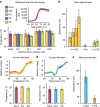
The effects of protein-ligand interactions on protein stability are typically monitored by a number of established solution-phase assays. Few translate readily to membrane proteins. We have developed an ion-mobility mass spectrometry approach, which discerns ligand binding to both soluble and membrane proteins directly via both changes in mass and ion mobility, and assesses the effects of these interactions on protein stability through measuring resistance to unfolding. Protein unfolding is induced through collisional activation, which causes changes in protein structure and consequently gas-phase mobility. This enables detailed characterization of the ligand-binding effects on the protein with unprecedented sensitivity. Here we describe the method and software required to extract from ion mobility data the parameters that enable a quantitative analysis of individual binding events. This methodology holds great promise for investigating biologically significant interactions between membrane proteins and both drugs and lipids that are recalcitrant to characterization by other means.
Figures


Similar articles
-
Collision induced unfolding of protein ions in the gas phase studied by ion mobility-mass spectrometry: the effect of ligand binding on conformational stability.J Am Soc Mass Spectrom. 2009 Oct;20(10):1851-8. doi: 10.1016/j.jasms.2009.06.010. Epub 2009 Jul 1. J Am Soc Mass Spectrom. 2009. PMID: 19643633
-
Characterization of Membrane Protein-Lipid Interactions by Mass Spectrometry Ion Mobility Mass Spectrometry.J Am Soc Mass Spectrom. 2017 Apr;28(4):579-586. doi: 10.1007/s13361-016-1555-1. Epub 2016 Dec 6. J Am Soc Mass Spectrom. 2017. PMID: 27924494
-
Mass spectrometry of protein complexes: from origins to applications.Annu Rev Phys Chem. 2015 Apr;66:453-74. doi: 10.1146/annurev-physchem-040214-121732. Epub 2015 Jan 14. Annu Rev Phys Chem. 2015. PMID: 25594852 Review.
-
Collisional unfolding of multiprotein complexes reveals cooperative stabilization upon ligand binding.Protein Sci. 2015 Aug;24(8):1272-81. doi: 10.1002/pro.2699. Epub 2015 May 27. Protein Sci. 2015. PMID: 25970849 Free PMC article.
-
Ion mobility-mass spectrometry as a tool to investigate protein-ligand interactions.Anal Bioanal Chem. 2017 Jul;409(18):4305-4310. doi: 10.1007/s00216-017-0384-9. Epub 2017 May 13. Anal Bioanal Chem. 2017. PMID: 28500372 Review.
Cited by
-
High-Performance Molecular Dynamics Simulations for Native Mass Spectrometry of Large Protein Complexes with the Fast Multipole Method.Anal Chem. 2024 Sep 17;96(37):15023-15030. doi: 10.1021/acs.analchem.4c03272. Epub 2024 Sep 4. Anal Chem. 2024. PMID: 39231152 Free PMC article.
-
Assembly and regulation of the chlorhexidine-specific efflux pump AceI.Proc Natl Acad Sci U S A. 2020 Jul 21;117(29):17011-17018. doi: 10.1073/pnas.2003271117. Epub 2020 Jul 7. Proc Natl Acad Sci U S A. 2020. PMID: 32636271 Free PMC article.
-
Protein Complex Heterogeneity and Topology Revealed by Electron Capture Charge Reduction and Surface Induced Dissociation.ACS Cent Sci. 2024 Jul 26;10(8):1537-1547. doi: 10.1021/acscentsci.4c00461. eCollection 2024 Aug 28. ACS Cent Sci. 2024. PMID: 39220701 Free PMC article.
-
Effects of Nano-Electrospray Ionization Emitter Position on Unintentional In-Source Activation of Peptide and Protein Ions.J Am Soc Mass Spectrom. 2024 Mar 6;35(3):498-507. doi: 10.1021/jasms.3c00371. Epub 2024 Feb 19. J Am Soc Mass Spectrom. 2024. PMID: 38374644
-
Negative Ions Enhance Survival of Membrane Protein Complexes.J Am Soc Mass Spectrom. 2016 Jun;27(6):1099-104. doi: 10.1007/s13361-016-1381-5. Epub 2016 Apr 22. J Am Soc Mass Spectrom. 2016. PMID: 27106602 Free PMC article.
References
-
- Marcoux J. & Robinson Carol V. Twenty years of gas phase structural biology. Structure 21, 1541–1550 (2013) . - PubMed
-
- Lanucara F., Holman S. W., Gray C. J. & Eyers C. E. The power of ion mobility-mass spectrometry for structural characterization and the study of conformational dynamics. Nat. Chem. 6, 281–294 (2014) . - PubMed
-
- Niu S., Rabuck J. N. & Ruotolo B. T. Ion mobility-mass spectrometry of intact protein–ligand complexes for pharmaceutical drug discovery and development. Curr. Opin. Chem. Biol. 17, 809–817 (2013) . - PubMed
-
- Benesch J. L. P. Collisional activation of protein complexes: picking up the pieces. J. Am. Soc. Mass. Spectrom. 20, 341–348 (2009) . - PubMed
-
- Ruotolo B. T. et al.. Ion mobility-mass spectrometry reveals long-lived, unfolded intermediates in the dissociation of protein complexes. Angew. Chem. Int. Ed. Engl. 46, 8001–8004 (2007) . - PubMed
Publication types
MeSH terms
Substances
Grants and funding
LinkOut - more resources
Full Text Sources
Other Literature Sources

