. 2015 Oct 2;11(10):e1005195.
doi: 10.1371/journal.ppat.1005195. eCollection 2015 Oct.

Differentiation-Dependent KLF4 Expression Promotes Lytic Epstein-Barr Virus Infection in Epithelial Cells

Affiliations

Differentiation-Dependent KLF4 Expression Promotes Lytic Epstein-Barr Virus Infection in Epithelial Cells

Dhananjay M Nawandar et al. PLoS Pathog. .

Abstract

Epstein-Barr virus (EBV) is a human herpesvirus associated with B-cell and epithelial cell malignancies. EBV lytically infects normal differentiated oral epithelial cells, where it causes a tongue lesion known as oral hairy leukoplakia (OHL) in immunosuppressed patients. However, the cellular mechanism(s) that enable EBV to establish exclusively lytic infection in normal differentiated oral epithelial cells are not currently understood. Here we show that a cellular transcription factor known to promote epithelial cell differentiation, KLF4, induces differentiation-dependent lytic EBV infection by binding to and activating the two EBV immediate-early gene (BZLF1 and BRLF1) promoters. We demonstrate that latently EBV-infected, telomerase-immortalized normal oral keratinocyte (NOKs) cells undergo lytic viral reactivation confined to the more differentiated cell layers in organotypic raft culture. Furthermore, we show that endogenous KLF4 expression is required for efficient lytic viral reactivation in response to phorbol ester and sodium butyrate treatment in several different EBV-infected epithelial cell lines, and that the combination of KLF4 and another differentiation-dependent cellular transcription factor, BLIMP1, is highly synergistic for inducing lytic EBV infection. We confirm that both KLF4 and BLIMP1 are expressed in differentiated, but not undifferentiated, epithelial cells in normal tongue tissue, and show that KLF4 and BLIMP1 are both expressed in a patient-derived OHL lesion. In contrast, KLF4 protein is not detectably expressed in B cells, where EBV normally enters latent infection, although KLF4 over-expression is sufficient to induce lytic EBV reactivation in Burkitt lymphoma cells. Thus, KLF4, together with BLIMP1, plays a critical role in mediating lytic EBV reactivation in epithelial cells.


Conflict of interest statement

The authors have declared that no competing interests exist.

Figures
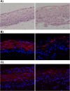
Fig 1
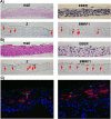
Fig 1. EBV-infected NOKs cells retain ability to be partially differentiated.
Uninfected NOKs (left panel) and EBV-infected NOKs-Akata (right panel) cells were grown in organotypic raft culture, samples formalin fixed, embedded in paraffin and 5-micron thick sections analyzed. A). Images from hematoxylin and eosin (H&E) stained sections are shown. Note the presence of squames at the top of the NOKs-Akata raft indicative of terminal differentiation. Also note that the basal compartment of the NOKs-Akata raft is much less well organized with signs of migration/invasion of the epithelial cells into the underlying dermal equivalent (poorly stained portion of the image) at the bottom of the image. Immunofluorescent images from sections of rafts stained with antibody against the epithelial cell differentiation markers, cytokeratin 10 (B), and involucrin (C) are shown. Cytokeratin 10 (K10)- and involucrin- positive cells are stained red. Blue nuclear counter stain is DAPI. Note the paucity of K10- and involucrin- positive cells in the NOKs-Akata raft indicating a perturbation of normal differentiation by EBV, even though morphologically terminal differentiation does occur as evidenced by the presence of squames.
Fig 2
Fig 2. Lytic EBV protein expression in NOKs-Akata cells is restricted to the more differentiated cell layers.
Two different independently generated NOKs-Akata cell lines (panels A and B) were grown in organotypic air-interface raft culture, and in situ hybridization or immunohistochemistry was performed to detect expression of the EBV EBERs or lytic EBV proteins (Z and BMRF1) as indicated. Examples of Z and BMRF1 stained cells are indicated by red arrows. C) NOKs-Akata cells grown in organotypic air-interface raft culture were examined by immunofluorescence using both anti-K10 (red) and anti-Z (green) antibodies. An example of a Z and K10 co-staining cell is shown in the left panel, and a Z-positive/K10 negative cell is shown in the right panel.
Fig 3
Fig 3. Treatment of NOKs-Akata cells with the differentiating agents, TPA and calcium chloride/serum, results in lytic EBV reactivation.
A) NOKs-Akata cells were treated with either TPA (20 ng/mL in K-SFM) or calcium chloride plus serum (1.2 mM CaCl in RPMI + 10% FBS) for 48 hours. Immunoblot analysis was performed to compare the levels of lytic viral proteins, Z and BMRF1, a differentiation-dependent cellular protein, involucrin, and the cellular protein, GAPDH (a loading control). B) NOKs-Akata cells were treated with TPA in the presence or absence of a ROCK inhibitor (Y27632) (10 μM) for 48 hours and immunoblot analysis was performed to compare the levels of Z, BMRF1 and involucrin. The cellular protein tubulin was used as a loading control.
Fig 4
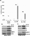
Fig 4. KLF4 activates both the Zp and Rp IE EBV promoters in reporter gene assays and induces lytic EBV reactivation when over-expressed in latently infected epithelial cells.
A) Reporter gene constructs containing either the BZLF1 promoter (Zp-668), the BRLF1 promoter (Rp-1068), or no promoter sequences upstream of the luciferase gene were co-transfected into EBV-negative NOKs cells with either control vector or a KLF4 expression vector as indicated, and luciferase assays was performed 2 days after transfection. Total luciferase activity for each of the conditions from a representative experiment is shown (average +/- the standard deviation of results from three replicates), as well as the fold-increase in activity induced by KLF4. Similar results were obtained in three separate experiments. B) NOKs-Akata cells and C) HONE-Akata cells were transfected with either control vector or a KLF4 expression vector and immunoblot analysis was performed to compare the levels of transfected KLF4 and lytic viral proteins Z, R and BMRF1. Actin or tubulin served as a loading control.
Fig 5
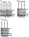
Fig 5. Endogenous KLF4 is required for TPA and sodium butyrate mediated lytic EBV reactivation in epithelial cell lines.
EBV-positive CNE-2 Akata cells were transfected with either control siRNA or two different KLF4 siRNAs, and then treated with TPA (20 ng/uL) (A) or sodium butyrate (3mM) (B) for 48 hours. Immunoblot analysis was performed to detect expression of endogenous KLF4, and lytic viral proteins Z, R and BMRF1. Actin served as a loading control. C) NOKs-Akata cells, transfected with either control siRNA or KLF4 siRNAs, were treated with TPA (20 ng/uL) for 48 hours and immunoblot analysis was performed to compare the levels of endogenous KLF4 and lytic viral proteins Z, R and BMRF1. Tubulin served as a loading control.
Fig 6
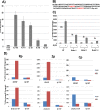
Fig 6. KLF4 binds to both the Zp and Rp IE EBV promoters, and enhances their association with activated RNA polymerase II.
A) Various 5’ Rp deletion luciferase constructs were co-transfected into EBV-negative NOKs cells with either control vector or a KLF4 expression vector, and luciferase assay was performed 2 days after transfection. The KLF4-induced fold-change in luciferase activity for each construct is shown relative to the activity of the promoter in presence of control vector (set to 1). Values shown are the average +/- the standard deviation of results from two replicates. B) The EBV Rp sequence located between -551 and -441 relative to the transcriptional start site is shown. KLF4 consensus binding sites are highlighted in red. C) The Rp -551 construct (with or without KLF4 consensus site mutants) were co-transfected into EBV negative NOKs cells with control vector or KLF4 expression vector. Luciferase activity for each of the conditions is shown. Values are given as average +/- the standard deviations of results from two replicates. KLF4 mutant 1 alters the CACCC motif and mutant 2 alters the GGGTG motif. D) HONE-Akata cells were transfected with either control vector or a KLF4 expression vector, and ChIP assay was performed 48 hours after transfection. Cross-linked DNA-protein complexes were immunoprecipitated using anti-KLF4 antibody (top panel), or anti-phospho-RNA polymerase II antibody (bottom panel) and control IgG antibody in each case. Quantitative PCR was performed to quantitate the amount of DNA pulled down for the IE Rp (left panel), Zp (middle panel) and negative control Cp (right panel) EBV promoters.
Fig 7
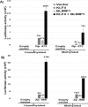
Fig 7. KLF4 synergizes with BLIMP1 to activate both Rp and Zp, irrespective of the promoter methylation state.
A) Mock treated or methylated promoterless (empty) and Rp-673 pCpGL luciferase constructs were co-transfected into EBV-negative NOKs cells with either control vector, KLF4 vector alone, BLIMP1 vector alone, or the combination of KLF4 and BLIMP1 vectors. Luciferase activity for each of the conditions is shown; values are given as average +/- the standard deviations of results from two replicates. B) Mock treated or methylated promoterless vector and Zp-668 pCpGL luciferase constructs were co-transfected into EBV-negative NOKs cells with control vector, KLF4 vector, BLIMP1 vector or KLF4 and BLIMP1. Luciferase activity for each of the conditions is shown; values are given as average +/- the standard deviations of results from two replicates.
Fig 8
Fig 8. KLF4 synergizes with BLIMP1 to induce lytic EBV reactivation and replication in latently infected epithelial cells.
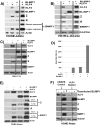
Control vector or KLF4 and BLIMP1 expression vectors (either alone or in combination) were transfected into A) HONE-Akata cells, B) NOKs-Akata cells, or C) SNU.719 gastric carcinoma cells and immunoblot analysis was performed to compare the levels of transfected KLF4 and BLIMP1, and induction of EBV lytic viral proteins Z, R and BMRF1. Actin or GAPDH served as a loading control. D) An infectious viral titer assay was performed, as described in materials and methods, using the supernatant of CNE2-Akata cells transfected with vector control, KLF4, BLIMP1, and KLF4 and BLIMP1, five days post transfection. E) Immunoblot analysis for the samples shown in Fig 8D. F) HONE-Akata cells in which KLF4 was knocked-out using CRISPR-Cas9 technology or control HONE-Akata cells were transfected with either control vector or a BLIMP1 expression vector and immunoblot analysis was performed to compare the levels of endogenous KLF4, transfected BLIMP1, and lytic viral proteins Z, R and BMRF1. Tubulin served as a loading control.
Fig 9
Fig 9. KLF4 and BLIMP1 expression is differentiation-dependent in normal tongue tissue.
Immunohistochemistry analysis was performed on paraffin-embedded formalin-fixed normal tongue tissue (A), or rafted NOKs-Akata cells (B), using antibodies directed against KLF4 and BLIMP1 as indicated. Examples of positively-staining cells are indicated by red arrows (Images:40x).
Fig 10
Fig 10. OHL cells express both KLF4 and BLIMP1.
H&E analysis (Image: 40X), and immunohistochemistry analysis was performed on a paraffin-embedded, formalin-fixed biopsy of an oral hairy leukoplakia (OHL) lesion using antibodies directed against Z, BMRF1, KLF4 and BLIMP1 as indicated (Images: 100x of region boxed in H & E stain). Examples of OHL cells, positively staining for each of these proteins, are indicated by black arrows.
Fig 11
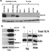
Fig 11. KLF4 is not expressed in B cells but can reactivate lytic EBV gene expression in this cell type.
A) Immunoblot analysis was performed to compare the levels of endogenous KLF4 expression in a series of EBV-infected epithelial cell lines and EBV-infected B cell lines. Actin served as a loading control. B) Raji Burkitt lymphoma cells were transfected with either control vector or a KLF4 expression vector and immunoblot analysis was performed to compare the levels of transfected KLF4 and lytic viral proteins Z, R and BMRF1. Actin served as a loading control. C) The levels of transfected KLF4 into Raji cells (from 11B) was compared to the endogenous levels of KLF4 in AGS-Akata cells.

Similar articles

Cited by

References

    1. Henle G, Henle W, Diehl V. Relation of Burkitt’s tumor-associated herpes-y type virus to infectious mononucleosis. Proc Natl Acad Sci U S A. 1968;59: 94–101. - PMC - PubMed
    1. Rickinson A. B., Kieff Elliot. Epstein-Barr Virus Fields Virology. 5th ed. Philadelphia: Wolters Kluwer Health/ Lippincott Williams & Wilkins; 2007. pp. 2655–2700.
    1. Zur Hausen H, Schulte-Holthausen H, Klein G, Henle W, Henle G, Clifford P, et al. EBV DNA in biopsies of Burkitt tumours and anaplastic carcinomas of the nasopharynx. Nature. 1970;228: 1056–1058. - PubMed
    1. Kieff Elliot D, Rickinson A. B. Epstein-Barr Virus and Its Replication Fields Virology. 5th ed. Philadelphia: Wolters Kluwer Health/ Lippincott Williams & Wilkins; 2007. pp. 2603–2654.
    1. Souza TA, Stollar BD, Sullivan JL, Luzuriaga K, Thorley-Lawson DA. Peripheral B cells latently infected with Epstein-Barr virus display molecular hallmarks of classical antigen-selected memory B cells. Proc Natl Acad Sci U S A. 2005;102: 18093–18098. - PMC - PubMed

Publication types

MeSH terms

Substances