Targeting of HPV-16+ Epithelial Cancer Cells by TCR Gene Engineered T Cells Directed against E6
- PMID: 26429982
- PMCID: PMC4603283
- DOI: 10.1158/1078-0432.CCR-14-3341
Targeting of HPV-16+ Epithelial Cancer Cells by TCR Gene Engineered T Cells Directed against E6
Abstract
Purpose: The E6 and E7 oncoproteins of HPV-associated epithelial cancers are in principle ideal immunotherapeutic targets, but evidence that T cells specific for these antigens can recognize and kill HPV(+) tumor cells is limited. We sought to determine whether TCR gene engineered T cells directed against an HPV oncoprotein can successfully target HPV(+) tumor cells.
Experimental design: T-cell responses against the HPV-16 oncoproteins were investigated in a patient with an ongoing 22-month disease-free interval after her second resection of distant metastatic anal cancer. T cells genetically engineered to express an oncoprotein-specific TCR from this patient's tumor-infiltrating T cells were tested for specific reactivity against HPV(+) epithelial tumor cells.
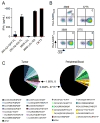
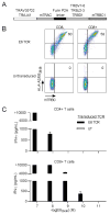
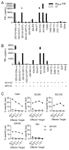
Results: We identified, from an excised metastatic anal cancer tumor, T cells that recognized an HLA-A*02:01-restricted epitope of HPV-16 E6. The frequency of the dominant T-cell clonotype from these cells was approximately 400-fold greater in the patient's tumor than in her peripheral blood. T cells genetically engineered to express the TCR from this clonotype displayed high avidity for an HLA-A*02:01-restricted epitope of HPV-16, and they showed specific recognition and killing of HPV-16(+) cervical, and head and neck cancer cell lines.
Conclusions: These findings demonstrate that HPV-16(+) tumors can be targeted by E6-specific TCR gene engineered T cells, and they provide the foundation for a novel cellular therapy directed against HPV-16(+) malignancies, including cervical, oropharyngeal, anal, vulvar, vaginal, and penile cancers.
©2015 American Association for Cancer Research.
Conflict of interest statement
Conflict of interest statement: Drs. Hinrichs and Rosenberg are inventors on an NIH patent related to this work. This research was funded in part by a cooperative research and development agreement with Kite Pharma.
Figures


Similar articles
-
A conserved E7-derived cytotoxic T lymphocyte epitope expressed on human papillomavirus 16-transformed HLA-A2+ epithelial cancers.J Biol Chem. 2010 Sep 17;285(38):29608-22. doi: 10.1074/jbc.M110.126722. Epub 2010 Jul 8. J Biol Chem. 2010. PMID: 20615877 Free PMC article.
-
Identification of an HLA-A24-restricted cytotoxic T lymphocyte epitope from human papillomavirus type-16 E6: the combined effects of bortezomib and interferon-gamma on the presentation of a cryptic epitope.Int J Cancer. 2007 Feb 1;120(3):594-604. doi: 10.1002/ijc.22312. Int J Cancer. 2007. PMID: 17096336
-
Identification of a naturally processed HLA-A*0201 HPV18 E7 T cell epitope by tumor cell mediated in vitro vaccination.Int J Cancer. 2003 Apr 10;104(3):345-53. doi: 10.1002/ijc.10940. Int J Cancer. 2003. PMID: 12569558
-
Genetically modified cellular vaccines for therapy of human papilloma virus type 16 (HPV 16)-associated tumours.Curr Cancer Drug Targets. 2008 May;8(3):180-6. doi: 10.2174/156800908784293596. Curr Cancer Drug Targets. 2008. PMID: 18473731 Review.
-
Immunotherapy in Anal Cancer.Curr Oncol. 2023 Apr 27;30(5):4538-4550. doi: 10.3390/curroncol30050343. Curr Oncol. 2023. PMID: 37232801 Free PMC article. Review.
Cited by
-
Immune Activation in Patients with Locally Advanced Cervical Cancer Treated with Ipilimumab Following Definitive Chemoradiation (GOG-9929).Clin Cancer Res. 2020 Nov 1;26(21):5621-5630. doi: 10.1158/1078-0432.CCR-20-0776. Epub 2020 Aug 18. Clin Cancer Res. 2020. PMID: 32816895 Free PMC article.
-
T-Cell Receptor Gene Therapy for Human Papillomavirus-Associated Epithelial Cancers: A First-in-Human, Phase I/II Study.J Clin Oncol. 2019 Oct 20;37(30):2759-2768. doi: 10.1200/JCO.18.02424. Epub 2019 Aug 13. J Clin Oncol. 2019. PMID: 31408414 Free PMC article. Clinical Trial.
-
Immunotherapy in cervical cancer: From the view of scientometric analysis and clinical trials.Front Immunol. 2023 Feb 3;14:1094437. doi: 10.3389/fimmu.2023.1094437. eCollection 2023. Front Immunol. 2023. PMID: 36817443 Free PMC article. Clinical Trial.
-
The common HLA class I-restricted tumor-infiltrating T cell response in HPV16-induced cancer.Cancer Immunol Immunother. 2023 Jun;72(6):1553-1565. doi: 10.1007/s00262-022-03350-x. Epub 2022 Dec 16. Cancer Immunol Immunother. 2023. PMID: 36526910 Free PMC article.
-
Human Papillomavirus E6 and E7: The Cervical Cancer Hallmarks and Targets for Therapy.Front Microbiol. 2020 Jan 21;10:3116. doi: 10.3389/fmicb.2019.03116. eCollection 2019. Front Microbiol. 2020. PMID: 32038557 Free PMC article. Review.
References
Publication types
MeSH terms
Substances
Grants and funding
LinkOut - more resources
Full Text Sources
Other Literature Sources
Molecular Biology Databases
Research Materials

