Dual-Affinity Re-Targeting proteins direct T cell-mediated cytolysis of latently HIV-infected cells

Julia A M Sung et al. J Clin Invest. .

Abstract

Enhancement of HIV-specific immunity is likely required to eliminate latent HIV infection. Here, we have developed an immunotherapeutic modality aimed to improve T cell-mediated clearance of HIV-1-infected cells. Specifically, we employed Dual-Affinity Re-Targeting (DART) proteins, which are bispecific, antibody-based molecules that can bind 2 distinct cell-surface molecules simultaneously. We designed DARTs with a monovalent HIV-1 envelope-binding (Env-binding) arm that was derived from broadly binding, antibody-dependent cellular cytotoxicity-mediating antibodies known to bind to HIV-infected target cells coupled to a monovalent CD3 binding arm designed to engage cytolytic effector T cells (referred to as HIVxCD3 DARTs). Thus, these DARTs redirected polyclonal T cells to specifically engage with and kill Env-expressing cells, including CD4+ T cells infected with different HIV-1 subtypes, thereby obviating the requirement for HIV-specific immunity. Using lymphocytes from patients on suppressive antiretroviral therapy (ART), we demonstrated that DARTs mediate CD8+ T cell clearance of CD4+ T cells that are superinfected with the HIV-1 strain JR-CSF or infected with autologous reservoir viruses isolated from HIV-infected-patient resting CD4+ T cells. Moreover, DARTs mediated CD8+ T cell clearance of HIV from resting CD4+ T cell cultures following induction of latent virus expression. Combined with HIV latency reversing agents, HIVxCD3 DARTs have the potential to be effective immunotherapeutic agents to clear latent HIV-1 reservoirs in HIV-infected individuals.


Figures
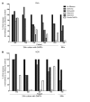
Figure 8
Figure 8. LCA to assess HIVxCD3 DART–redirected CD8+ T cell activity.
Resting CD4+ T cells from HIV-infected, ART-suppressed patients were incubated with PHA (A) or VOR (B), plated in 12–36 replicate wells depending on the size of the patient’s latent reservoir, and cocultured with autologous CD8+ T cells at an E/T ratio of 1:10 in the absence or presence of HIVxCD3 or control DARTs at 100 ng/ml for 24 hours (or up to 96 hours where indicated), after which DARTs were washed off and CD8-depleted PBMCs from a seronegative donor were added to amplify residual virus. Wells were assessed for the presence or absence of p24 by ELISA at day 15. Combo indicates a 1:1 cocktail of 7B2xCD3 and A32xCD3 at a total concentration of 100 ng/ml. Results are shown as percent viral recovery (number of positive wells/total number plated), normalized to a control in which no CD8+ T cells are added. Dashed lines indicate undetectable viral recovery following incubation with combo DARTs, except for patient 408 in panel B, where they indicate undetectable viral recovery after 7B2xCD3 or A32xCD3 individually. NT indicates conditions not tested due to low cell availability. NT generally refers to 2 conditions not tested (7B2xCD3 or A32xCD3 individually), except for patient 408 in panel B, where NT refers to combo DARTs not tested. Refer to Supplemental Table 5 for the individual experimental values for each of the conditions tested.
Figure 7
Figure 7. Viral clearance assay detects HIVxCD3 DART–redirected CD8+ T cell clearance of JR-CSF– or AR virus–infected CD4+ cells using lymphocytes from HIV-infected ART suppressed patients.
CD4+ depleted T cells from HIV-infected, ART-suppressed patients were activated with PHA and infected with HIV-1 subtype B clone JR-CSF (AC) or AR virus isolates (DF), and then incubated without (A and D) or with autologous CD8+ T effector cells at E/T ratios of 1:10 (B and E) or 1:1 (C and F) in the absence (No DART) or presence of HIVxCD3 (A32xCD3, 7B2xCD3) or control (7B2x4420, 4420xCD3) DARTs at a concentration of 100 ng/ml for 7 days. Combo indicates a 1:1 cocktail of 7B2xCD3 and A32xCD3 at a total concentration of 100 ng/ml. Each bar represents the log-fold reduction of p24 detected in culture supernatants, calculated as the log (p24 levels of infected target cells divided by p24 levels of the test condition). (G) Schematic of gating strategy to identify live/CD3+CD4+CD107+ effector (TFL4) T cells after their incubation with HIV-1 JR-CSF–infected target cells in presence of DARTs for 6 hours. (H) The percentage of live/effector cells (TFL4)/CD3+/CD4+/107a+ cells following a 6-hour incubation with the indicated DARTs and JR-CSF–infected targets in n = 4 patients. Error bars represent ± SEM of n = 8 (AC, except for combo [n = 5] and 7B2x4420 [n = 6]), n = 5 (DF), and n = 4 (G and H). *P < 0.05 with Dunnett’s test for multiple comparisons.
Figure 6
Figure 6. Viral clearance assay to assess HIVxCD3 DART redirected CD8+ T cell killing of autologous JR-CSF–infected CD4+ T cells from healthy HIV seronegative donors.
Activated CD4+ T cells from HIV seronegative donors were infected with HIV-1 clone JR-CSF and then incubated with autologous resting CD8+ T effector cells at an E/T ratio of 1:1 in the absence (No DART) or presence of HIVxCD3 or control DARTs at a concentration of 100 ng/ml for 7 days. Results are shown for 2 healthy donors (A and B), as well as for healthy donor 2 in the presence of integrase and nonnucleoside reverse transcriptase inhibitors during the coculture period to inhibit virus replication (C). Each bar represents the absolute p24 concentration detected in culture supernatants. Error bars represent standard error mean (± SEM) of n = 3. *P < 0.05 with Dunnett’s test for multiple comparisons.
Figure 5
Figure 5. HIVxCD3 DARTs induce specific degranulation of CD8+ T cells.
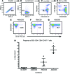
(AD) Schematic of gating strategy to identify live/CD3+CD8+CD107+ T cells after their incubation with HIV-1 BaL–infected target cells in the presence of DARTs for 6 hours. (EG) Dot plots represent the percentage of live/CD3+CD8+CD107+ cells observed in the presence of 1 ng/ml of 4420xCD3 (E), 7B2xCD3 (F), or A32xCD3 (G). (H) Frequency of the CD3+CD4CD8+CD107+ T cells observed in each of the 5 HIV-1 seronegative healthy donors after 6 hours of incubation with the autologous infected CD4+ T cells using the E/T ratio of 33:1. Each symbol represents the average of duplicate stimulations performed for each donor. The lines represent the mean ± SD. *P < 0.05 after Dunnett’s test for multiple comparisons.
Figure 4
Figure 4. HIVxCD3 DARTs redirect T cell cytotoxicity against CD4+ cells infected with HIV-1 IMCs of different subtypes.
(AC) DART-concentration dependence. Activated CD4+ T cells from a HIV-1 seronegative donor were infected with HIV-1 subtype B BaL (A), subtype AE CM235 (B), or subtype C 1086.C (C) IMC and incubated for 48 hours with A32xCD3 (red circles), 7B2xCD3 (blue squares), or 4420xCD3 (black diamonds) in the presence of autologous resting CD8+ T cells at an E/T ratio of 33:1 (filled symbols) or in the absence of effector cells (E/T ratio of 0:1) (open symbols). The data are reported as %SL. DART concentrations ranged from 0.001–1,000 ng/ml. (DF) Time course. The data represent the maximal %SL observed at 6, 24, and 48 hours for each DART against CD4+ T cells infected infected with HIV-1 subtype B BaL (D), subtype AE CM235 (E), or subtype C 1086.C (F) IMC and incubated with autologous resting CD8+ T cells at an E/T ratio of 33:1.
Figure 3
Figure 3. HIVxCD3 DART–redirected T cell killing of Env+ target cells.
(A) DART concentration–dependent killing of Env+ Jurkat-522 F/Y cells in the presence of human T cells at an E/T ratio of 10:1 for 48 hours with cytolysis measured by LDH release assay; EC50 values were 230 and 160 pg/ml for A32xCD3 and 7B2xCD3, respectively. The control DARTs (A32x4420, 7B2x4420, 4420xCD3) were inactive. (B) Lack of DART-mediated killing of Env+ Jurkat-522 F/Y cells in the absence of effector T cells with cytolysis measured by LDH release assay. (C) Lack of DART-redirected T cell killing of Env Jurkat ΔKS cells at an E/T ratio of 10:1 for 48 hours with cytolysis measured by LDH release assay. (D) DART concentration–dependent killing of Env+ Jurkat-522 F/Y GF cells in the presence of human T cells at an E/T ratio of 10:1 for 48 hours with cytolysis measured by LUM assay; EC50 values were 172 and 147 pg/ml for A32xCD3 and 7B2xCD3, respectively. (EG) 7B2xCD3 DART concentration–dependent redirected T cell killing of Env+ Jurkat-522 F/Y GF cells at different E/T ratios (10:1, 5:1, 1:1) and incubation times (24, 48, 72 hours) with cytolysis measured by LUM assay. (H) Time course of maximal cytolytic activity with 7B2xCD3 at different E/T ratios (data from EG).
Figure 2
Figure 2. HIVxCD3 DART binding properties.
(AC) Antigen binding by ELISA. DART binding to human CD3 protein (A), to JR-FL gp140 protein (B), or simultaneously to both JR-FL gp140 and human CD3 proteins (C). (DF) Cell-surface binding by flow cytometry analysis. DART binding to primary human T cells expressing CD3 (D); to HEK293-D371 cells expressing HIV-1 Env, CM244, subtype AE (E); or to Jurkat-522 F/Y cells expressing CD3 and HIV-1 Env, HXBC2, subtype B (F). Data are reported as mean fluorescence intensity (MFI). CD3 and Env expression characteristics of the cells are reported in parenthesis. A32 and 7B2 are targeting arms that recognize HIV-1 gp120 and gp41, respectively; CD3 is the effector arm that recognizes CD3ε; and hBU12, RSV, and 4420 are irrelevant, negative control arms.
Figure 1
Figure 1. HIVxCD3 DART structure.
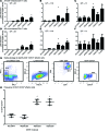
(A and B) These DART molecules contain an anti–HIV-1 binding arm (A32 or 7B2) combined with an anti-CD3 binding arm (hXR32). They are composed of 2 polypeptide chains: one with the VL of anti-CD3 linked to the VH of anti-HIV; the second with the VL of anti-HIV linked to the VH of anti-CD3. The carboxy termini of the chains have an interchain disulfide bond and paired oppositely charged E-coil/K-coil dimerization domains. Control DARTs have one of the arms replaced by an irrelevant one derived from an anti-FITC antibody (4420) or from an anti-RSV antibody, palivizumab (RSV) sequence. (C) Schematic representation of HIVxCD3 DART binding to 2 distinct antigens simultaneously and redirecting the cytotoxic T cells (effectors) to lyse the Env-expressing, HIV-1–infected cells (targets).

Similar articles

Cited by

References

    1. Chun TW, Finzi D, Margolick J, Chadwick K, Schwartz D, Siliciano RF. In vivo fate of HIV-1-infected T cells: quantitative analysis of the transition to stable latency. Nat Med. 1995;1(12):1284–1290. doi: 10.1038/nm1295-1284. - DOI - PubMed
    1. Chun TW, et al. Quantification of latent tissue reservoirs and total body viral load in HIV-1 infection. Nature. 1997;387(6629):183–188. doi: 10.1038/387183a0. - DOI - PubMed
    1. Finzi D, et al. Identification of a reservoir for HIV-1 in patients on highly active antiretroviral therapy. Science. 1997;278(5341):1295–1300. doi: 10.1126/science.278.5341.1295. - DOI - PubMed
    1. Wong JK, et al. Recovery of replication-competent HIV despite prolonged suppression of plasma viremia. Science. 1997;278(5341):1291–1295. doi: 10.1126/science.278.5341.1291. - DOI - PubMed
    1. Pierson TC, Zhou Y, Kieffer TL, Ruff CT, Buck C, Siliciano RF. Molecular characterization of preintegration latency in human immunodeficiency virus type 1 infection. J Virol. 2002;76(17):8518–8531. doi: 10.1128/JVI.76.17.8518-8513.2002. - DOI - PMC - PubMed

Publication types

MeSH terms

Substances