. 2015 Dec;35(23):4043-52.
doi: 10.1128/MCB.00742-15. Epub 2015 Sep 21.

NSun2 Promotes Cell Growth via Elevating Cyclin-Dependent Kinase 1 Translation

Affiliations

NSun2 Promotes Cell Growth via Elevating Cyclin-Dependent Kinase 1 Translation

Junyue Xing et al. Mol Cell Biol. 2015 Dec.

Abstract

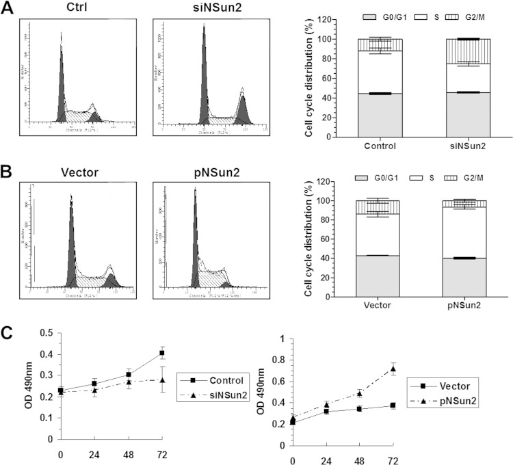
The tRNA methytransferase NSun2 promotes cell proliferation, but the molecular mechanism has not been elucidated. Here, we report that NSun2 regulates cyclin-dependent kinase 1 (CDK1) expression in a cell cycle-dependent manner. Knockdown of NSun2 decreased the CDK1 protein level, while overexpression of NSun2 elevated it without altering CDK1 mRNA levels. Further studies revealed that NSun2 methylated CDK1 mRNA in vitro and in cells and that methylation by NSun2 enhanced CDK1 translation. Importantly, NSun2-mediated regulation of CDK1 expression had an impact on the cell division cycle. These results provide new insight into the regulation of CDK1 during the cell division cycle.


Figures

FIG 1
FIG 1
NSun2 regulates CDK1 expression without influencing CDK1 mRNA levels. (A) At 48 h after transfection of HeLa cells with a vector expressing NSun2 (pNSun2) or with a siRNA targeting NSun2 (shSun2), lysates were prepared to assess the levels of NSun2, cyclin A, cyclin B1, PCNA, CDK1, CDC25A, CDC25C, and GAPDH by Western blotting. Ctrl., control. (B) The protein levels of CDK1 and CDC25C were quantified by densitometry and are represented as means ± standard deviations (SD) of the results of 3 independent experiments; statistical significance is indicated. V, vector. (C) RNA isolated from the cells described for panel A was subjected to real-time qPCR analysis (normalized to GAPDH mRNA) to assess the levels of CDK1 mRNA. The real-time qPCR data are represented as the means ± SEM of the results of 3 independent experiments.
FIG 2
FIG 2
NSun2 methylates the CDK1 CR and 3′UTR. (A) Schematic presentation of the fragments of CDK1 mRNA used for in vitro methylation assays. (B) Incorporation of 3H-labeled SAM into CDK1 fragments 5′UTR, CR, CR-A, CR-B, CR-C, 3′UTR, 3′UTR-A, 3′UTR-B, and 3′UTR-C. The incorporation of 3H-labeled SAM into CDK1 cDNA and the p16 3′UTR served as a negative control and a positive control, respectively. CPM, counts per minute. (C) U2OS cells were transfected with the pGL3 reporter vector or with the pGL3 reporter bearing the CDK1 5′UTR (pGL3-5′UTR), the CDK1 CR (pGL3-CR), or the CDK1 3′UTR (pGL3-3′UTR) plus the pRL-CMV control reporter. At 24 h later, cells were further transfected with a vector expressing NSun2 or with an NSun2 siRNA and cultured for an additional 48 h. Firefly luciferase activity relative to Renilla luciferase activity was analyzed. Data represent the means ± SEM of the results of 3 independent experiments; significance was analyzed by Student's t test.
FIG 3
FIG 3
NSun2 regulates CDK1 expression by methylating the CDK1 3′UTR. (A) The CDK1 CR and 3′UTR fragments were methylated in vitro by NSun2 using nonisotopic SAM. The fragments were then subjected to MS-HPLC analysis to assess the presence of m5C, as described in Materials and Methods. (B) Fragments 3′UTR-C, 3′UTR-Ca, 3′UTR-Cb, and 3′UTR-Cc (left, schematic) were used for in vitro methylation assays (right). (C) Fragment 3′UTR-Cb was methylated in vitro using NSun2 and nonisotopic SAM and subjected to bisulfate RNA sequencing analysis to identify the methylation site (m5C). The proportions of methylation at C1733, C1757, and C1780 are indicated. (D) Fragment 3′UTR or 3′UTR-C mutating C1733 (C-G) (3′UTRΔ or 3′UTR-CΔ) was used for in vitro methylation assays as described in the Fig. 2B legend. (E) In vitro-methylated CDK1 3′UTR was subjected to methylation-specific RT-PCR to assess the levels of methylated and unmethylated 3′UTR fragments (left), as described in Materials and Methods. The levels of total 3′UTR served as a loading control. RNA isolated from U2OS cells in which NSun2 was overexpressed or silenced was subjected to methylation-specific RT-PCR to assess the levels of methylated or unmethylated CKD1 mRNA (right). The levels of total CDK1 mRNA served as loading controls. (F) U2OS cells were transfected with pGL3-derived reporters bearing the 3′UTR (pGL3-3′UTR) or the 3′UTR mutating C1733 (pGL3-3′UTRΔ) together with a pRL-CMV control reporter. At 24 h later, cells were further transfected with a vector expressing NSun2 or with an NSun2 siRNA and cultured for an additional 48 h. Firefly luciferase activity was assayed relative to Renilla luciferase activity. Data represent the means ± SEM of the results of 3 independent experiments; significance was analyzed by Student's t test.
FIG 4
FIG 4
Methylation by NSun2 enhances the translation of CDK1. (A) U2OS cells were transfected with a siRNA targeting NSun2. At 48 h later, polysomes were fractionated by centrifugation through sucrose gradients and RNA was prepared from each of 11 fractions for analysis of the distribution of CDK1 and β-actin mRNAs. (B) In vitro-methylated (Met.) or unmethylated (Non-met.) linear pGL3, pGL3-CR, pGL3-3′UTR, and pGL3-3′UTRΔ reporter transcripts were used for in vitro translation assays. Firefly luciferase activity was measured to reflect the translation efficiency. Data represent the means ± SEM of the results of 3 independent experiments; significance was analyzed by Student's t test.
FIG 5
FIG 5
Cell cycle-dependent expression of NSun2 and CDK1. (A) U2OS cells were synchronized by serum starvation for 3 days or were left unsynchronized (Asyn.). Cells released from arrest by addition of 10% fetal bovine serum (FBS) at the times indicated were subjected to FACS analysis to monitor the cell cycle distribution. (B) Cell lysates prepared from the cells described for panel A were subjected to Western blot analysis to assess NSun2, CDK1, and GAPDH proteins. (C) RNA isolated from the cells described for panel A at times 0 and 18 h was subjected to bisulfate RNA sequencing to assess the methylation levels of endogenous CDK1 mRNA. Open boxes indicate cytosine-to-uracil conversion, read as thymidine levels in the cDNA (nonmethylated [Unmet.]), and filled boxes indicate a retained cytosine (methylated [Met.]). The numbers below the columns refer to cytosine positions in the CDK1 3′UTR.
FIG 6
FIG 6
The NSun2-CDK1 regulatory process impacts the cell division cycle. (A and B) U2OS cells were transfected with a siRNA targeting NSun2 (A) or with a vector expressing NSun2 (B). At 48 h later, cells were collected and subjected to FACS analysis to assess the cell cycle distribution. Data represent the means ± SD of the results of 3 independent experiments. (C) U2OS cells transfected with a siRNA targeting NSun2 (left) or with a vector expressing NSun2 (right). After transfection, the cell numbers were determined by MTT assays at the times indicated. OD, optical density.
FIG 7
FIG 7
Methylation of CDK1 3′UTR is required for the function of NSun2 in regulating CDK1 in the cell division cycle. (A) U2OS cells were transfected with a pGL3 or pGL3-3′UTR reporter vector. At 24 h later, cells were further transfected with a vector expressing NSun2 (pNSun2) or an empty vector (V) and cultured for an additional 48 h. The levels of NSun2, CDK1, luciferase, and GAPDH proteins were assessed by Western blotting (left). The fold increases in the level of the CDK1 3′UTR were determined by real-time qPCR (right). (B and C) The cells described for panel A were subjected to FACS analysis (B) and MTT assays (C) to assess cell cycle distribution and cell growth, respectively. Data represent the means ± SD of the results of 3 independent experiments.

Similar articles

Cited by

References

    1. Malumbres M, Barbacid M. 2005. Mammalian cyclin-dependent kinases. Trends Biochem Sci 30:630–641. doi:10.1016/j.tibs.2005.09.005. - DOI - PubMed
    1. Malumbres M, Barbacid M. 2009. Cell cycle, CDKs and cancer: a changing paradigm. Nat Rev Cancer 9:153–166. doi:10.1038/nrc2602. - DOI - PubMed
    1. Fisher D, Krasinska L, Coudreuse D, Novák B. 2012. Phosphorylation network dynamics in the control of cell cycle transitions. J Cell Sci 125(Pt 20):4703–4711. doi:10.1242/jcs.106351. - DOI - PubMed
    1. Shimada M, Niida H, Zineldeen DH, Tagami H, Tanaka M, Saito H, Nakanishi M. 2008. Chk1 is a histone H3 threonine 11 kinase that regulates DNA damage-induced transcriptional repression. Cell 132:221–232. doi:10.1016/j.cell.2007.12.013. - DOI - PubMed
    1. Marais A, Ji Z, Child ES, Krause E, Mann DJ, Sharrocks AD. 2010. Cell cycle-dependent regulation of the forkhead transcription factor FOXK2 by CDK·cyclin complexes. J Biol Chem 285:35728–35739. doi:10.1074/jbc.M110.154005. - DOI - PMC - PubMed

Publication types

LinkOut - more resources