

Genes in the GABA Pathway Increase in the Lateral Thalamus of Sprague-Dawley Rats During the Proestrus/Estrus Phase
- PMID: 26388520
- PMCID: PMC4728001
- DOI: 10.1002/jcp.25198
Genes in the GABA Pathway Increase in the Lateral Thalamus of Sprague-Dawley Rats During the Proestrus/Estrus Phase
Abstract
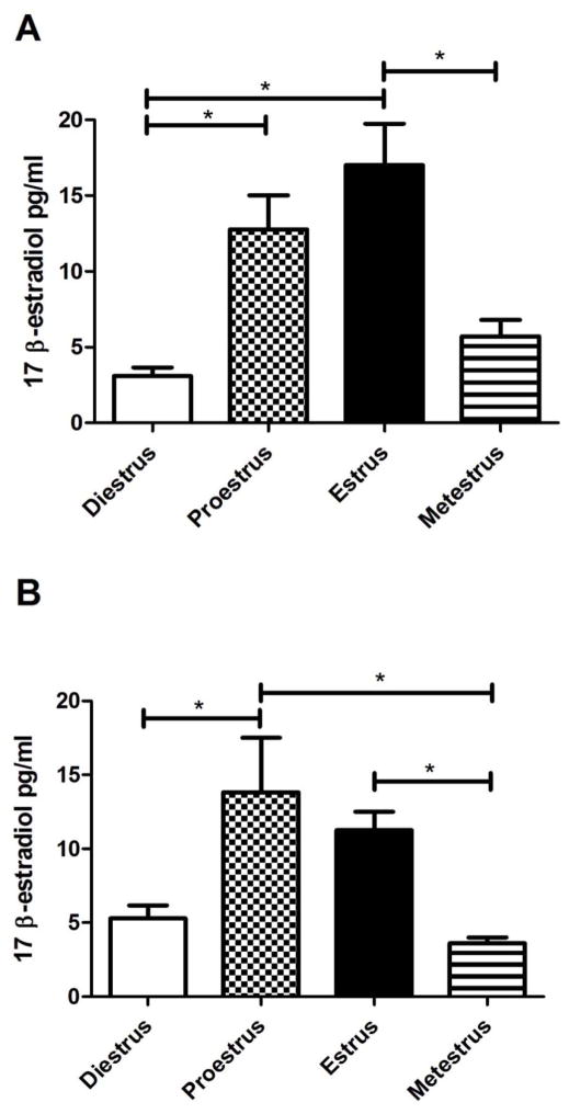
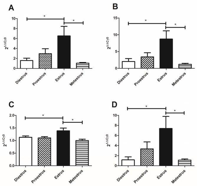
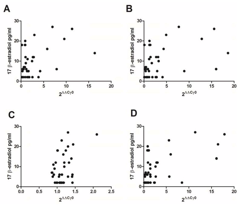
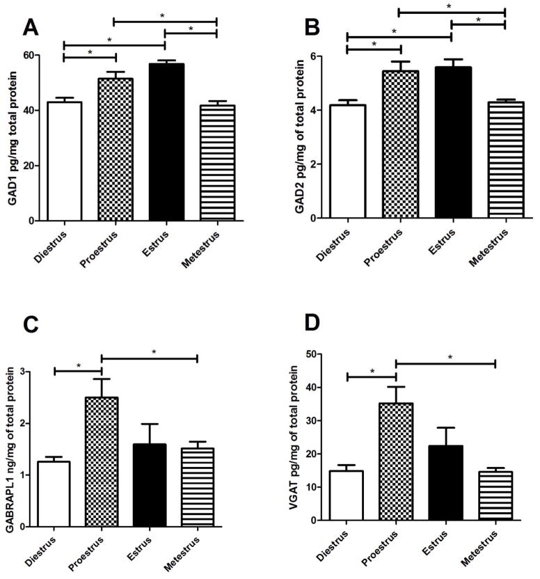
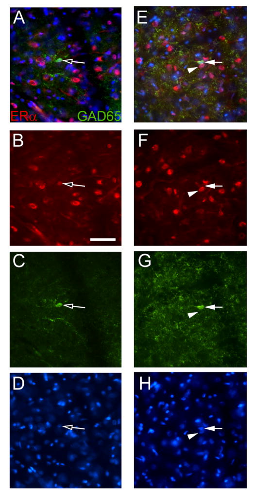
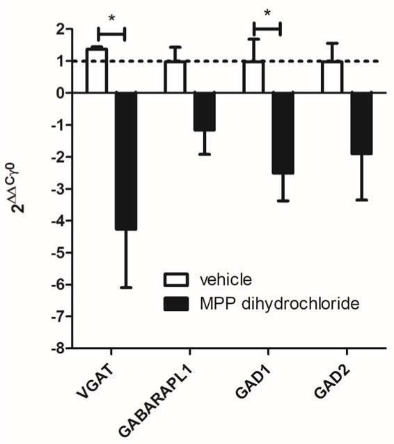
Pain can vary over the estrous cycle as a result of changes in estradiol concentration but the mechanism causing this variation is unclear. Because the thalamus is important in pain control, gene expression in the lateral thalamus (ventral posteromedial, ventral posterolateral, reticular thalamic nuclei) was screened at different phases of the estrous cycle. Gene expression changes in Sprague-Dawley rats were further analyzed by real-time PCR and ELISA and plasma estradiol levels were measured by RIAs at different phases of the estrous cycle. Our results indicated that both the RNA and protein expression of glutamate decarboxylase 1 and 2 (GAD1, GAD2), GABA(A) receptor-associated protein like 1 (GABARAPL1), and vesicular GABA transporter (VGAT) significantly increased in the lateral thalamus when plasma estradiol levels were elevated. Estradiol levels were elevated during the proestrus and estrus phases of the estrous cycle. Estrogen receptor α (ERα) was observed to be co-localized in thalamic cells and thalamic infusion of an ERα antagonist significantly reduced GAD1 and VGAT transcript. GAD1, GAD2, GABARAPL1, and VGAT have been shown to effect neuronal responses suggesting that attenuation of pain during the estrous cycle can be dependent, in part, through estradiol induced changes in thalamic gene expression.
© 2015 Wiley Periodicals, Inc.
Figures
Similar articles
-
Estrogen-induced calbindin-D 9k gene expression in the rat uterus during the estrous cycle: late antagonistic effect of progesterone.Endocrinology. 1993 Feb;132(2):489-95. doi: 10.1210/endo.132.2.8425470. Endocrinology. 1993. PMID: 8425470
-
Reduced activity of GAD67 expressing cells in the reticular thalamus enhance thalamic excitatory activity and varicella zoster virus associated pain.Neurosci Lett. 2020 Sep 25;736:135287. doi: 10.1016/j.neulet.2020.135287. Epub 2020 Aug 4. Neurosci Lett. 2020. PMID: 32763361 Free PMC article.
-
Regulation of Akt expression and phosphorylation by 17beta-estradiol in the rat uterus during estrous cycle.Reprod Biol Endocrinol. 2003 Jun 12;1:47. doi: 10.1186/1477-7827-1-47. Reprod Biol Endocrinol. 2003. PMID: 12816542 Free PMC article.
-
In vitro Autoradiographic Analysis of Regional Changes in Estrogen Receptor Alpha in the Brains of Cycling Female Rats.Neuroendocrinology. 2016;103(5):538-51. doi: 10.1159/000441077. Epub 2015 Oct 1. Neuroendocrinology. 2016. PMID: 26422138
-
Changes in mRNA levels of a pituitary-specific trans-acting factor, Pit-1, and prolactin during the rat estrous cycle.Eur J Endocrinol. 1995 Jun;132(6):771-6. doi: 10.1530/eje.0.1320771. Eur J Endocrinol. 1995. PMID: 7788020
Cited by
-
Effect of Pregnancy on TMJ Nociception in Rats.Kou Qiang Yi Xue Yan Jui. 2018 Mar;34(3):332-338. doi: 10.13701/j.cnki.kqyxyj.2018.03.029. Kou Qiang Yi Xue Yan Jui. 2018. PMID: 31404459 Free PMC article.
-
Sex difference in the contribution of GABAB receptors to tibial neuromodulation of bladder overactivity in cats.Am J Physiol Regul Integr Comp Physiol. 2017 Mar 1;312(3):R292-R300. doi: 10.1152/ajpregu.00401.2016. Epub 2016 Dec 14. Am J Physiol Regul Integr Comp Physiol. 2017. PMID: 27974317 Free PMC article.
-
Lateral thalamic control of nociceptive response after whisker pad injection of varicella zoster virus.Neuroscience. 2017 Jul 25;356:207-216. doi: 10.1016/j.neuroscience.2017.05.030. Epub 2017 May 24. Neuroscience. 2017. PMID: 28549561 Free PMC article.
-
Estradiol Acts in Lateral Thalamic Region to Attenuate Varicella Zoster Virus Associated Affective Pain.Neuroscience. 2019 Aug 21;414:99-111. doi: 10.1016/j.neuroscience.2019.06.029. Epub 2019 Jul 2. Neuroscience. 2019. PMID: 31271831 Free PMC article.
-
Local Synthesis of Estradiol in the Rostral Ventromedial Medulla Protects against Widespread Muscle Pain in Male Mice.eNeuro. 2024 Aug 28;11(8):ENEURO.0332-24.2024. doi: 10.1523/ENEURO.0332-24.2024. Print 2024 Aug. eNeuro. 2024. PMID: 39111835 Free PMC article.
References
-
- Amandusson A, Blomqvist A. Estrogenic influences in pain processing. Frontiers in neuroendocrinology. 2013;34(4):329–349. - PubMed
-
- Demling J, Fuchs E, Baumert M, Wuttke W. Preoptic catecholamine, GABA, and glutamate release in ovariectomized and ovariectomized estrogen-primed rats utilizing a push-pull cannula technique. Neuroendocrinology. 1985;41(3):212–218. - PubMed
-
- Erlander MG, Tillakaratne NJ, Feldblum S, Patel N, Tobin AJ. Two genes encode distinct glutamate decarboxylases. Neuron. 1991;7(1):91–100. - PubMed
-
- Fillingim RB, Maixner W. Sex, Gender and Pain. Seattle: IASP Press; 2000.
-
- Fischer L, Torres-Chavez KE, Clemente-Napimoga JT, Jorge D, Arsati F, de AV, Tambeli CH. The influence of sex and ovarian hormones on temporomandibular joint nociception in rats. J Pain. 2008;9(7):630–638. - PubMed
Publication types
MeSH terms
Substances
Grants and funding
LinkOut - more resources
Full Text Sources
Other Literature Sources
