. 2015 Oct;17(10):1270-81.
doi: 10.1038/ncb3236. Epub 2015 Sep 7.

An interconnected hierarchical model of cell death regulation by the BCL-2 family

Affiliations

An interconnected hierarchical model of cell death regulation by the BCL-2 family

Hui-Chen Chen et al. Nat Cell Biol. 2015 Oct.

Abstract

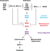
Multidomain pro-apoptotic BAX and BAK, once activated, permeabilize mitochondria to trigger apoptosis, whereas anti-apoptotic BCL-2 members preserve mitochondrial integrity. The BH3-only molecules (BH3s) promote apoptosis by either activating BAX-BAK or inactivating anti-apoptotic members. Here, we present biochemical and genetic evidence that NOXA is a bona fide activator BH3. Using combinatorial gain-of-function and loss-of-function approaches in Bid(-/-)Bim(-/-)Puma(-/-)Noxa(-/-) and Bax(-/-)Bak(-/-) cells, we have constructed an interconnected hierarchical model that accommodates and explains how the intricate interplays between the BCL-2 members dictate cellular survival versus death. BID, BIM, PUMA and NOXA directly induce stepwise, bimodal activation of BAX-BAK. BCL-2, BCL-XL and MCL-1 inhibit both modes of BAX-BAK activation by sequestering activator BH3s and 'BH3-exposed' monomers of BAX-BAK, respectively. Furthermore, autoactivation of BAX and BAK can occur independently of activator BH3s through downregulation of BCL-2, BCL-XL and MCL-1. Our studies lay a foundation for targeting the BCL-2 family for treating diseases with dysregulated apoptosis.


Figures
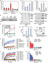
Figure 1
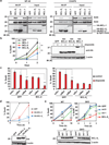
Figure 1. NOXA directly activates BAX and BAK independently of BID, BIM and PUMA
(a) Mitochondria isolated from wild-type or Bax−/−Bak−/− MEFs were incubated with the indicated IVTT proteins generated using reticulocyte lysates or wheat germ extract (WGE) at 30°C for 30 min, after which the release of cytochrome c was quantified by ELISA assays (mean ± s.d., n = 3 independent experiments). (b) Isolated wild-type mitochondria were incubated with the indicated amounts of IVTT mouse NOXA protein (WGE) and the release of cytochrome c was quantified (mean ± s.d., n = 3 independent experiments). (c) Isolated wild-type mitochondria were incubated with the indicated IVTT proteins (WGE) and the release of cytochrome c was quantified (mean ± s.d., n = 3 independent experiments). (d) Radiolabeled IVTT NOXA or tBID protein was incubated with GST, GST-BAXΔC or GST-BAKΔC protein immobilized on glutathione beads. The precipitates and input were analyzed by Nu-PAGE and autoradiography. (e) Isolated wild-type mitochondria were incubated with the indicated IVTT proteins for 30 min then treated with BMH crosslinker. The BAX and BAK homo-oligomers were detected by anti-BAX and anti-BAK immunoblots, respectively. (f) Mitochondria isolated from wild-type or Bid−/−Bim−/−Puma−/− MEFs were incubated with IVTT NOXA (WGE) at 30°C for the indicated times and release of cytochrome c was quantified (mean ± s.d., n = 3 independent experiments). (g) Bid−/−Bim−/−Puma−/− MEFs were infected with the indicated retrovirus. Cell death was quantified by annexin-V staining at 30 h (mean ± s.d., n = 3 independent experiments). The expression of the indicated proteins was detected by an anti-HA immunoblot. (h and i) ANTS/DPX (fluorophore/quencher)-encapsulated liposomes were incubated with recombinant BAX protein plus the indicated IVTT proteins generated using WGE. The release of entrapped fluorophore monitored with time is shown in (h) (mean ± s.d., n = 3 independent experiments). The release of entrapped fluorophore at 60 min is shown in (i) (mean ± s.d., n = 3 independent experiments). (j and k) ANTS/DPX-encapsulated liposomes were incubated with recombinant BAX protein plus the indicated IVTT proteins (WGE). The release of entrapped fluorophore monitored with time is shown in (j) (mean ± s.d., n = 3 independent experiments). The release of entrapped fluorophore at 60 min is shown in (k) (mean ± s.d., n =3 independent experiments). **, P < 0.01; ***, P < 0.001 (Student’s t-test). Uncropped images of blots are shown in Supplementary Fig. S7.
Figure 2
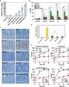
Figure 2. Noxa deficiency further protects Bid−/−Bim−/−Puma−/− mouse embryonic fibroblasts or small intestine from apoptosis
(a) The mRNA levels of Noxa in the indicated tissues or cells were assessed by qRT-PCR. Data are normalized against 18S rRNA (mean ± s.d., n = 3 independent experiments). (b) Primary MEFs generated from E13.5 wild-type, Bid−/−Bim−/−Puma−/− TKO, Bid−/−Bim−/−Puma−/−Noxa−/− QKO, or Bax−/−Bak−/− DKO mouse embryos were untreated, or cultured in the absence of serum or glucose for 3 days, or in the presence of tunicamycin (TC) or thapsigargin (TG) for 2 days. Cell death was quantified by annexin-V staining (mean ± s.d., n = 3 independent experiments). (c and d) Apoptosis in the small intestinal crypts of wild-type (n = 3), Bid−/−Bim−/−Puma−/− TKO (n = 3), Bid−/−Bim−/−Puma−/−Noxa−/− QKO (n = 2), or conditional Bax and Bak DKO (n = 2) mice at 8 to 17 weeks of age at 4 h after 18 Gy whole body irradiation was assessed by TUNEL staining (brown, magnification 400×). 300 small intestinal crypts from each mouse were analyzed. Representative light microscopy images are shown in (c). Scale bars, 50 µm. The number of TUNEL positive cells in the crypts was quantified and summarized in (d) (mean ± s.d.). (e) CD4+ T cells purified from the spleens of wild-type (n = 3), Bid−/−Bim−/−Puma−/− TKO (n = 3), or Bid−/−Bim−/−Puma−/−Noxa−/− QKO mice (n = 3) at 8 to 10 weeks of age were cultured in the absence of cytokine, in the presence of etoposide, in the presence of dexamethasone, or after exposure to 2.5 Gy γ-irradiation. Cell death was quantified by annexin-V staining at the indicated times (mean ± s.d.). **, P < 0.01; ***, P < 0.001 (Student’s t-test).
Figure 3
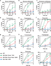
Figure 3. Quadruple deficiency of Bid, Bim, Puma and Noxa abrogates apoptosis in transformed mouse embryonic fibroblasts triggered by growth factor deprivation and ER stress but not genotoxic stress
(a–g) E1A/Ras-transformed wild-type, Bid−/−Bim−/−Puma−/− TKO, Bid−/−Bim−/−Puma−/−Noxa−/− QKO, or Bax−/−Bak−/− DKO MEFs were untreated, or cultured in the absence of serum (a), glucose (b) or glutamine (c), or in the presence of tunicamycin (d), thapsigargin (e) or etoposide (f), or irradiated with UV-C (g). Cell death was quantified by annexin-V staining at the indicated times (mean ± s.d., n = 3 independent experiments). (h) SV40-transformed wild-type, Bid−/−Bim−/−Puma−/− TKO, Bid−/−Bim−/−Puma−/−Noxa−/− QKO, or Bax−/−Bak−/− DKO MEFs were untreated, or cultured in in the presence of tunicamycin, thapsigargin or etoposide, or irradiated with UV-C. Cell death was quantified by annexin-V staining at the indicated times (mean ± s.d., n = 3 independent experiments). *, P < 0.05; **, P < 0.01; ***, P < 0.001 (Student’s t-test).
Figure 4
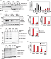
Figure 4. DNA damage activates BAX and BAK-dependent mitochondrial apoptosis in transformed Bid−/−Bim−/−Puma−/−Noxa−/− QKO mouse embryonic fibroblasts
(a) SV40-transformed wild-type or Bid−/−Bim−/−Puma−/−Noxa−/− QKO MEFs, untreated or treated with etoposide for the indicated times, were subjected to subcellular fractionation. Cytosolic and mitochondrial fractions were analyzed by anti-cytochrome c, anti-LDH and anti-VDAC1 immunoblots. LDH and VDAC1 serve as cytosolic and mitochondrial controls, respectively. (b) SV40-transformed wild-type or Bid−/−Bim−/−Puma−/−Noxa−/− QKO MEFs, untreated or treated with etoposide for the indicated times, were analyzed for Caspase-3/7 activities (mean ± s.d., n = 3 independent experiments). (c) SV40-transformed wild-type, Bid−/−Bim−/−Puma−/− TKO, or Bid−/−Bim−/−Puma−/−Noxa−/− QKO MEFs, untreated or treated with etoposide (etop) or tunicamycin (TC), were analyzed by anti-PARP, anti-cleaved Caspase-3, and anti-actin immunoblots. (d) SV40-transformed Bid−/−Bim−/−Puma−/−Noxa−/− QKO MEFs were infected with retrovirus expressing shRNA against luciferase or Apaf-1, or transfected with scrambled siRNA (siScr) or siRNA against Cytochrome c or Caspase-9. After 48 h, cells were untreated or treated with etoposide for 36 h. Cell death was quantified by annexin-V staining (mean ± s.d., n = 3 independent experiments). (e) Mitochondria isolated from SV40-transformed wild-type or Bid−/−Bim−/−Puma−/−Noxa−/− QKO MEFs untreated or treated with etoposide for 15 h (WT) or 36 h (QKO) were subjected to BMH crosslinking. The BAX and BAK homo-oligomers were detected by anti-BAX and anti-BAK immunoblots, respectively. (f) SV40-transformed Bid−/−Bim−/−Puma−/−Noxa−/− QKO MEFs were transfected with scrambled siRNA (siScr) or siRNA against Bax and/or Bak. After 48 h, cells were untreated or treated with etoposide for 36 h. Cell death was quantified by annexin-V staining (mean ± s.d., n = 3 independent experiments). *, P < 0.05; ***, P < 0.001 (Student’s t-test). Uncropped images of blots are shown in Supplementary Fig. S7.
Figure 5
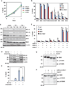
Figure 5. BAX and BAK can be autoactivated by DNA damage independently of activators BID, BIM, PUMA and NOXA through downregulation of BCL-2, BCL-XL and MCL-1
(a) SV40-transformed Bid−/−Bim−/−Puma−/−Noxa−/− QKO MEFs were infected with retrovirus expressing shRNA against luciferase, Bad, Bmf or Bik. After 48 h, cells were untreated or treated with etoposide for 36 h. Cell death was quantified by annexin-V staining (mean ± s.d., n = 3 independent experiments). (b) SV40-transformed wild-type, Bid−/−Bim−/−Puma−/− TKO, Bid−/−Bim−/−Puma−/−Noxa−/− QKO, or Bax−/−Bak−/− MEFs were infected with retrovirus expressing GFP or the indicated BH3-only proteins to induce spontaneous apoptosis. NOXA denotes human NOXA. Cell death was quantified by annexin-V staining at 30 h (mean ± s.d., n = 3 independent experiments). (c) SV40-transformed Bid−/−Bim−/−Puma−/−Noxa−/− QKO MEFs, untreated or treated with etoposide, tunicamycin (TC) or thapsigargin (TG), were subjected to immunoblot analysis using the indicated antibodies. (d) SV40-transformed Bid−/−Bim−/−Puma−/−Noxa−/− QKO MEFs were untreated or treated with etoposide and/or MG132 for 18h, and subjected to immunoblot analysis using the indicated antibodies. (e) SV40-transformed Bid−/−Bim−/−Puma−/−Noxa−/− QKO MEFs were untreated or treated with etoposide and/or MG132 for 36h. Cell death was quantified by annexin-V staining (mean ± s.d., n = 3 independent experiments). (f) SV40-transformed wild-type, Bid−/−Bim−/−Puma−/− TKO, Bid−/−Bim−/−Puma−/−Noxa−/− QKO, or Bax−/−Bak−/− MEFs were transfected with scrambled siRNA (siScr) or siRNA against Bcl-2, Bcl-xL and/or Mcl-1 to induce spontaneous apoptosis. After 2 days, cell death was quantified by annexin-V staining (mean ± s.d., n = 3 independent experiments). (g) SV40-transformed Bid−/−Bim−/−Puma−/−Noxa−/− QKO MEFs were untreated or treated with etoposide in the presence of the pancaspase inhibitor Q-VD-OPh to preserve cell integrity upon apoptosis induction. After 24 h, cells were permeabilized with digitonin and subjected to limited trypsin proteolysis. The BAK cleavage products were detected by an anti-BAK (G23) immunoblot. (h) SV40-transformed Bid−/−Bim−/−Puma−/−Noxa−/− QKO MEFs transfected with scrambled siRNA or siRNA against Bcl-xL and Mcl-1 were subjected to limited trypsin proteolysis. The BAK cleavage products were detected by an anti-BAK (G23) immunoblot. **, P < 0.01; ***, P < 0.001 (Student’s t-test). Uncropped images of blots are shown in Supplementary Fig. S7.
Figure 6
Figure 6. BCL-XL is superior to BCL-2 and MCL-1 in preventing DNA damage-induced apoptosis due to its dual inhibition of BAX and BAK as well as higher protein stability
(a) SV40-transformed Bid−/−Bim−/−Puma−/−Noxa−/− QKO MEFs stably expressing GFP, HA-BCL-2, HA-BCL-XL or HA-MCL-1 were subjected to anti-HA immunoprecipitation in 0.2% NP-40 or 1% CHAPS lysis buffer. The input (5%) and immunoprecipitates were analyzed by anti-BAX, anti-BAK, and anti-HA immunoblots. (b) SV40-transformed Bid−/−Bim−/−Puma−/−Noxa−/− QKO MEFs stably expressing GFP, HA-BCL-2, HA-BCL-XL or HA-MCL-1 were untreated or treated with etoposide. Cell death was quantified by annexin-V staining at the indicated times (mean ± s.d., n = 3 independent experiments). The expression of HA-BCL-2, HA-BCL-XL or HA-MCL-1 was detected by an anti-HA immunoblot with or without etoposide treatment for 12 h. (c) SV40-transformed Bid−/−Bim−/−Puma−/−Noxa−/− QKO MEFs stably expressing GFP, BCL-2 or BCL-XL were transfected with scrambled siRNA (siScr) or siRNA against Bax or Bak. After 48 h, cells were untreated or treated with etoposide for 36 h. Cell death was quantified by annexin-V staining (mean ± s.d., n = 3 independent experiments). (d) SV40-transformed wild-type MEFs stably expressing HA-tagged BCL-2 or BCL-XL were untreated or treated with etoposide. Cell death was quantified by annexin-V staining at the indicated times (mean ± s.d., n = 3 independent experiments). The expression of HA-tagged BCL-2 and BCL-XL was detected by an anti-HA immunoblot. (e) SV40-transformed wild-type or Bid−/−Bim−/−Puma−/−Noxa−/− QKO MEFs stably expressing GFP, wild-type BCL-XL, BCL-XL mutant 1 (F131V/D133A) or BCL-XL mutant 8 (G138E/R139L/I140N) were untreated or treated with etoposide. Cell death was quantified by annexin-V staining at the indicated times (mean ± s.d., n = 3 independent experiments). The expression of BCL-XL was analyzed by an anti-BCL-XL immunoblot. *, P < 0.05; **, P < 0.01; ***, P < 0.001 (Student’s t-test). Uncropped images of blots are shown in Supplementary Fig. S7.
Figure 7
Figure 7
A schematic depicts the interconnected hierarchical model in which the BCL-2 family proteins regulate mitochondrion-dependent cell death.

Similar articles

Cited by

References

    1. Gross A, McDonnell JM, Korsmeyer SJ. BCL-2 family members and the mitochondria in apoptosis. Genes Dev. 1999;13:1899–1911. - PubMed
    1. Youle RJ, Strasser A. The BCL-2 protein family: opposing activities that mediate cell death. Nat Rev Mol Cell Biol. 2008;9:47–59. - PubMed
    1. Czabotar PE, Lessene G, Strasser A, Adams JM. Control of apoptosis by the BCL-2 protein family: implications for physiology and therapy. Nat Rev Mol Cell Biol. 2014;15:49–63. - PubMed
    1. Wang X. The expanding role of mitochondria in apoptosis. Genes Dev. 2001;15:2922–2933. - PubMed
    1. Newmeyer DD, Ferguson-Miller S. Mitochondria: releasing power for life and unleashing the machineries of death. Cell. 2003;112:481–490. - PubMed

Publication types

MeSH terms