. 2015 Sep 3;17(3):273-86.
doi: 10.1016/j.stem.2015.07.022.

The Daxx/Atrx Complex Protects Tandem Repetitive Elements during DNA Hypomethylation by Promoting H3K9 Trimethylation

Affiliations

The Daxx/Atrx Complex Protects Tandem Repetitive Elements during DNA Hypomethylation by Promoting H3K9 Trimethylation

Quanyuan He et al. Cell Stem Cell. .

Abstract

In mammals, DNA methylation is essential for protecting repetitive sequences from aberrant transcription and recombination. In some developmental contexts (e.g., preimplantation embryos) DNA is hypomethylated but repetitive elements are not dysregulated, suggesting that alternative protection mechanisms exist. Here we explore the processes involved by investigating the role of the chromatin factors Daxx and Atrx. Using genome-wide binding and transcriptome analysis, we found that Daxx and Atrx have distinct chromatin-binding profiles and are co-enriched at tandem repetitive elements in wild-type mouse ESCs. Global DNA hypomethylation further promoted recruitment of the Daxx/Atrx complex to tandem repeat sequences, including retrotransposons and telomeres. Knockdown of Daxx/Atrx in cells with hypomethylated genomes exacerbated aberrant transcriptional de-repression of repeat elements and telomere dysfunction. Mechanistically, Daxx/Atrx-mediated repression seems to involve Suv39h recruitment and H3K9 trimethylation. Our data therefore suggest that Daxx and Atrx safeguard the genome by silencing repetitive elements when DNA methylation levels are low.

Keywords: ATRX; DAXX; DNA methylation; DNMTs; histone modification; mouse embryonic stem cells; repetitive sequences; telomeres.


Conflict of interest statement

Competing financial interests

The authors declare no competing financial interests.

Figures
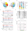
Figure 1
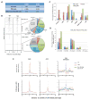
Figure 1. DAXX and ATRX exhibit distinct genome-wide distribution of binding sites in wildtype (WT) mES cells
See also supplemental Figure S1. (A–B) ChIP-seq analysis of wildtype J1 mES cells was carried out using anti-DAXX and ATRX antibodies. The different types of binding sites identified throughout the genome are summarized here. (C) The identified binding sites at different gene loci were divided into DAXX only (top), DAXX & ATRX co-binding (middle), and ATRX only (bottom) sites. The distance to signal peaks and number of sites in each group were plotted on the x- and y-axis respectively, to show representative signal peak distributions of various DAXX/ATRX binding sites. Green bar, CpG island. Orange bar, LTR region. (D) Genomic features of the three groups of binding sites from (C) were plotted and further compared. (E) Comparison of the DAXX and ATRX binding profiles at tRNA genes. The x-axis indicates distance to the transcriptional start site (TSS) of tRNA genes. (F) The binding profiles of ATRX and DAXX at IAPEz regions (including ±2kb from these regions). Top panel, relative intensities of the peaks were plotted on the y-axis. The blue and red lines denote the average binding profiles of ATRX and DAXX respectively. The grey line indicates the distribution of tandem repetitive sequences. The bottom two panels are heat maps of ATRX and DAXX binding profiles.
Figure 2
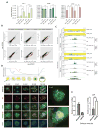
Figure 2. Loss of DNA methylation in TKO cells leads to preferential targeting of both DAXX and ATRX to repeat sequences
See also supplemental Figure S1–3. TKO cells were used for ChIP-seq analysis using anti-DAXX and ATRX antibodies. (A) Comparison of DAXX and ATRX-binding sites in wildtype J1 (WT) vs. TKO cells. (B) A comparison of genome-wide DAXX and ATRX-binding sites in TKO vs. wildtype J1 (WT) cells. Left, all of the identified binding sites for DAXX and ATRX were plotted, where the x-axis is the distance to signal peaks and the y-axis represents the signaling intensity for each site. Right, the binding sites specific for TKO cells were classified and graphed as pie charts. (C) DAXX-binding sites that were found only in wildtype J1 (WT_Alone) or TKO (TKO_Alone) cells, or shared between the cells (Shared) were plotted based on various genomic features. (D) The DAXX-binding sites on LTR-containing sequences were further classified and similarly plotted as in (C). (E) The average distribution of DAXX, ATRX, and DNA methylation (±5kb from the binding peaks on LTRs) in wildtype J1 (WT) (blue) and TKO (red) cells was plotted and compared. The center vertical dotted lines indicate the summit of binding peaks.
Figure 3
Figure 3. DAXX and ATRX are important for repressing repetitive sequences in cells undergoing global hypomethylation
See also Figure S4. (A) RT-qPCR analysis of transcript levels of IAP-1, DAXX, and ATRX in wildtype J1 (WT) and TKO cells, as well as in WT and TKO cells knocked out for DAXX or ATRX. Error bars are standard deviation (n=3). The data were analyzed using the Student t-test. ** indicates p<0.01 and * indicates p<0.05. (B) Wildtype J1 (WT) and TKO cells as well as J1 and TKO cells knocked out for DAXX were grown in regular or ground-state (2i+vitamin C (VC)) media and examined for the transcriptional levels of five types of repetitive elements (color coded as indicated). RPKMs (Reads Per Kilobase of transcript per Million mapped reads) of various samples were plotted in pairs on log10 scale. In each paired comparison (plot), the expression of a particular repetitive element (represented by RPKMs) in different cells or conditions was plotted on the x- or y-axis as indicated. A shift from the diagonal line indicates differential expression between the two samples. IAPEz-int, intracisternal A particle interspersed repeats. ETn/MusD, the early transposon family of long repeated sequences, also known as MMET in Repbase database (Jurka et al., 2005). MERVL, mouse retroelement MuERV-L/MERVL. MMERVK, LTR family of mouse endogenous retrovirus K. RLTR4, Long Terminal Repeat for HERV3 endogenous retrovirus. (C) A snapshot of aligned ChIP-seq and RNA-seq data shows a representative case of IAP expression differences between samples. RNA-seq data from wildtype J1, wildtype DAXX KO, TKO, and TKO DAXX KO cells grown under regular vs. ground-state conditions were used. The relative coverage was normalized by the total number of aligned reads for each sample and the plot was drawn using GenomeBrowse (Golden Helix Inc.). Blue and green represent reads aligned to the plus and minus strand respectively. (D) Top, the global DNA demethylation process that occurs in early pre-implantation embryos. Bottom, different stages of mouse embryos were examined by immuno-FISH using a telomere FISH probe (red) and anti-ATRX antibodies (green). Arrowheads indicate co-localized foci. Magnified images of 2-cell and 16-cell embryos are shown on the right. (E) ATRX knockdown induces IAP de-repression in early mouse embryos. Mouse embryos were injected with control siRNA oligos or those against ATRX, and allowed to recover. Morula-stage embryos were then collected for quantitative RT-PCR analysis to determine the expression level of ATRX (left) and IAP (right) messages. Error bars indicate standard error. The data were analyzed using the Student t-test. ** indicates p<0.01.
Figure 4
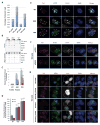
Figure 4. The DAXX/ATRX complex is targeted to subtelomeric/telomeric regions in response to genomic DNA demethylation in ES cells
See also supplemental Figure S5–6. (A) We calculated and plotted the percentages of telomeric reads (TTAGGG(6)) out of total reads that were aligned to the mouse genome using our ChIP-seq data from wildtype J1 (WT) and TKO cells. (B) Telomere ChIP experiments were performed using antibodies against endogenous DAXX and ATRX. The telomeric protein RAP1 served as a positive control, and rabbit IgG was used as a negative control. The blots were probed with a radiolabeled TTAGGG(3) (Tel(3)) probe or a major satellite DNA probe. (C) Quantification of data from (B). Three independent experiments were performed. Error bars indicate standard deviation. The data were analyzed using the Student t-test. ** indicates p<0.01. (D) The percentages of telomeres occupied by ATRX and DAXX. Immuno-FISH experiments were carried out in wildtype J1 (WT), DKO, and TKO cells. ATRX and DAXX were detected by immunostaining using anti-ATRX and DAXX antibodies, while telomeres were detected using a Tel-RNP FISH probe. Error bars indicate standard deviation (n=3). The data were analyzed using the Student t-test. ** indicates p<0.01. (E) Representative IF-FISH images from (D). The white arrows indicate ATRX/DAXX occupied telomeres. (F) TKO cells depleted of DAXX or ATRX (siDAXX_3 and siATRX_1) were immunostained with antibodies against ATRX (green), DAXX (gray), and the Tel-RNP FISH probe that marks telomeres (red). Dotted circles indicate cells depleted of ATRX or DAXX due to successful knockdown. (G) TKO cells stably expressing Myc-tagged wildtype or mutant (Dnmt3a_R716A and Dnmt3b_V725G) Dnmt3a and Dnmt3b proteins were immunostained with anti-ATRX (green) and Myc (gray) antibodies. Telomeres were marked with a Tel-RNP FISH probe (red). Dotted circles indicate cells not expressing ectopic Dnmt3a/3b proteins.
Figure 5
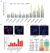
Figure 5. The DAXX/ATRX complex protects telomeres and ensures genome stability in response to DNA hypomethylation
(A) Wildtype J1 (WT) and TKO cells knocked down for DAXX or ATRX were analyzed by RT-PCR for TERRA expression. Two different siRNA oligos each were used for DAXX and ATRX. Error bars indicate standard deviation (n=3). (B) The levels of telomeric sister-chromatid exchange (T-SCE) in wildtype J1 (WT) and TKO cells and those knocked down for DAXX and ATRX individually or together (siDAXX_3 and siATRX_1) were determined by CO-FISH. Leading and lagging strands were labeled by G3-Cy3 (TTAGGGTTAGGGTTAGGG) and C3-FAM (CCCTAACCCTAACCCTAA) respectively. Representative images are shown on top. The percentages of T-SCE in different cells were quantitated and plotted below. Error bars indicate standard deviation (n=3). (C) Metaphase spreads from wildtype J1 (WT) and TKO cells and those knocked down for DAXX or ATRX were prepared for FISH hybridization using the G3-Cy3 and C3-FAM oligo probes. Representative images of fragile and super telomeres are shown on top. The percentages of defective telomeres in different cells were quantitated and summarized below.
Figure 6
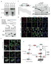
Figure 6. DNA hypomethylation leads to increased recruitment of H3K9me3 and HP1 on telomeres and LTR-containing repeat elements
(A) Telomere ChIP experiments were performed using wildtype J1 (WT), DKO, and TKO cells with the indicated antibodies. Precipitated DNA was dot-blotted and probed with the Tel(3) probe (TTAGGG(3)). IgG and anti-RAP1 antibodies were used as negative and positive controls respectively. Two different anti-H3K9me3 antibodies were used (H3K9me3-1 from Abcam and H3K9me3-2 from Upstate). *, non-relevant sample. (B) Three independent telomere ChIP experiments as described in (A) were performed and quantified. Error bars indicate standard deviation. The data were analyzed using the Student t-test. ** indicates p<0.01 and * indicates p<0.05. (C) Wildtype J1 (WT) and TKO cells were used for immunostaining analysis with an anti-H3K9me3 antibody (Abcam). Telomeres were marked with a telomere PNA probe. Arrows indicate co-staining of H3K9me3 with telomere foci. (D) Wildtype J1 (WT) and TKO cells were used for Immuno-FISH studies with antibodies against HP1α. Telomeres were marked with a Tel-RNP FISH probe. Arrows indicate co-staining of HP1α and telomere foci. (E) Wildtype J1 (WT) and TKO cells were used in ChIP-qPCR experiments with anti-H3K9me3 antibodies for LTR-containing repeat sites that are targeted by DAXX. Error bars indicate standard deviation (n=3). The data were analyzed using the Student t-test. ** indicates p<0.01 and * indicates p<0.05.
Figure 7
Figure 7. DAXX associates with SUV39H1 and facilitates H3K9me3 enrichment on telomeres in response to DNA demethylation
See also supplemental Figure S7. (A) SFB-tagged DAXX or ATRX were co-expressed in mES cells with GST-tagged SUV39H1 or SETDB1. The cells were then harvested for immunoprecipitations (IP) using anti-FLAG antibodies. Co-precipitated proteins were detected by anti-GST antibodies. SFB-Con, vector alone. (B) GST-tagged full-length SUV39H1 was co-expressed with FLAG-tagged full-length or truncation mutants of DAXX in mES cells. The cells were then harvested for immunoprecipitations (IP) using anti-FLAG antibodies and probed with anti-GST antibodies. (C) FLAG-tagged full-length DAXX was co-expressed with GST-tagged full-length or truncation mutants of SUV39H1 in mES cells. The cells were then harvested for GST pull down and probed with anti-FLAG antibodies. (D) Wildtype J1 (WT) and TKO cells were transfected with control (siNeg) or DAXX (siDAXX_3) siRNAs and then analyzed by Immuno-FISH using anti-DAXX and H3K9me3 antibodies along with the telomere FISH probe TelG3-Cy3. Arrows indicate co-stained foci. (E) Parental and DAXX-knockout TKO cells were immunostained using anti-ATRX and H3K9me3 antibodies. Telomeres were marked by the telomere FISH probe TelG3-Cy3. Arrows indicate co-stained foci. (F) DAXX may act as a transcriptional regulator on gene promoters and tRNA regions that are normally low in DNA methylation. Global DNA demethylation may promote the recruitment/translocation of the DAXX/ATRX complex to tandem repeats (e.g. IAP, RLTR, and telomeres) to silence transcription and prevent instability. Through interaction with SUV39H1, DAXX may facilitate H3K9me3 recruitment to repetitive sequences for heterochromatin maintenance in the absence of DNA methylation.

Comment in

Similar articles

Cited by

References

    1. Armour JA, Anttinen T, May CA, Vega EE, Sajantila A, Kidd JR, Kidd KK, Bertranpetit J, Paabo S, Jeffreys AJ. Minisatellite diversity supports a recent African origin for modern humans. Nat Genet. 1996;13:154–160. - PubMed
    1. Azzalin CM, Reichenbach P, Khoriauli L, Giulotto E, Lingner J. Telomeric repeat containing RNA and RNA surveillance factors at mammalian chromosome ends. Science. 2007;318:798–801. - PubMed
    1. Baumann C, Schmidtmann A, Muegge K, De La Fuente R. Association of ATRX with pericentric heterochromatin and the Y chromosome of neonatal mouse spermatogonia. BMC molecular biology. 2008;9:29. - PMC - PubMed
    1. Baumann C, Viveiros MM, De La Fuente R. Loss of maternal ATRX results in centromere instability and aneuploidy in the mammalian oocyte and pre-implantation embryo. PLoS Genet. 2010;6:e1001137. - PMC - PubMed
    1. Blaschke K, Ebata KT, Karimi MM, Zepeda-Martinez JA, Goyal P, Mahapatra S, Tam A, Laird DJ, Hirst M, Rao A, et al. Vitamin C induces Tet-dependent DNA demethylation and a blastocyst-like state in ES cells. Nature. 2013;500:222–226. - PMC - PubMed

Publication types

MeSH terms

Substances

Associated data

LinkOut - more resources