. 2015 Nov;35(22):3829-40.
doi: 10.1128/MCB.01497-14. Epub 2015 Aug 31.

BRCA1 Is Required for Maintenance of Phospho-Chk1 and G2/M Arrest during DNA Cross-Link Repair in DT40 Cells

Affiliations

BRCA1 Is Required for Maintenance of Phospho-Chk1 and G2/M Arrest during DNA Cross-Link Repair in DT40 Cells

Margarethe Draga et al. Mol Cell Biol. 2015 Nov.

Abstract

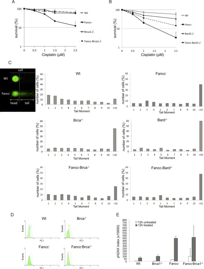
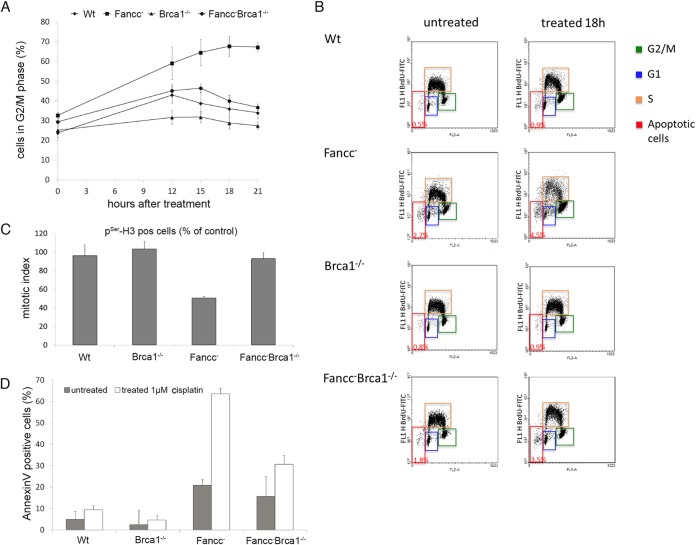
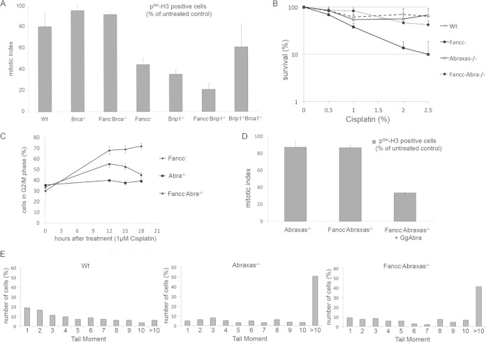
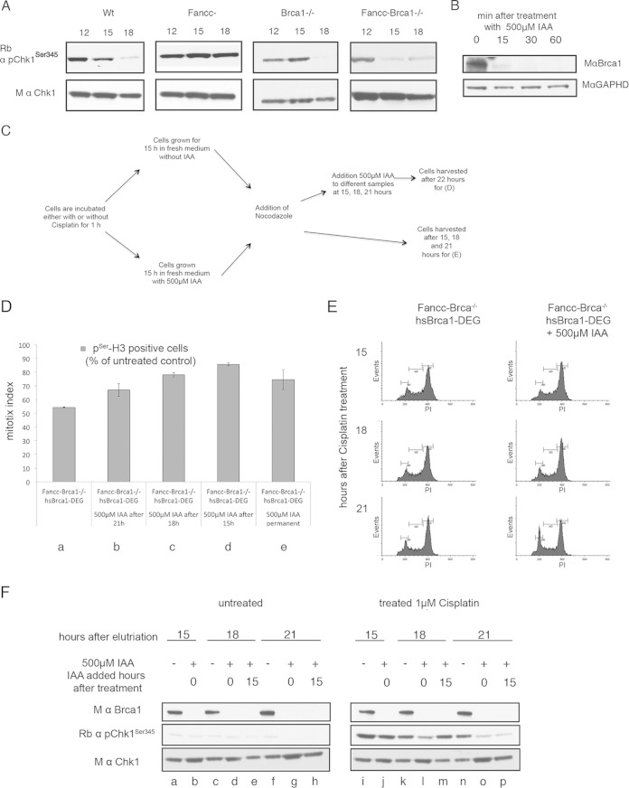
The Fanconi anemia DNA repair pathway is pivotal for the efficient repair of DNA interstrand cross-links. Here, we show that FA-defective (Fancc(-)) DT40 cells arrest in G2 phase following cross-link damage and trigger apoptosis. Strikingly, cell death was reduced in Fancc(-) cells by additional deletion of the BRCA1 tumor suppressor, resulting in elevated clonogenic survival. Increased resistance to cross-link damage was not due to loss of toxic BRCA1-mediated homologous recombination but rather through the loss of a G2 checkpoint. This proapoptotic role also required the BRCA1-A complex member ABRAXAS (FAM175A). Finally, we show that BRCA1 promotes G2 arrest and cell death by prolonging phosphorylation of Chk1 on serine 345 after DNA damage to sustain arrest. Our data imply that DNA-induced cross-link death in cells defective in the FA pathway is dependent on the ability of BRCA1 to prolong cell cycle arrest in G2 phase.


Figures

FIG 1
FIG 1
Defects in BRCA1 ameliorate the sensitivity of Fancc DT40 cells to cisplatin treatment. (A and B) Graphs showing clonogenic survival curves for indicated DT40 mutant cells after treatment with different concentrations of cisplatin. Fancc Brca1−/− and Fancc Bard1−/− are less sensitive to treatment with low doses of cisplatin compared to Fancc cells. (A) Fifty percent inhibitory concentration values (IC50): wild-type cells, 3.5 μM; Fancc cells, 0.8 μM; Brca1−/− cells, 3.4 μM; and Fancc Brca1−/− cells, 3.8 μM. (B) IC50 values: wild-type cells, 3.2 μM; Fancc cells, 0.47 μM; Bard1−/− cells, 1.1 μM; and Fancc Bard1−/− cells, 0.7 μM. The data presented are means from three experiments; error bars indicate one standard deviation. (C) A comet assay showed the increased tail moment and therefore the persistence of DNA breaks in wild-type and mutant DT40 cells, as indicated. Cells were treated with 1 μM cisplatin for 1 h, allowed to recover in fresh medium for 15 h, and then analyzed by alkaline comet assay. Tail moments (TM) were calculated for >100 comets from each sample, and the percentage of cells showing a specific TM was plotted. (D) Mutant cells were treated with 1 μM cisplatin for 1 h, allowed to recover in fresh medium for 12 h, and then stained with primary antibody against γH2AX and secondary antibody conjugated to fluorescein isothiocyanate. γH2AX was quantified by fluorescence-activated cell sorting (FACS), where the FL1 channel corresponds to fluorescein. A green histogram represents untreated cells, and a white histogram represents cells treated with 1 μM cisplatin. (E) Quantification of γH2AX staining in panel D measured in arbitrary units. Further information is contained in Fig. S1 and S3 in the supplemental material.
FIG 2
FIG 2
Fancc mutant cells, but not Brca1−/− or Fancc Brca1−/− cells, arrest with 4C DNA content and undergo apoptotic cell death after treatment with cisplatin. (A and B) Wild-type and mutant cells were damaged by 1 μM cisplatin, pulse-labeled with BrdU, and harvested at the times indicated. Cells were analyzed for BrdU incorporation and propidium iodide staining by FACS to determine the DNA content. The percentages of cells with near 4C DNA content and therefore in G2/M phase (green boxes) of the cell cycle are shown at different times after DNA damage treatment. (C) Fancc mutant cells, but not Brca1−/− or Fancc Brca1−/− cells, arrest prior to entering mitosis after treatment with cisplatin. The mitotic index of cells treated with 1 μM cisplatin is shown. Nocodazole was added 15 h after damage treatment to trap cells entering mitosis. Mitotic cells were quantified 24 h after treatment by staining for pSer-H3 and measured by FACS. The mitotic index is calculated as the ratio of treated to untreated cells staining positive for pSer-H3. (D) Loss of BRCA1 function reduces apoptotic cell death in Fancc mutant cells. The percentage of cells that stained for annexin V, 22 h after treatment 1 μM cisplatin, was used as a measurement of apoptosis. The data shown in panels A, C, and E show the means from three experiments; error bars indicate the standard deviations. See also Fig. S1 in the supplemental material.
FIG 3
FIG 3
Loss of ABRAXAS but not BRIP1 abrogates G2/M arrest in FANCC-defective cells. (A) Mitotic index of mutant cells treated and not treated with 1 μM cisplatin and stained for pSer-H. (B) Clonogenic survival curves for indicated DT40 mutant cells after treatment with different concentrations of cisplatin. Fancc Abra−/− mutants are less sensitive to treatment with low doses of cisplatin than Fancc cells. (C) Fancc cells, but not Abra1−/− or Fancc Abra1−/− cells, arrest in G2/M with near 4C DNA content after cisplatin treatment. DNA content was quantified by using FACS after incorporation of BrdU into cells and after staining with propidium iodide. The percentage of cells with 4C content and therefore in G2/M phase are shown. (D) Mitotic index of cells treated or not treated with 5 μM cisplatin. The percentage of cells positive for pSer-H3 is shown. The experiments from panels C and D were performed at the same time as those depicted in Fig. 2A and C. The wild-type control in the latter experiments can be used for comparison. (E) A comet assay of mutant cells (as indicated) shows the persistence of DNA strand breaks 15 h after treatment with 1 μM cisplatin. Tail moments were calculated for >100 comets and are shown as a percentage in the total cell population. The data shown in panels A to D are the means from three experiments; error bars indicate the standard deviations.
FIG 4
FIG 4
Caffeine treatment improves survival in Fancc DT40 mutant cells treated with cisplatin. (A and B) Clonogenic survival assays were performed for mutant cells treated with different concentrations of cisplatin and in the presence or absence of 2 mM caffeine. The inclusion of 2 mM caffeine in the media improves survival in Fancc cells treated with different concentrations of cisplatin. (C) Mitotic index of cells treated and not treated with 5 μM cisplatin with or without 2 mM caffeine. The percentage of cells positive for pSer-H3 is shown. Caffeine improves cell cycle progression into mitosis in Fancc cells. The data presented are the mean of three experiments; error bars indicate one standard deviation. See also Fig. S4 in the supplemental material.
FIG 5
FIG 5
BRCA1 is required to maintain CHK1 activation in response to DNA damage. (A) CHK1 is activated through phosphorylation at serine 345 in response to cisplatin-induced DNA damage in wild-type and mutant cell lines (as indicated). A G1 population of cells for each mutant cell line was collected by centrifugal elutriation and treated with cisplatin for 1 h. The cells were incubated for the indicated times and analyzed for the presence of unmodified and phosphorylated (activated) CHK1 by Western blotting. (B) BRCA1-tagged with an auxin-inducible degron sequence (BRCA1-DEG) was expressed as a transgene in Fancc Brca1−/− cells. Depletion of BRCA1-DEG with time after addition of 500 μM IAA was measured by Western blotting. Approximately 90% of the BRCA1-DEG protein was depleted within 15 min. (C) Schematic diagram depicting the experiments described in panels D and F. A 1 μM concentration of cisplatin was added to Fancc Brca1−/− cells for 1 h at time zero, and the cells were grown for a further 22 h before harvesting them for analysis as described in panels D and F. BRCA1 was depleted in different samples of cells at 0, 15, 18, and 21 h by the addition of 500 μM IAA. (D) Depletion of BRCA1 correlates with impaired cell cycle arrest and progression into mitosis. Fancc Brca1−/− cells expressing a Brca1-DEG transgene arrest in G2, as indicated by a decrease in the mitotic index. Depletion of BRCA1-DEG at 0, 15, 18, or 21 h after DNA damage results correlates with progression into mitosis as indicated by increased mitotic index. G1-synchronized Fancc Brca1−/− cells plus BRCA1-DEG cells were treated with cisplatin for 1 h and then incubated for 22 h in fresh media. Next, 500 μM IAA was added to different samples. Cells were harvested after 22 h, and the mitotic index was determined (as described above). The data presented are the means from three experiments; error bars indicate one standard deviation. (E) Cell cycle profile of cisplatin-treated cells in panel D after the addition of IAA. Cells were fixed and stained with propidium iodide and analyzed by FACS to determine the DNA content. (F) Phosphorylation of CHK1 at Ser345 was determined in cell populations treated as for panel D. Loss of pCHK1 correlates with progression of cells into mitosis as determined by mitotic index (described above).

Similar articles

Cited by

References

    1. Kim H, D'Andrea AD. 2012. Regulation of DNA cross-link repair by the Fanconi anemia/BRCA pathway. Genes Dev 26:1393–1408. doi:10.1101/gad.195248.112. - DOI - PMC - PubMed
    1. Rosado IV, Langevin F, Crossan GP, Takata M, Patel KJ. 2011. Formaldehyde catabolism is essential in cells deficient for the Fanconi anemia DNA-repair pathway. Nat Struct Mol Biol 18:1432–1434. doi:10.1038/nsmb.2173. - DOI - PubMed
    1. Langevin F, Crossan GP, Rosado IV, Arends MJ, Patel KJ. 2011. Fancd2 counteracts the toxic effects of naturally produced aldehydes in mice. Nature 475:53–58. doi:10.1038/nature10192. - DOI - PubMed
    1. Garcia-Higuera I, Taniguchi T, Ganesan S, Meyn MS, Timmers C, Hejna J, Grompe M, D'Andrea AD. 2001. Interaction of the Fanconi anemia proteins and BRCA1 in a common pathway. Mol Cell 7:249–262. doi:10.1016/S1097-2765(01)00173-3. - DOI - PubMed
    1. Smogorzewska A, Matsuoka S, Vinciguerra P, McDonald ER III, Hurov KE, Luo J, Ballif BA, Gygi SP, Hofmann K, D'Andrea AD, Elledge SJ. 2007. Identification of the FANCI protein, a monoubiquitinated FANCD2 paralog required for DNA repair. Cell 129:289–301. doi:10.1016/j.cell.2007.03.009. - DOI - PMC - PubMed

Publication types

MeSH terms

LinkOut - more resources