. 2015 Sep 15;43(3):475-87.
doi: 10.1016/j.immuni.2015.07.021. Epub 2015 Aug 25.

MCPIP1 Endoribonuclease Activity Negatively Regulates Interleukin-17-Mediated Signaling and Inflammation

Affiliations

MCPIP1 Endoribonuclease Activity Negatively Regulates Interleukin-17-Mediated Signaling and Inflammation

Abhishek V Garg et al. Immunity. .

Abstract

Interleukin-17 (IL-17) induces pathology in autoimmunity and infections; therefore, constraint of this pathway is an essential component of its regulation. We demonstrate that the signaling intermediate MCPIP1 (also termed Regnase-1, encoded by Zc3h12a) is a feedback inhibitor of IL-17 receptor signal transduction. MCPIP1 knockdown enhanced IL-17-mediated signaling, requiring MCPIP1's endoribonuclease but not deubiquitinase domain. MCPIP1 haploinsufficient mice showed enhanced resistance to disseminated Candida albicans infection, which was reversed in an Il17ra(-/-) background. Conversely, IL-17-dependent pathology in Zc3h12a(+/-) mice was exacerbated in both EAE and pulmonary inflammation. MCPIP1 degraded Il6 mRNA directly but only modestly downregulated the IL-6 promoter. However, MCPIP1 strongly inhibited the Lcn2 promoter by regulating the mRNA stability of Nfkbiz, encoding the IκBζ transcription factor. Unexpectedly, MCPIP1 degraded Il17ra and Il17rc mRNA, independently of the 3' UTR. The cumulative impact of MCPIP1 on IL-6, IκBζ, and possibly IL-17R subunits results in a biologically relevant inhibition of IL-17 signaling.

Keywords: IL-17; Regnase-1; autoimmunity; fungal immunity; negative regulation; signal transduction.


Figures

Figure 1
Figure 1. MCPIP1 is a negative feedback inhibitor of IL-17 signaling
a. RNA from WT or Il17ra−/− tongues (n=3) collected 48 h after oral infection with PBS (Sham) or C. albicans. Expression of Zc3h12a was assessed by qPCR normalized to Gapdh. *p<0.05 vs. Sham. b. OKF6/TERT2 oral keratinocytes were treated ± IL-17 and mRNA analyzed for ZC3H12A by qPCR. Data presented as fold change vs. unstimulated (0 h) *p<0.05 vs. untreated. c. ST2 cells were incubated ± IL-17 and mRNA was analyzed by qPCR, normalized to Gapdh. Inset: Lysates from ST2 cells treated ± IL-17 were immunoblotted for MCPIP1 or β-Tubulin. Data presented as fold change vs. unstimulated. d–e. ST2 cells were transfected with siRNAs against Act1, MCPIP1 or scrambled control. Cells were treated ± IL-17 for 3 h. Supernatants were assessed by ELISA (left). Il6 and Lcn2 were assessed by qPCR. *p<0.05 vs. IL-17-treated siRNA control. ‡ p<0.05 vs. untreated. *p<0.05 vs. IL-17-treated siRNA control. ‡ p<0.05 vs. untreated. Data presented as fold change relative to untreated siRNA control. f. Zc3h12a−/− fibroblasts were transfected with an empty vector or a plasmid encoding murine MCPIP1. Cells were treated with IL-17 for 4 h, and IL-6 assessed by ELISA. *p<0.05 vs. IL-17-treated controls. g. Primary fibroblasts from Zc3h12a+/− or WT littermates were treated with IL-17 for 24 h and IL-6 assessed by ELISA.*p<0.05 vs. IL-17-treated controls. ‡ p<0.05 vs. untreated. Data presented as mean ± SEM. All experiments were performed a minimum of twice.
Figure 2
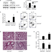
Figure 2. MCPIP1 inhibits IL-17-mediated pulmonary inflammation in MCPIP1+/− mice
a. Fibroblasts from Zc3h12a+/− mice or WT littermates were treated with LPS or IL-17 for 4 h and MCPIP1 and β-Tubulin assessed by immunoblotting (left) or qPCR (right). Data expressed as fold-change vs. untreated WT. *p<0.05 vs. IL-17-treated WT sample. b–c. WT or MCPIP1+/− mice (n=5) were treated intranasally with 300 ng recombinant IL-17. 24 h later BALF was stained for Gr-1 and F4/80 and quantified by FACS. C=control unchallenged. *p<0.05 compared to IL-17-treated WT mice. d. H&E stained lung sections from the indicated mice are shown. Scale bars indicate 200 µM. e. WT or Zc3h12a+/− mice (n=2 for control, n=5 for IL-17-treated) were treated intranasally with 500 ng IL-17. After 8 h, levels of CXCL1 and CXCL5 in BALF were assessed by ELISA. *p<0.05 vs. IL-17-treated WT mice. n.s., not significant. Data are presented as mean ± SEM. All experiments were performed a minimum of twice.
Figure 3
Figure 3. MCPIP1 restrains IL-17-dependent responses to Candida albicans infection
a. Zc3h12a+/− mice crossed to Il17ra−/− mice (n=5–8) and littermates were subjected to candidiasis by i.v. injection of C. albicans. After 2 d, fungal burdens in kidney were assessed by plating and colony enumeration. Data presented as mean ± SEM. *p<0.05 vs. WT. Data pooled from 2 independent experiments. b. Survival curve of WT (solid line, n=11) and Zc3h12a+/− mice (dashed line, n=10) following candidiasis. Data are pooled from 2 independent experiments. c. Periodic acid Schiff (PAS) stained kidney sections. Yellow boxes indicate location of higher magnification images. Scale bars indicate 1000 µM (top) or 200 µM (bottom). d. WT or Zc3h12a+/− mice (n=4–6) were infected i.v. with C. albicans after administration of α-IL-6R or isotype Abs. Fungal loads in kidney were assessed 2 d post infection. Data presented as mean ± SEM. *p<0.05 vs. α-IL-6R-treated WT mice. ‡ p<0.05 vs. isotype-treated WT mice. All experiments were performed a minimum of twice.
Figure 4
Figure 4. MCPIP1 limits IL-17-dependent autoimmune CNS pathology
a. The indicated mice (n=13–17) were subjected to EAE and clinical scores assessed daily (left). The percentage of mice exhibiting EAE symptoms is indicated (right). Data are pooled from 2 experiments. Data are presented as mean clinical score of all mice. *p<0.05 by ANOVA and student’s t-test. b. Gene expression in spinal cords was measured by qPCR normalized to Gapdh. Data presented as mean ± SEM.
Figure 5
Figure 5. MCPIP1 differentially regulates IL-17 target promoters and transcription factors
a. ST2 cells were transfected with varying doses of MCPIP1 plasmid (0–300 ng) togehter with Luc reporters driven by the IL-6 or Lcn2 promoters. Cells were treated ± IL-17 for 8 h and Luc activity assessed in triplicate. Data presented relative to the unstimulated control without MCPIP1. *p<0.05 vs. unstimulated sample of the corresponding condition. b. ST2 cells were transfected with siRNAs against MCPIP1, stimulated ± IL-17 for 3 h, and analyzed for expression of the indicated genes by qPCR. *p<0.05 vs. unstimulated. ‡ p<0.05 vs. IL-17-treated control siRNA. Data presented as mean ± SEM. c. HEK293T cells were transfected with MCPIP1 ± constructs expressing IκBζ with or without its 3’ UTR. Whole cell lysates were analyzed by immunoblotting for IκBζ (top), MCPIP1 (middle) or β-Tubulin (bottom). d. Top: Murine Lcn2 promoter construct. ST2 cells were transfected with siRNAs targeting IκBζ, MCPIP1 or a scrambled control, treated ± IL-17 for 3 h, and the indicated genes evaluated by qPCR. Data expressed as fold-change relative to the untreated control siRNA. *p<0.05 vs. control siRNA sample treated with IL-17. ‡p<0.05 vs. MCPIP1 siRNA treated with IL-17. Experiments were performed a minimum of twice.
Figure 6
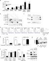
Figure 6. MCPIP1 induces degradation of IL-17R subunit but not Act1 mRNA transcripts
a. ST2 cells were transfected with indicated siRNAs, treated ± IL-17 for 3 h, and analyzed for IL-6 by ELISA. *p<0.05 vs. IL-17-treated control siRNA. ‡p<0.05 vs. unstimulated control. Data presented as mean ± SEM. b. HEK293T cells were transfected with murine Flag-tagged MCPIP1 together with Myc-tagged murine IL-17RA, IL-17RC and Act1. After 24 h, lysates were immunoprecipitated with anti-Myc Abs and immunoblotted for Myc and Flag. Arrows indicate IL-17RA, IL-17RC or Act1. c. HEK293T cells were transfected with varying concentrations of Flag-MCPIP1 with Myc-IL-17RA. Lysates were immunoblotted for Myc or Flag. d. Left: HEK293T cells were transected with mIL-17RA and the indicated genes (vector, A20, TRAF6, MCPIP1, MCPIP1ΔZF). After 24 h, cells were stained with APC-tagged α-IL-17RA Abs and APC-tagged 2° Ab. Right: MFI within IL-17RA+ gates, depicted as the ratio of the MFI of each sample relative to vector control. Data for 2 independent experiments is shown. e. HEK293T cells were transfected with MCPIP1 and the indicated constructs. After 24 h, mRNA was assessed by qPCR. Data are expressed as fold-change relative to vector-transfected controls. *p<0.05 vs. corresponding samples without MCPIP1. f. HEK293T cells were transfected with MCPIP1 and the indicated IL-17RA mutants. After 24 h, mRNA was assessed by qPCR. Data are expressed as fold-change relative to Il17ra(1–775)-transfected control. *p<0.05 vs. corresponding samples without MCPIP1. HEK293T cells were transfected with the indicated constructs. Left: After 24 h mRNA was assessed as in panel e. Right: Whole cell lysates were analyzed by immunoblotting as in panel b. g. In vitro transcribed mRNAs encoding Il6 3’ UTR or Il17ra (nucleotides 1–775) were incubated with water, buffer or recombinant MCPIP1 for 1 h at 30°C. Transcripts were analyzed on a denaturing agarose gel. Panels are derived from the same gel image.
Figure 7
Figure 7. Degradation of IL-17RA by MCPIP1 requires RNase but not DUB activity
a. Diagram of MCPIP1 subdomains. Residue designations are for the human homologue. UBA, Ubiquitin Association Domain; NYN, Nedd4-BP1, YacP nuclease; ZF, Zinc Finger; DUB, deubiquitinase domain; PRR, Proline rich region. b–c. HEK293T cells were transfected with plasmids encoding human MCPIP1 or mutants with Myc-tagged murine IL-17RA. After 24 h, whole cell lysates were immunoblotted for Myc (top) and MCPIP1 (bottom). d. HEK293T cells were transfected with IκBζ with a 3’ UTR with MCPIP1 and the indicated mutants. Lysates were analyzed by immunoblotting for IκBζ (top), MCPIP1 (middle) or β-Tubulin (bottom). Note that the ΔDUB mutant (Δ371–385) is GFP-tagged and migrates as a larger band. e. MCPIP1−/− cells were transfected with vector, MCPIP1 or the MCPIP1.C306R mutant. Cells were treated with IL-17 for 3 h. IL-6 was assessed by ELISA and Il6 assessed by qPCR. *p<0.05 vs. IL-17-treated vector control. ‡ p<0.05 vs. unstimulated vector control. n.s., not significant. Data presented as mean ± SEM. Data expressed as fold-change relative to untreated vector control. Experiments were performed a minimum of twice.

Similar articles

Cited by

References

    1. Basu S, Quilici C, Zhang HH, Grail D, Dunn AR. Mice lacking both G-CSF and IL-6 are more susceptible to Candida albicans infection: critical role of neutrophils in defense against Candida albicans . Growth Fac. 2008;26:23–34. - PubMed
    1. Bulek K, Liu C, Swaidani S, Wang L, Page RC, Gulen MF, Herjan T, Abbadi A, Qian W, Sun D, et al. The inducible kinase IKKi is required for IL-17-dependent signaling associated with neutrophilia and pulmonary inflammation. Nat Immunol. 2011;12:844–852. - PMC - PubMed
    1. Camporeale A, Poli V. IL-6, IL-17 and STAT3: A holy trinity in auto-immunity? Front Biosci. 2012;17:2306–2326. - PubMed
    1. Carpenter S, Ricci EP, Mercier BC, Moore MJ, Fitzgerald KA. Post-transcriptional regulation of gene expression in innate immunity. Nat Rev Immunol. 2014;14:361–376. - PubMed
    1. Conti H, Shen F, Nayyar N, Stocum E, JN S, Lindemann M, Ho A, Hai J, Yu J, Jung J, et al. Th17 cells and IL-17 receptor signaling are essential for mucosal host defense against oral candidiasis. J Exp Med. 2009;206:299–311. - PMC - PubMed

Publication types

MeSH terms