Review
. 2015 Aug 21;7(8):4800-25.
doi: 10.3390/v7082844.

Cloak and Dagger: Alternative Immune Evasion and Modulation Strategies of Poxviruses

Affiliations
Review

Cloak and Dagger: Alternative Immune Evasion and Modulation Strategies of Poxviruses

Susanna R Bidgood et al. Viruses. .

Abstract

As all viruses rely on cellular factors throughout their replication cycle, to be successful they must evolve strategies to evade and/or manipulate the defence mechanisms employed by the host cell. In addition to their expression of a wide array of host modulatory factors, several recent studies have suggested that poxviruses may have evolved unique mechanisms to shunt or evade host detection. These potential mechanisms include mimicry of apoptotic bodies by mature virions (MVs), the use of viral sub-structures termed lateral bodies for the packaging and delivery of host modulators, and the formation of a second, "cloaked" form of infectious extracellular virus (EVs). Here we discuss these various strategies and how they may facilitate poxvirus immune evasion. Finally we propose a model for the exploitation of the cellular exosome pathway for the formation of EVs.

Keywords: exocytosis; exosome; immune evasion; vaccinia virus; virus entry.


Figures
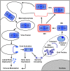
Figure 1
Figure 1
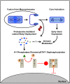
Vaccinia virus (VACV) replication cycle. The lifecycle of VACV begins when either, a single-membrane mature virion (MV) or double-membrane extracellular virus (EV), containing the genome, lateral bodies (LBs), and early transcription machinery, enters the host cell by inducing their own macropinocytic uptake. Upon fusion of the viral and the cellular limiting membrane of the macropinosome, the LBs dissociate from the core and are deposited in the cytoplasm. The LBs disperse, releasing virus host modulatory factors. Cores undergo activation concomitant with the initiation of early gene expression. Approximately one half of the proteins encoded by early genes serve an immunomodulatory function, while the remainder are required for genome uncoating and subsequent genome replication. Genome replication occurs in cytoplasmic viral factories where MVs are also assembled. Assembly is a highly complex multi-step process involving the formation of several non-infectious virus intermediates (crescents/immature virions). Once formed, MVs either exit cells by lysis or become wrapped by two additional cell derived membranes (red) which direct their exocytosis and thereby formation of EVs.
Figure 2
Figure 2
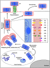
VACV LBs as Immunomodulatory Delivery Packets. After internalisation via macropinocytosis VACV particles undergo fusion with the limiting membrane of the macropinosome releasing the viral core into the cytoplasm. The released viral cores are “activated” as indicated by morphological changes and the initiation of early gene expression from within. Upon fusion, the LBs detach from the core and remain associated with the viral membrane. Once exposed to the cytoplasm, LBs are rapidly disassembled, with the major LB structural protein, F17, undergoing proteasome dependent degradation. Disassembly of the LB appears to facilitate release of other LB proteins and, in the case of the viral dual specificity phosphatase H1, is required for their action. Release of H1 from LBs, serves to shunt cellular antiviral transcription prior to the expression of early viral genes. To do this, H1 dephosphorylates phospho-STAT1 preventing its homodimerisation and nuclear translocation. To date only three LB components F17, H1, and a viral disulfide oxidoreductase G4 have been identified.
Figure 3
Figure 3
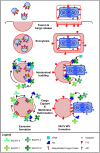
VACV MV to EV. After assembly within the cytoplasmic virus factory, a subset of MVs are transported on microtubules to the site of wrapping in the region of the microtubule organising center. The wrapping membranes are thought to be derived from the trans-Golgi network (TGN), or potentially LEs, and EEs after recycling of EV proteins from the plasma membrane. MV wrapping results in the formation of a triple membrane WV. WVs contain 9 additional proteins (illustrated in the zoom box) not found in MVs that direct the wrapping, post-wrapping transport, exocytosis and virus spread. Once formed, WVs are transported to the cell surface on microtubules. Upon reaching the plasma membrane the outermost WV membrane undergoes fusion thereby exocytosing the underlying double-membrane enveloped EV. EVs can either remain associated with the producer cell, detach from the cell surface to mediate long distance spread, or by the action of actin tails be propelled away from the cell surface to facilitate cell-to-cell spread. VACV: vaccinia virus; MV: mature virion; WV: wrapped virion; EV: extracellular virion; LE: late endosome; EE: early endosome.
Figure 4
Figure 4
Model for exosome-like extracellular virus (EV) formation. The process of vaccinia virus (VACV) EV formation is highly reminiscent of cellular exosome formation. Both processes proceed through four major steps: Cargo capture and membrane deformation, intraluminal budding, exocytosis and finally fusion with the plasma membrane to release the membrane bound cargo. Canonical exosome formation (left) is regulated by the ESCRTs. ESCRT-0 acts to recognise membrane-bound ubiquitinated cargo proteins and direct them into distinct late endosome (LE) membrane regions. ESCRT-I/ESCRT-II drive membrane deformation. After recruitment of ESCRT-III via ESCRT-II or the accessory protein Alix, ESCRT-I/ESCRT-II depart, and ESCRT-III drives invagination and subsequent membrane fission with assistance of the AAA+ ATPase complex, Vps4. The newly formed multivesicular body (MVB) is transported to the cell surface on microtubules and the intralumenal vesicles are released from the cell when the limiting membrane of the MVB fuses with the plasma membrane, thereby forming exosomes. Based on the evidence described in the text, we propose a model of VACV wrapped virion (WV) formation akin to exosome formation (right). As the EV protein F13 is essential for wrapping, contains a late domain, is present in LEs during infection, and interacts with late endosomal factors, we suggest that F13 acts as an ESCRT-0 mimic that serves to recognise mature viruses (MVs) as cargo for wrapping. While it is unknown what F13 recognises on the MV; both A27, an MV membrane protein required for EV formation, and ubiquitin on the VACV membrane could serve as F13 recognition targets. As an ESCRT-0 mimic, F13 could also serve to recruit ESCRT-I/II and/or ESCRT-III via the accessory protein Alix. This would initiate wrapping, a process topologically analogous to intralumenal budding during MVB formation. In support of this both the ESCRT-1 component, TSG101, and the accessory protein Alix are required for EV formation. To complete WV formation, the Vps4 complex could be recruited to facilitate the sealing of the protective EV membrane. Like exosome release, fully formed WVs require microtubules for transport to the plasma membrane where they fuse, releasing the membrane-bound MV cargo, thus forming the double-membrane EV.

Similar articles

Cited by

References

    1. Mercer J., Schelhaas M., Helenius A. Virus entry by endocytosis. Annu. Rev. Biochem. 2010;79:803–833. doi: 10.1146/annurev-biochem-060208-104626. - DOI - PubMed
    1. Tam J.C., Jacques D.A. Intracellular immunity: Finding the enemy within—How cells recognize and respond to intracellular pathogens. J. Leukoc. Biol. 2014;96:233–244. doi: 10.1189/jlb.4RI0214-090R. - DOI - PMC - PubMed
    1. Randow F., MacMicking J.D., James L.C. Cellular self-defense: How cell-autonomous immunity protects against pathogens. Science. 2013;340:701–706. doi: 10.1126/science.1233028. - DOI - PMC - PubMed
    1. Blasius A.L., Beutler B. Intracellular toll-like receptors. Immunity. 2010;32:305–315. doi: 10.1016/j.immuni.2010.03.012. - DOI - PubMed
    1. Janssens S., Beyaert R. Role of toll-like receptors in pathogen recognition. Clin. Microbiol. Rev. 2003;16:637–646. doi: 10.1128/CMR.16.4.637-646.2003. - DOI - PMC - PubMed

Publication types

LinkOut - more resources