. 2015 Sep;22(9):712-9.
doi: 10.1038/nsmb.3075. Epub 2015 Aug 24.

Ubp6 deubiquitinase controls conformational dynamics and substrate degradation of the 26S proteasome

Affiliations

Ubp6 deubiquitinase controls conformational dynamics and substrate degradation of the 26S proteasome

Charlene Bashore et al. Nat Struct Mol Biol. 2015 Sep.

Abstract

Substrates are targeted for proteasomal degradation through the attachment of ubiquitin chains that need to be removed by proteasomal deubiquitinases before substrate processing. In budding yeast, the deubiquitinase Ubp6 trims ubiquitin chains and affects substrate processing by the proteasome, but the underlying mechanisms and the location of Ubp6 within the holoenzyme have been elusive. Here we show that Ubp6 activity strongly responds to interactions with the base ATPase and the conformational state of the proteasome. Electron microscopy analyses reveal that ubiquitin-bound Ubp6 contacts the N ring and AAA+ ring of the ATPase hexamer and is in proximity to the deubiquitinase Rpn11. Ubiquitin-bound Ubp6 inhibits substrate deubiquitination by Rpn11, stabilizes the substrate-engaged conformation of the proteasome and allosterically interferes with the engagement of a subsequent substrate. Ubp6 may thus act as a ubiquitin-dependent 'timer' to coordinate individual processing steps at the proteasome and modulate substrate degradation.


Figures
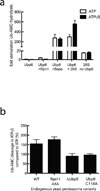
Figure 1
Figure 1. Ubp6 deubiquitination activity responds to the conformational state of the proteasome
Ub-AMC cleavage activity of Ubp6 was measured in response to interactions with the proteasomes holoenzyme or isolated subcomplexes. (a) Deubiquitination assays using proteasomes reconstituted with heterologously expressed base subcomplex purified from E. coli as well as core and lid subcomplexes purified from yeast, in the presence of ATP or the non-hydrolyzable ATPγS that induces the engaged state of the proteasome. (b) Deubiquitination assays using proteasomes purified from yeast strains with either wild-type, deleted, or inactive (C118A) Ubp6, or with wild-type Ubp6 and an inactive Rpn11 (AXA). Shown are the relative activities in the presence of ATPγS compared to ATP. Data in a and b are means and s.e.m.of three independent experiments.
Figure 2
Figure 2. Ubp6 allostery is connected to substrate engagement
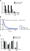
To separate ubiquitin processing from substrate engagement and translocation, we designed an ubiquitin-independent recruitment system by fusing a linked permutant of the bacterial dimeric substrate adapter SspB2 to Rpt2 of the base. (a) Schematic of a SspB2-fused proteasome recruiting an ssrA tagged substrate and SDS-PAGE of E. coli-expressed base subcomplex with either wild-type or SspB2-fused Rpt2. (b) Degradation of a GFP model substrate containing the ssrA recognition motif was measured using proteasomes reconstituted with either wild-type or SspB2-fused base complexes. (c) Ubiquitin-bound Ubp6 (Ubp6 C118A with di-ubiquitin or ubiquitin-vinylsulfone-fused Ubp6, Ubp6-UbVS) and substrate translocation stimulate the ATPase rate of SspB2-fused proteasomes. Data shown in b and c are means and s.e.m. of three technical replicates.
Figure 3
Figure 3. Ubiquitin-bound Ubp6 interacts with the Rpt hexamer of the base
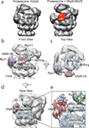
(a) 3D reconstructions of the proteasome holoenzyme in complex with ATPγS and ubiquitin-free (left) or permanently ubiquitin-bound Ubp6 shown in red (right). (b–e) PDB models of various RP subunits were docked into the 3D electron density map obtained from negatively stained samples. Rpn1 is shown in purple (PDB 4CR4 Chain Z), Rpn11 in green (PDB 4O8X), and the ATPase ring in blue (PDB 4CR4, Chains H-M). The Ubp6-Ub homology model was docked into the corresponding density of the 22.3-Å resolution map (Ubp6 is shown in red, ubiquitin in blue). PDB models for all Rpt proteins of the base are alternately colored in two different shades of blue. (b) Front view of the RP. Connecting density is observed between Rpn1 and the catalytic domain of Ubp6, which contacts the Rpt ring directly in front of Rpn11. (c) Top view of the RP. Ubp6 makes specific contacts with the N-terminal domain of Rpt1. (d) Side view of the RP. Ubp6 bridges the N-ring and the AAA+ ring in their coaxially stacked, engaged conformation. The N-domain residues of Rpt1 appear to interact with surface loops of Ubp6, while the AAA+ domain of Rpt1 contacts two C-terminal helices of Ubp6. This architecture places Ubp6 in close proximity to Rpn11 (~20 Å), with its bound ubiquitin only ~30 Å from the Rpn11 active site. (e) Zoomed-in view of (d), highlighting the Ubp6-base interface and proximity to Rpn11.
Figure 4
Figure 4. Ubiquitin-bound Ubp6 stabilizes the substrate-engaged conformation of the proteasome
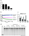
Ubiquitin-independent substrate delivery to the proteasome reveals that ubiquitin-bound Ubp6 allosterically inhibits multiple- but not single-turnover degradation. (a) Multiple-turnover degradation of a permanently unfolded model substrate and a GFP fusion substrate by reconstituted SspB2-Rpt2 proteasomes in the absence or presence of Ubp6 C118A and di-ubiquitin (Ub2). Data shown are means and s.e.m of three technical replicates. Representative gels are shown in Supplementary Data Set 1. (b) Single-turnover degradation of the GFP fusion substrate by saturating amounts of reconstituted SspB2-Rpt2 proteasomes in the absence or presence of Ubp6 C118A and Ub2. Curves shown are representative of three individual experiments. (c) Rate constants for degradation of the GFP fusion substrate under multiple- and single-turnover conditions shown in (a) and (b). Rate constants for single turnover degradations were determined from a single exponential regression of data; error bars represent s.e.m. of three individual experiments.
Figure 5
Figure 5. Ubp6 affects ubiquitin-dependent degradation
(a) Ubiquitin binding to Ubp6 strongly inhibits Rpn11 deubiquitination activity. Ubp6 (wild type or C118A) was treated with ubiquitin aldehyde (Ub-H) and added to Ubp6-free holoenzymes purified from yeast. Rpn11 deubiquitination activity was measured by Ub-AMC cleavage. Data are means and s.e.m. of 3 independent experiments. (b,c) The same GFP fusion substrate used for ubiquitin-independent turnover in figure 4 was ubiquitinated in vitro at an engineered single lysine residue, to examine the effects of Ubp6 on ubiquitin-dependent substrate degradation. (b) Single-turnover degradation of the polyubiquitinated GFP fusion substrate were measured with saturating amounts of proteasome holoenzyme purified from an Ubp6-knockout yeast strain, with no Ubp6, wild-type Ubp6, Ubp6 C118A, or Ubp6-UbVS added back. (c) Rate constants for single- and multiple-turnover degradation of the ubiquitinated GFP model substrate. Data shown are means and s.e.m. from three technical replicates. (d) Degradation of a poly-ubiquitinated EGFP, substrate was assessed by SDS-PAGE and in-gel GFP fluorescence detection. Used proteasomes were purified from ubp6Δ yeast cells and incubated with either buffer, WT, C118A or UbVS-treated Ubp6.
Figure 6
Figure 6. Model for Ubp6 acting as an ubiquitin-dependent timer to allosterically control proteasome conformational changes, Rpn11 deubiquitination, and substrate degradation
(a) Ubiquitin-chain binding to an intrinsic receptor (e.g. Rpn10) tethers a substrate to the proteasome. Ubp6 is bound to the proteasome via its Ubl domain interacting with Rpn1. (b) Engagement of the unstructured initiation region of the substrate by the ATPase hexamer induces a conformational switch of the regulatory particle to a substrate-engaged, translocation competent state, characterized by a coaxial alignment of Rpn11, N-ring, AAA+ ring and 20S core. If ubiquitin-bound, for instance during debranching or trimming of ubiquitin chains, Ubp6 interacts with and stabilizes the engaged state of the ATPase hexamer by bridging the N-ring and AAA+ ring. In this state, ubiquitin-bound Ubp6 inhibits Rpn11-mediated deubiquitination and consequently substrate degradation. (c) Translocation moves the ubiquitin-modified lysines of the substrate into the Rpn11 active site for co-translocational ubiquitin-chain removal. (d) Even after complete substrate translocation, ubiquitin-bound Ubp6 stabilizes the engaged conformation of the proteasome, prevents switching back to the engagement-competent state, and thus interferes with the degradation of the subsequent substrate. Such trapping of the engaged state would allow Ubp6 to clear ubiquitin chains from proteasomal receptors before the next substrate is engaged and degradation is initiated. (e) As soon as it is no longer occupied with ubiquitin, the catalytic domain of Ubp6 releases from the N-ring and AAA+ ring, and allows the regulatory particle to return to the pre-engaged state for the next round of substrate degradation.

Comment in

Similar articles

Cited by

References

    1. Finley D. Recognition and Processing of Ubiquitin-Protein Conjugates by the Proteasome. Annu. Rev. Biochem. 2009;78:477–513. - PMC - PubMed
    1. Goldberg AL. Protein degradation and protection against misfolded or damaged proteins. Nature. 2003;426:895–899. - PubMed
    1. Goldberg AL. Functions of the proteasome: from protein degradation and immune surveillance to cancer therapy. Biochem. Soc. Trans. 2007;35:12–17. - PubMed
    1. Verma R, et al. Role of Rpn11 metalloprotease in deubiquitination and degradation by the 26S proteasome. Science. 2002;298:611–615. - PubMed
    1. Yao T, Cohen RE. A cryptic protease couples deubiquitination and degradation by the proteasome. Nature. 2002;419:403–407. - PubMed

Publication types

MeSH terms

Substances