. 2015 Sep 15;112(37):11660-5.
doi: 10.1073/pnas.1514026112. Epub 2015 Aug 19.

Structural characterization of muropeptides from Chlamydia trachomatis peptidoglycan by mass spectrometry resolves "chlamydial anomaly"

Affiliations

Structural characterization of muropeptides from Chlamydia trachomatis peptidoglycan by mass spectrometry resolves "chlamydial anomaly"

Mathanraj Packiam et al. Proc Natl Acad Sci U S A. .

Abstract

The "chlamydial anomaly," first coined by James Moulder, describes the inability of researchers to detect or purify peptidoglycan (PG) from pathogenic Chlamydiae despite genetic and biochemical evidence and antibiotic susceptibility data that suggest its existence. We recently detected PG in Chlamydia trachomatis by a new metabolic cell wall labeling method, however efforts to purify PG from pathogenic Chlamydiae have remained unsuccessful. Pathogenic chlamydial species are known to activate nucleotide-binding oligomerization domain-containing protein 2 (NOD2) innate immune receptors by as yet uncharacterized ligands, which are presumed to be PG fragments (muramyl di- and tripeptides). We used the NOD2-dependent activation of NF-κB by C. trachomatis-infected cell lysates as a biomarker for the presence of PG fragments within specific lysate fractions. We designed a new method of muropeptide isolation consisting of a double filtration step coupled with reverse-phase HPLC fractionation of Chlamydia-infected HeLa cell lysates. Fractions that displayed NOD2 activity were analyzed by electrospray ionization mass spectrometry, confirming the presence of muramyl di- and tripeptides in Chlamydia-infected cell lysate fractions. Moreover, the mass spectrometry data of large muropeptide fragments provided evidence that transpeptidation and transglycosylation reactions occur in pathogenic Chlamydiae. These results reveal the composition of chlamydial PG and disprove the "glycanless peptidoglycan" hypothesis.

Keywords: NOD2 receptor; chlamydia; mass spectrometry; muropeptide; peptidoglycan.


Conflict of interest statement

The authors declare no conflict of interest.

Figures
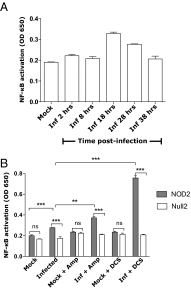
Fig. 1.
Fig. 1.
Kinetics of Chlamydia NOD2 ligand production during the developmental cycle and enhancement of activation by antibiotics that inhibit PG synthesis. (A) Chlamydia-infected and mock-infected HeLa cell lysates were harvested at various time points during the developmental cycle, and their ability to induce NOD2-dependent NF-κB activity was assessed in a HEK-Blue NOD2 reporter cell line carrying a NF-κB–inducible alkaline phosphatase reporter gene. Peak NOD2-dependent activity was observed for Chlamydia-infected cell lysates harvested at 18 h PI. (B) The ability of Chlamydia-infected HeLa cell lysates harvested at 18 h PI to induce NF-κB activity was tested in HEK-Blue cell lines expressing (gray bars) or devoid of (white bars) NOD2 innate immune receptors and carrying a NF-κB–inducible alkaline phosphatase reporter gene. Alkaline phosphatase activity was measured at an OD of 650 nm following addition of infected and mock-infected cell lysates. Chlamydia-infected cell lysates induced higher NF-κB activity over mock-infected cell lysates in a NOD2-dependent manner. Treatment with PG synthesis inhibitors ampicillin (Amp, 1 μg/mL) and DCS (30 μg/mL) greatly enhanced the ability of Chlamydia-infected cell lysates to induce NF-κB activity in a NOD2-dependent manner over the corresponding untreated infected cell lysates. **P < 0.01, ***P < 0.005 unpaired t test; ns, nonsignificant. OD650 values are mean ± SEM; n = 3.
Fig. S1.
Fig. S1.
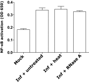
Nature of Chlamydia NOD2 ligand production during the developmental cycle. Chlamydia-infected cell lysates harvested at 18 h PI were subjected to heat treatment at 95 °C for 6 min or RNase A (40 μg/mL) treatment, and their ability to induce NOD2-dependent NF-κB activity was assessed. OD650 values are mean ± SEM; n = 3.
Fig. 2.
Fig. 2.
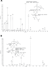
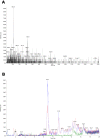
Molecular detection of chlamydial MDP and MTP in Chlamydia-infected cell lysates. Mass and product ion scans were performed on 3 kDa flow-through of Chlamydia-infected HeLa cell lysates specifically looking for ions with an m/z of 494.4 and 666.2. (A) Tandem mass spectrum of the molecular ion [M+H]+ = 494.4 m/z. The spectral interpretation of the daughter ions generated is provided in the Inset; fragmentation is indicated by dashed lines. (B) Tandem mass spectrum of the molecular ion [M+H]+ = 666.2 m/z. The spectral interpretation of the daughter ions generated is provided in the Inset; fragmentation is indicated by dashed lines.
Fig. S2.
Fig. S2.
Validation of the PG isolation method by cell lysate fractionation by detection of Shigella anhydro MTP. Mass and product ion scans were performed on 3 kDa flow-through cell lysates of midlog phase grown Shigella. Tandem mass spectrometry was performed on molecular ion m/z 648.2, which corresponds to Shigella anhydro MTP. Fragmentation is indicated by dashed lines.
Fig. S3.
Fig. S3.
Tandem mass spectrum of synthetic NOD2 ligands MDP and MTP. Mass and product ion scans were performed on chemically synthesized (A) MDP and (B) MTP. The structures of the synthetic ligands are provided in the Inset; fragmentation is indicated by dashed lines. The MS/MS spectra generated by the synthetic NOD2 ligands were used in the interpretation of Chlamydia MDP and MTP species. Synthetic MDP differs from Chlamydia MDP in that it contains isoglutamine instead of glutamate.
Fig. 3.
Fig. 3.
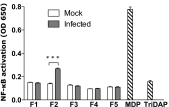
NOD2 stimulating activity of F2 fraction of Chlamydia-infected cells collected from a reverse-phase C18 HPLC column. HPLC fractionation was performed, and 1.2-mL fractions were collected every minute. Mock-infected cell lysates were also fractionated, and NOD2 activities induced by them were considered as background. NOD2-induced NF-κB activity (measured at OD650) for the various fractions collected from Chlamydia-infected (gray bar) or mock-infected lysates (open bar) was tested in HEK-Blue NOD2 cell lines as in Fig. 1. MDP (positive) and Tri-DAP (l-Ala-γ-d-Glu-mDAP) (negative) control ligands were used at 1 μg/mL. ***P < 0.005 unpaired t test. OD650 values are mean ± SEM; n = 3.
Fig. S4.
Fig. S4.
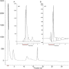
C18 column interacting properties of Chlamydia muropeptides as well as synthetic NOD2 ligands. The elution characteristics of Chlamydia-infected HeLa cell lysate (<3 kDa filtrate) from the reverse-phase C18 column are shown. The fraction from Chlamydia-infected cell lysates that eluted from the column in the second minute (F2) contains NOD2 ligands as assessed by NOD2-dependent NF-κB activity. The chromatograms of synthetic ligands (A) MDP and (B) MTP (Inset) show the synthetic ligands also elute from the C18 column in the second minute as measured by UV absorbance at 215 nm. The fraction highlighted by red lines in the three chromatograms had maximal NOD2 activity. The results suggest that Chlamydia NOD2 ligands have column interacting properties very similar to those of synthetic ligands.
Fig. 4.
Fig. 4.
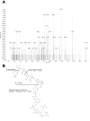
Molecular detection of a muropeptide fragment that serves as evidence for transpeptidation in chlamydial PG. A high-throughput IDA method of mass spectrometry was performed on a NOD2-activating (F2) fraction, and an ion with an m/z of 653.0 was selected for tandem MS/MS analysis. (A) Structural interpretation of the major ions of the MS/MS spectrum is given in B. Fragmentation is indicated by dashed lines.
Fig. S5.
Fig. S5.
(A) EMSs of NOD2-activating (F2) fraction. EMS revealed two prominent ions 653.0 m/z and 763.9 m/z at an Rt of 8.8 min, which were determined to be fragments of the polymerized PG complex. (B) Effect of ampicillin treatment on stem peptide cross-linked fragment ion 653.0 m/z. Extracted ion current of 653.0 m/z was compared among uninfected (green line), infected untreated (blue line), and infected ampicillin-treated (red line) NOD2-activating (F2) fractions. A decrease in intensity of ion 653.0 m/z seen in the ampicillin-treated cell lysates and the ion was found at background levels in uninfected cell lysates.
Fig. S6.
Fig. S6.
Molecular detection of a muropeptide fragment that serves as evidence for transglycosylation in chlamydial PG. A high-throughput IDA method of mass spectrometry was performed on a NOD2-activating (F2) fraction, and an ion with a m/z of 763.9 was selected for tandem MS/MS analysis (A). This PG fragment is similar to the parent presented in Fig. 4 with the addition of a second MurNAc; the inferred structure with fragments unique to this parent is presented in B. Fragmentation is indicated by dashed lines.

Similar articles

Cited by

References

    1. Moulder JW, Novosel DL, Officer JE. Inhibition of the growth of agents of the psittacosis group by D-cycloserine and its specific reversal by D-alanine. J Bacteriol. 1963;85:707–711. - PMC - PubMed
    1. Tamura A, Manire GP. Effect of penicillin on the multiplication of meningopneumonitis organisms (Chlamydia psittaci) J Bacteriol. 1968;96(4):875–880. - PMC - PubMed
    1. Stephens RS, et al. Genome sequence of an obligate intracellular pathogen of humans: Chlamydia trachomatis. Science. 1998;282(5389):754–759. - PubMed
    1. Fox A, et al. Muramic acid is not detectable in Chlamydia psittaci or Chlamydia trachomatis by gas chromatography-mass spectrometry. Infect Immun. 1990;58(3):835–837. - PMC - PubMed
    1. Hatch TP. Disulfide cross-linked envelope proteins: The functional equivalent of peptidoglycan in chlamydiae? J Bacteriol. 1996;178(1):1–5. - PMC - PubMed

Publication types

LinkOut - more resources