. 2015 Aug 18;10(8):e0135951.
doi: 10.1371/journal.pone.0135951. eCollection 2015.

Dexamethasone-Mediated Activation of Fibronectin Matrix Assembly Reduces Dispersal of Primary Human Glioblastoma Cells

Affiliations

Dexamethasone-Mediated Activation of Fibronectin Matrix Assembly Reduces Dispersal of Primary Human Glioblastoma Cells

Stephen Shannon et al. PLoS One. .

Abstract

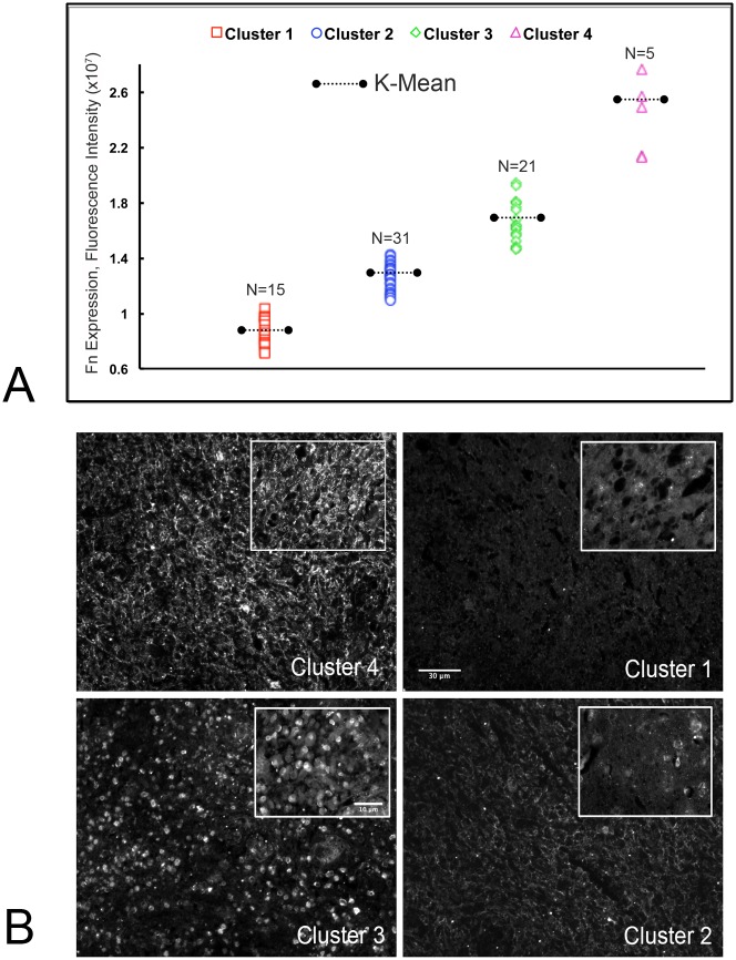
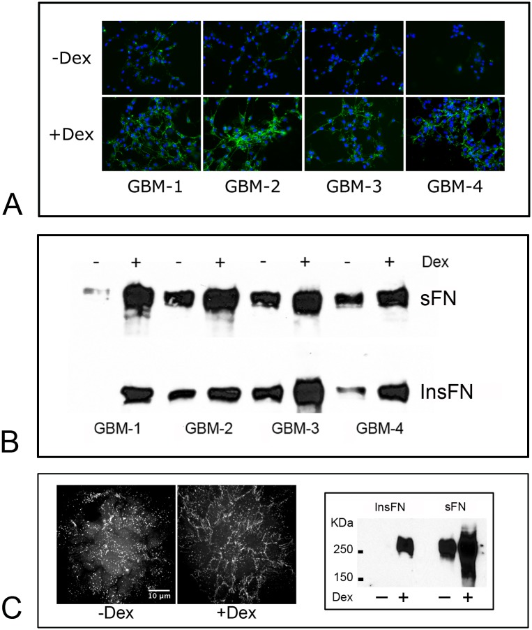
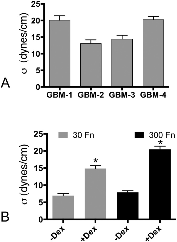
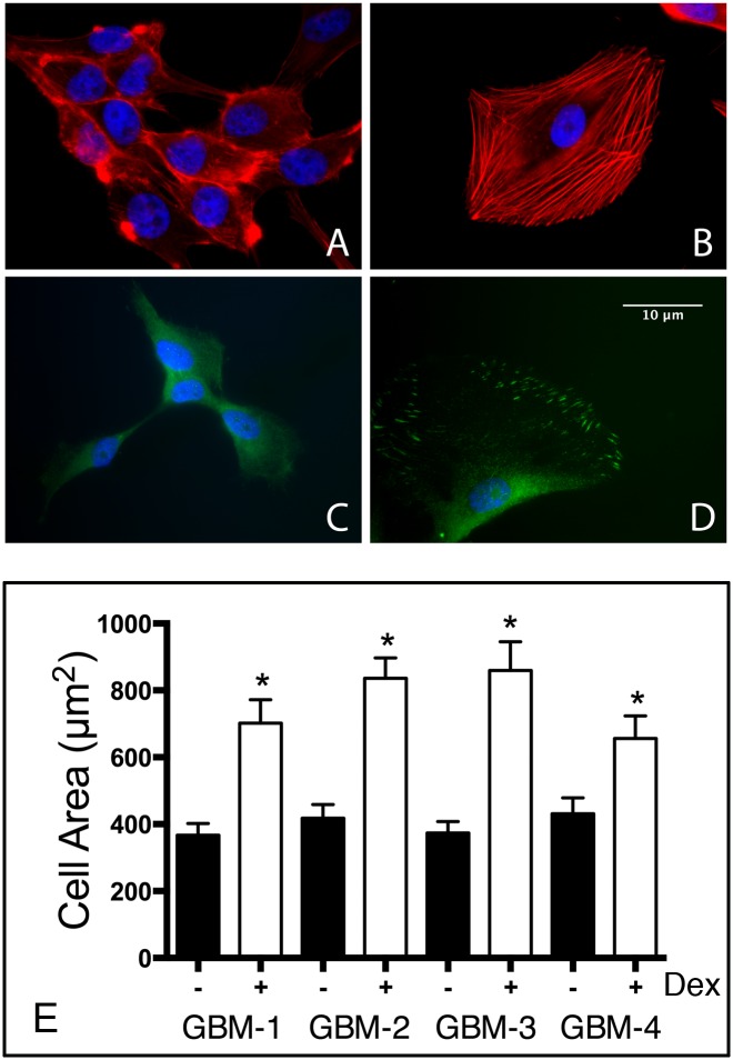
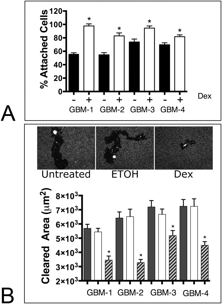
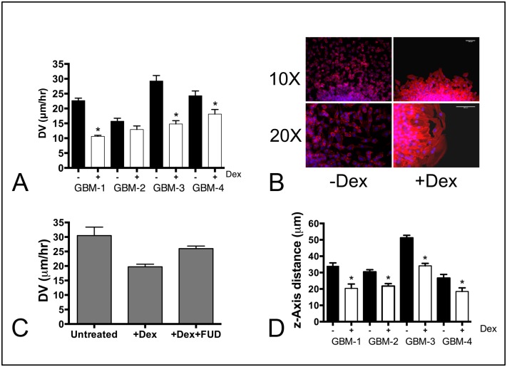
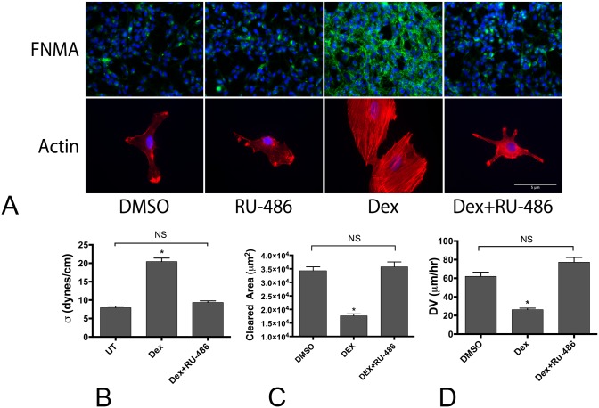
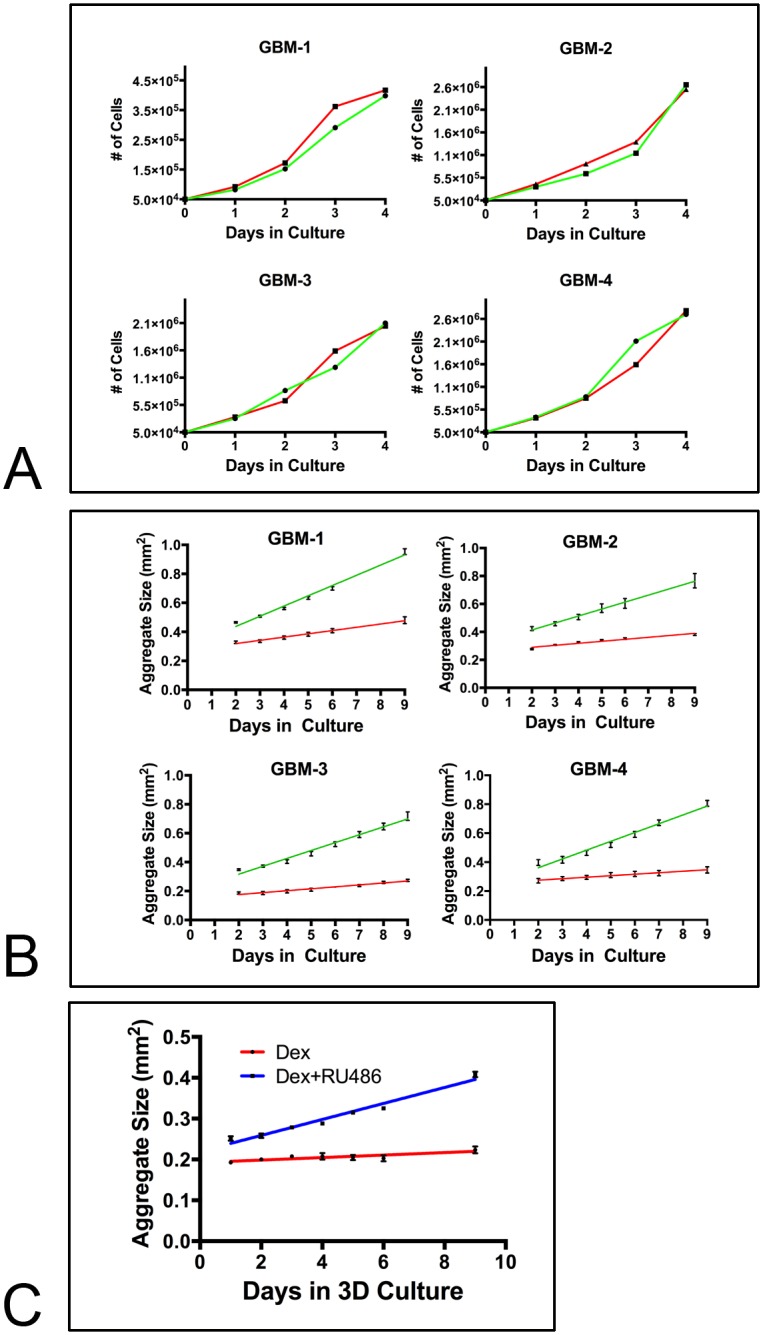
Despite resection and adjuvant therapy, the 5-year survival for patients with Glioblastoma multiforme (GBM) is less than 10%. This poor outcome is largely attributed to rapid tumor growth and early dispersal of cells, factors that contribute to a high recurrence rate and poor prognosis. An understanding of the cellular and molecular machinery that drive growth and dispersal is essential if we are to impact long-term survival. Our previous studies utilizing a series of immortalized GBM cell lines established a functional causation between activation of fibronectin matrix assembly (FNMA), increased tumor cohesion, and decreased dispersal. Activation of FNMA was accomplished by treatment with Dexamethasone (Dex), a drug routinely used to treat brain tumor related edema. Here, we utilize a broad range of qualitative and quantitative assays and the use of a human GBM tissue microarray and freshly-isolated primary human GBM cells grown both as conventional 2D cultures and as 3D spheroids to explore the role of Dex and FNMA in modulating various parameters that can significantly influence tumor cell dispersal. We show that the expression and processing of fibronectin in a human GBM tissue-microarray is variable, with 90% of tumors displaying some abnormality or lack in capacity to secrete fibronectin or assemble it into a matrix. We also show that low-passage primary GBM cells vary in their capacity for FNMA and that Dex treatment reactivates this process. Activation of FNMA effectively "glues" cells together and prevents cells from detaching from the primary mass. Dex treatment also significantly increases the strength of cell-ECM adhesion and decreases motility. The combination of increased cohesion and decreased motility discourages in vitro and ex vivo dispersal. By increasing cell-cell cohesion, Dex also decreases growth rate of 3D spheroids. These effects could all be reversed by an inhibitor of FNMA and by the glucocorticoid receptor antagonist, RU-486. Our results describe a new role for Dex as a suppressor of GBM dispersal and growth.


Conflict of interest statement

Competing Interests: The authors have declared that no competing interests exist.

Figures

Fig 1
Fig 1
(A) k-means cluster analysis of FN expression in a 40-sample, 80-core, human GBM tissue microarray. k-means clustering partitions a given data set into clusters by grouping each n, into the cluster with the nearest mean, serving as a prototype of the cluster. This results in a partitioning of the data space into Voronoi cells. In this case, cluster analysis was performed for 3 and 4 Voronoi cells (3k and 4k). A k of 4 provided the maximum difference in center means (ANOVA, p<0.0001). (B) Representative immunofluorescence images of the predominant patterns of FN expression in a human GBM microarray; high FN with FNMA (Cluster 4), high intracellular FN with no FNMA (Cluster 3), low FN with reduced FNMA (Cluster 2), and low to absent FN with no FNMA (Cluster 1).
Fig 2
Fig 2
(A) Dex treatment activates FNMA in GBM cells. Immunofluorescence was used to compare FNMA in untreated and Dex treated GBM cells. Dex was added to a final concentration of 1x10⁻⁷M. Cells were treated for 24 hours prior to fixation. Dex treatment significantly increased FNMA in all lines. (B) Dex-mediated upregulation of FNMA was confirmed by immunoblot analysis. Lysates of untreated and Dex-treated cells were prepared by DOC differential solubilization and soluble (sFN) and insoluble (InsFN) fractions of FN were separated by SDS-PAGE. Note the increase in both soluble and insoluble fibronectins in response to Dex treatment. (C) 3D spheroids of GBM-3 were generated in the presence or absence of Dex and 50 μg/ml Rhodamine-conjugated FN. Dex treatment resulted in a marked re-organization of FN from punctate to fibrous (left panel). FNMA by spheroids was also assessed by DOC/SDS-PAGE and immunoblot analysis. Note the presence of insoluble fibronectin in response to Dex treatment (right panel).
Fig 3
Fig 3
(A) The four GBM lines differ in surface tension. ANOVA and Tukey’s MCT revealed that the four GBM lines clustered into 2 groups, with GBM-1 (n = 19) and GBM-4 (n = 20) being more cohesive than GBM-2 (n = 16) and GBM-3 (n = 18). (B) Dex treatment significantly increases aggregate surface tension. The low cohesivity line GBM-3 was used to generate aggregates in the absence and presence of Dex and 30 or 300 μg/ml of FN. Dex treatment significantly increased aggregate surface tension in 30 μg/ml FN (n = 22). This increase was more pronounced when aggregates were incubated with 300 μg/ml FN (n = 19). * represents significance for pair-wise comparison by Student’s t-test, p<0.0001.
Fig 4
Fig 4. Dex treatment results in a change in cell shape, actin organization and pFAK localization.
GBM-3 cells were either left untreated (A, C) or were treated with 10−7 M Dex for 24 hours (B, D), whereupon they were fixed and permeabilized and labeled with either rhodamine-phalloidin or with a pFAK antibody, and DAPI as counterstain. Note that Dex treatment resulted in a change in cell shape and a significant reorganization of actin from cortical (A) into stress fibers (B). This was accompanied by a change in the localization of pFAK from cytoplasmic (C) to sites of cell-ECM adhesion (D). ImageJ was used to measure cell area in the absence or presence of Dex for the 4-GBM lines. Pair-wise comparison by Student t-test showed a significant increase in cell size in response to Dex treatment (p<0.05).
Fig 5
Fig 5. Dex enhances resistance to shear-induced detachment and reduces cell motility.
(A) Untreated and Dex-treated GBM cells attached to PET membranes were subjected to 30 dynes/cm of shear flow for 3 hours, whereupon the number of cells retained on the membranes was quantified. For all lines, Dex treatment resulted in a significant retention of cells (t-test pairwise comparison, p<0.0001 for GBM-1,2, p = 0.0003 for GBM-3, and p = 0.0042 for GBM-4). Cell motility assays were also conducted in order to determine whether Dex, could also impact cell locomotion. A fluorescent microbead phagokinetic track assay revealed that Dex treated cells were less motile since treated cells appeared to essentially remain in place (B, upper panel). Motility was quantified by measuring cleared area of 20 cells for each untreated (solid bars), carrier-treated (clear bars), and Dex treated (hashed bars) groups and by comparing means by ANOVA and Tukey’s MCT. For all GBM lines, Dex significantly decreased cleared area (B, lower panel, ANOVA, p<0.0001, asterisk denotes significance, p<0.05).
Fig 6
Fig 6. Dex decreases dispersal of GBM cells.
(A) Aggregates, ranging in size from 50–70 μm in diameter were plated onto tissue culture plastic in complete medium. DV was measured by calculating the change in diameter as a function of time. Regression analysis generated a slope and R2 value that were used to calculate velocity. The DV of untreated aggregates (black bars) was significantly higher than that of Dex-treated aggregates (clear bars), for GBM-1,3, and 4. GBM-2 dispersal was reduced, but not significantly (p = 0.12). (B) Dex increases cohesion and promotes actin stress fiber formation between cells at the advancing cell front. Note single cell dispersal from untreated aggregates, in contrast to a higher level of cell-cell contact and actin stress fibers in response to Dex treatment. (C) One of the high motility lines (GBM-3) was used to demonstrate a functional role for FNMA in reducing DV. Inclusion of the FUD fragment blocked the effects of Dex and restored DV of Dex-treated aggregates to levels similar to those of untreated aggregates (Tukey’s MCT, p>0.05). (D) Dex also decreased dispersal of aggregates of GBM cells through a normal human astrocyte seeded 3D scaffold (* represent pair-wise comparison, t-test p<0.05).
Fig 7
Fig 7. RU-486 blocks the effects of Dex.
(A) RU-486 blocks Dex-mediated effects on FNMA and actin organization. GBM-3 cells incubated in the presence of Dex and RU-486 fail to assemble a fibronectin matrix and do not reorganize actin into stress fibers. (B). Aggregates incubated in Dex and RU-486 have surface tensions similar to those measured for untreated aggregates. (C) RU-486 appears to block the effects of Dex on cell motility. Combination treatment restores cell motility to levels similar to those of DMSO treated cells. (D) Dex-mediated decrease in aggregate DV can be blocked by RU-486. Dex treatment resulted in a significant decrease in DV compared to DMSO treated aggregates. RU-486 increased DV to levels similar to those of DMSO-treated aggregates (ANOVA, p<0.0001, *** signifies statistical difference by Tukey’s MCT, NS = not significant).
Fig 8
Fig 8. Dex treatment significantly reduces the growth rate of GBM spheroids.
(A) Untreated (green line) or Dex-treated (red line) GBM were plated as single cells and proliferation was monitored over a 4-day period. Under such conditions, Dex did not appear to influence proliferation. (B) When untreated (green lines) or Dex-treated (red lines) GBM were cultured as 3D spheroids for 9-days, growth rate was significantly reduced as demonstrated by a significant shallowing of the slope of the line (ANCOVA, p<0.0001). (C) RU-486 blocks the effects of Dex on 3D growth. Spheroids of GBM-3 were incubated for 9 days either in the presence of Dex or in a combination of Dex and RU-486. Note the increase in the slope of the line of the Dex and RU-486 treated spheroids (blue line) as compared to aggregates treated with Dex alone (red line). The difference in the slope equates to a 6.4-fold increase in growth rate.

Similar articles

Cited by

References

    1. Stupp R, Hegi ME, Mason WP, van den Bent MJ, Taphoorn MJ, Janzer RC, et al. Effects of radiotherapy with concomitant and adjuvant temozolomide versus radiotherapy alone on survival in glioblastoma in a randomised phase III study: 5-year analysis of the EORTC-NCIC trial. The Lancet Oncology. 2009;10(5):459–66. 10.1016/S1470-2045(09)70025-7 . - DOI - PubMed
    1. Sampetrean O, Saga I, Nakanishi M, Sugihara E, Fukaya R, Onishi N, et al. Invasion precedes tumor mass formation in a malignant brain tumor model of genetically modified neural stem cells. Neoplasia. 2011;13(9):784–91. - PMC - PubMed
    1. Clarke J, Butowski N, Chang S. Recent advances in therapy for glioblastoma. Archives of neurology. 2010;67(3):279–83. Epub 2010/03/10. 10.1001/archneurol.2010.5 . - DOI - PubMed
    1. Sabari J, Lax D, Connors D, Brotman I, Mindrebo E, Butler C, et al. Fibronectin matrix assembly suppresses dispersal of glioblastoma cells. PloS one. 2011;6(9):e24810 Epub 2011/10/08. 10.1371/journal.pone.0024810 - DOI - PMC - PubMed
    1. Ryan PL, Foty RA, Kohn J, Steinberg MS. Tissue spreading on implantable substrates is a competitive outcome of cell-cell vs. cell-substratum adhesivity. Proceedings of the National Academy of Sciences of the United States of America. 2001;98(8):4323–7. Epub 2001/03/29. - PMC - PubMed

MeSH terms