. 2015 Sep;17(9):1218-27.
doi: 10.1038/ncb3216. Epub 2015 Aug 10.

Genome-wide association between YAP/TAZ/TEAD and AP-1 at enhancers drives oncogenic growth

Affiliations

Genome-wide association between YAP/TAZ/TEAD and AP-1 at enhancers drives oncogenic growth

Francesca Zanconato et al. Nat Cell Biol. 2015 Sep.

Abstract

YAP/TAZ are nuclear effectors of the Hippo pathway regulating organ growth and tumorigenesis. Yet, their function as transcriptional regulators remains underinvestigated. By ChIP-seq analyses in breast cancer cells, we discovered that the YAP/TAZ transcriptional response is pervasively mediated by a dual element: TEAD factors, through which YAP/TAZ bind to DNA, co-occupying chromatin with activator protein-1 (AP-1, dimer of JUN and FOS proteins) at composite cis-regulatory elements harbouring both TEAD and AP-1 motifs. YAP/TAZ/TEAD and AP-1 form a complex that synergistically activates target genes directly involved in the control of S-phase entry and mitosis. This control occurs almost exclusively from distal enhancers that contact target promoters through chromatin looping. YAP/TAZ-induced oncogenic growth is strongly enhanced by gain of AP-1 and severely blunted by its loss. Conversely, AP-1-promoted skin tumorigenesis is prevented in YAP/TAZ conditional knockout mice. This work highlights a new layer of signalling integration, feeding on YAP/TAZ function at the chromatin level.


Conflict of interest statement

The authors declare no competing financial interest.

Figures
Figure 1
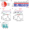
Figure 1. Genome-wide co-localization of YAP, TAZ and TEAD on enhancers.
(a) Overlap of peaks identified with YAP and TAZ antibodies. See Supplementary Figure 1a for specificity controls of the antibodies, and Table S1 for the results of de novo motif finding in YAP/TAZ peaks. (b) Overlap of YAP/TAZ peaks and TEAD4 peaks. See Supplementary Figure 1g for specificity controls of TEAD4 antibody, and Supplementary Figure 1h for ChIP-seq profiles at positive control loci. (c) Position of TEAD4 peak summits relative to the summits of the overlapping YAP/TAZ peaks, in a 500 bp window surrounding the summit of YAP/TAZ peaks. (d) Linear correlation between the signal of YAP or TAZ and TEAD4 peaks in the 5522 shared binding sites. r2 is the coefficients of determination of the two correlations. (e) ChIP-qPCR showing YAP binding to the indicated sites in MDA-MB-231 cells transfected with control (siCO) or TEAD siRNAs (siTEAD A). Relative DNA binding was calculated as fraction of input and normalized to IgG (IgG bars are omitted); data from 2 biological replicates from one representative experiment are shown. (f) Absolute distance of YAP peaks (n=7709), TAZ peaks (n=9798), TEAD4 peaks (n=8406) or overlapping YAP/TAZ/TEAD peaks (n=5522) to the nearest TSS. (g-h) Association of YAP/TAZ/TEAD peaks to promoters and enhancers according to ChIP-seq data for histone modifications. (g) Scheme illustrating peak classification. (h) Fraction of YAP/TAZ/TEAD peaks associated with each category. See Supplementary Figure 1j for validation of the enhancer/promoter status of a set of YAP/TAZ/TEAD-bound regions. (i) Heatmap representing YAP/TAZ/TEAD binding sites located on promoters (top) and enhancers (bottom). YAP, TAZ and TEAD4 peaks are ranked from the strongest to weakest signal in TAZ ChIP, in a window of ±1kb centered on the summit of TAZ peaks. H3K4me1 and H3K4me3 signal in the corresponding genomic regions is shown on the right. (j) Bimodal distribution of H3K4me1 signal around the summit of YAP/TAZ and TEAD4 peaks. (k) Distance between YAP/TAZ/TEAD binding sites and the TSS of the direct target genes they are associated to. Overall, 635 peaks were associated to 379 genes positively regulated by YAP/TAZ. See Methods for reproducibility of experiments.
Figure 2
Figure 2. YAP/TAZ/TEAD transcriptional program.
(a) Biological functions associated to YAP/TAZ direct positive targets, identified by GO terms. (b) YAP/TAZ or TEAD depletion impairs the expression of YAP/TAZ/TEAD direct target genes involved in cell proliferation, as evaluated by qRT-PCR (siCO=control siRNA, siYT=YAP/TAZ siRNA, siTEAD=TEAD siRNA). For a subset of genes, the downregulation of the corresponding proteins was also verified by Western blot (Supplementary Fig. 2b). See Supplementary Figure 2a for validation of TEAD siRNAs. (c-d) Validation of the long-range interaction between YAP/TAZ-occupied enhancers and the promoters of MYC (c) and TOP2A (d) by DNA looping, using 3C assays in MDA-MB-231 cells. TAZ and TEAD4 ChIP-seq profiles show the position of YAP/TAZ/TEAD binding sites upstream of MYC or TOP2A genes (here named "MYC enhancer 1", “TOP2A enhancer2” “TOP2A enhancer3”), whereas no YAP/TAZ/TEAD binding sites were detected close to their TSS. The chart shows the frequency of interaction (measured as cross-linking frequency) between MYC or TOP2A promoter ("anchor") and the indicated sites surrounding YAP/TAZ/TEAD (YTT) peaks (green lines). Interaction frequency is higher close to YAP/TAZ peak. Data points are mean+SEM from n=3 biological replicates. See also Supplementary Figs. 2d-e for the additional interactions between MYC and TOP2A promoters and a different set of YAP/TAZ/TEAD-bound enhancers. (e) ChIP-qPCR comparing the levels of H3K27ac (normalized to total H3 levels) in MDA-MB-231 cells transfected with control (siCO) or combined YAP/TAZ siRNAs (siYT1+2). Data from 2 biological replicates from one representative experiment are shown. See Methods for reproducibility of experiments.
Figure 3
Figure 3. Control of cell proliferation by YAP/TAZ/TEAD.
(a) Growth curve of MDA-MB-231 cells transfected with control siRNA (siCO) or two different combinations of siRNAs targeting YAP and TAZ (siYT). Data are mean+SD of n=8 biological replicates. Individual depletion of YAP or TAZ has no effect on cell growth (Supplementary Fig. 3a). (b) Percentage of MDA-MB-231 cells in G1, S and G2/M phases of cell cycle, as determined by flow-cytometric analysis of DNA content. Cells were transfected with control (siCO) or YAP/TAZ siRNAs (siYT) 48hr before fixation. Data are mean+SD of n=3 biological replicates. (c) Sustained expression of TAZ, but not of TEAD-binding deficient TAZS51A, rescues cell proliferation in YAP/TAZ-depleted cells. Empty-vector-, wild-type TAZ- (wt) or TAZS51A-transduced MDA-MB-231 cells were transfected with control (siCO) or YAP/TAZ (siYT) siRNAs, as indicated. Proliferation was evaluated as in (f). Data are mean+SD of n=8 biological replicates. (d) MDA-MB-231 cells were transduced with lentiviral vectors encoding rtTA and doxycycline-inducible MYC (MDA TetON MYC) and transfected with control or YAP/TAZ siRNAs. Where indicated, MYC expression was induced with 0.1 μg/ml doxycycline at the time of transfection. Cell growth was evaluated as in (f). Data are mean+SD of n=8 biological replicates. A control experiment with doxycycline-inducible EGFP is shown in Supplementary Figure 3h. (e) Average gene expression values of validated YAP/TAZ/TEAD direct target genes (listed in Fig. 2b) in invasive breast cancer samples, classified according to their histological grade. Individual data points and the mean value (black line) of each group are shown. (f) Kaplan-Meier graph representing the probability of cumulative metastasis-free survival in breast cancer patients stratified according to the expression of validated YAP/TAZ/TEAD direct target gene signature. High expression of the signature is associated with shorter metastasis-free survival (log-rank p-value < 0.0001). See Methods for reproducibility of experiments.
Figure 4
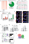
Figure 4. Co-occupancy of YAP/TAZ/TEAD and AP-1 at the same genomic loci.
(a) Density maps of YAP, TAZ, TEAD4 and JUN ChIP-seq reads at YAP/TAZ/TEAD-bound loci. See Supplementary Figures 4a-b for specificity controls of JUN antibody. (b) Percentage of YAP/TAZ/TEAD4 peaks (n=5522) overlapping with JUN ChIP-seq peaks. (c) Representative examples of YAP/TAZ/TEAD-bound enhancers co-occupied by JUN in the genome of MDA-MB-231 cells. (d) Co-presence of YAP and JUN at the same genomic regions. Results are fold enrichment relative to FLAG IP in control (empty vector-transduced) cells (ChIP 1) or relative to IgG negative control (ChIP 2). Data from one representative experiment are shown; experiments were repeated twice with similar results. (e) In situ PLA detection of endogenous YAP/AP-1 and TEAD1/AP-1 interactions in MDA-MB-231 cells. Nuclei were counterstained with DAPI (blue). The detected dimers are represented by fluorescent dots (red). The specificity of the interactions is revealed by the reduced number of dots detected after depletion of either of the partners with siRNAs. See Supplementary Figure 5a for different combinations of antibodies and Supplementary Table 5 for details about antibodies. Magnification is the same for all pictures. (f) AP-1 proteins co-precipitate with FLAG-TEAD1 in protein lysates of MDA-MB-231 cells. Input and IP were run on different gels. (g) TEAD1 co-precipitates with FOSL1 at endogenous protein levels in MDA-MB-231 cells. IP was performed with FOSL1 (N-17) antibodies. All samples were run on the same gel. JUN is a positive control. (h) TEAD1 co-precipitates with JUND at endogenous protein levels in MDA-MB-231 cells. FOSL1 is a positive control. All samples were run on the same gel. (i) CTGF and ANKRD1 promoters (either wild-type, or carrying mutations in AP-1 or TEAD binding sites) were cloned upstream of the luciferase coding sequence and their activity was measured in MDA-MB-231 cells. Data are normalized to wild-type promoter sequences. Data are presented as mean+SD of n=4 biological replicates from 2 independent experiments. Binding of TEAD and AP-1 proteins to their respective binding sites was verified by DNA pull down; mutations abolished binding (Supplementary Fig. 5j). (j) Model of the complex formed by YAP/TAZ/TEAD and AP-1 on DNA. See Supplementary Figure 7 for uncropped Western blots, and Methods for reproducibility of experiments.
Figure 5
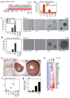
Figure 5. AP-1 factors synergize with YAP/TAZ/TEAD to promote oncogenic growth.
(a) The expression of YAP/TAZ/TEAD target genes involved in cell growth depends on AP-1. MDA-MB-231 cells were transduced with rtTA and doxycycline-inducible JUN-DN. Cells were left untreated (CO) or treated with 1 μg/ml doxycycline for 48h. As control, JUN-DN reduces the expression of FOSL1, CTGF and ANKRD1 (see Supplementary Fig. 6a). All expression levels are normalized to GAPDH. (b) Control and TAZS89A-overexpressing MII cells were transfected with the indicated siRNAs and tested for mammosphere formation. Data are mean+SD of n=6 biological replicates from a representative experiment. See Supplementary Figure 6c for a comparison with control MII cells. (c) Quantification and representative pictures of colonies formed by the indicated MCF10A derivatives in soft agar assays. Only background/not growing cell clusters were formed by control and MCF10A+AP-1 cells, and were not counted as colonies. Data are presented as mean+SD of n=3 biological replicates from a representative experiment. Magnification is the same for all pictures. (d) Quantification and representative pictures of primary mammospheres formed by the indicated MCF10A derivatives. Data are presented as mean+SD of n=6 biological replicates from a representative experiment. Magnification is the same for all pictures. (e-g) YAP and AP-1 synergize to promote tumor growth. (e) Representative IHC pictures of xenografts formed by the indicated cell lines. MCF10A cells were stained with a human-specific pan-cytokeratin antibody. (f) Tumor volumes at harvesting (individual tumors are plotted, line is the mean). (g) Quantification of Ki67-positive cells in tumor sections; data are mean + SEM of at least n=8 different samples. (h) YAP5SA and AP-1 cooperate to activate YAP/TAZ/TEAD proliferative program in MCF10A cells. MCF10A derivatives were grown on a thick Matrigel coating for one week before harvesting for RNA extraction. mRNA levels of the indicated genes were evaluated by qRT-PCR and normalized to GAPDH. See Methods for reproducibility of experiments.
Figure 6
Figure 6. YAP/TAZ are required for AP-1-driven skin tumorigenesis.
(a) K14-CreER and K14-CreER;Yapfl/fl;Tazfl/+ mice were treated with tamoxifen to activate Cre in the skin basal layer; after two weeks, mice received a single DMBA administration, followed by repeated TPA treatments for 40 weeks. Left: representative picture of control (K14-CreER) and YAP/TAZ deficient mice at time of harvesting (40 weeks after the beginning of the experiment). Right: time course of tumor development in mice with the indicated genotypes (data are mean+SD, n=9 for both experimental groups). See Supplementary Figure 6d for the verification of Cre-mediated recombination of YAP and TAZ alleles. (b) Representative H&E -stained sections of tumors from control mice, or ostensibly normal skin from DMBA/TPA-treated YAP/TAZ conditional knockout mice. Lesions developed by control mice are characterized by skin folds integrated by a core of connective tissue and lined by an hyperplastic, hyperkeratotic, stratified squamous epithelium; some foci of squamous cell carcinoma were also observed (see Supplementary Fig. 6e). Magnification is the same for all pictures. Scale bar is 1mm. See Methods for reproducibility of experiments.

Similar articles

Cited by

References

    1. Sudol M. Yes-associated protein (YAP65) is a proline-rich phosphoprotein that binds to the SH3 domain of the Yes proto-oncogene product. Oncogene. 1994;9:2145–2152. - PubMed
    1. Kanai F, et al. TAZ: a novel transcriptional co-activator regulated by interactions with 14-3-3 and PDZ domain proteins. The EMBO journal. 2000;19:6778–6791. - PMC - PubMed
    1. Piccolo S, Dupont S, Cordenonsi M. The biology of YAP/TAZ: hippo signaling and beyond. Physiological reviews. 2014;94:1287–1312. - PubMed
    1. Ramos A, Camargo FD. The Hippo signaling pathway and stem cell biology. Trends in cell biology. 2012 - PMC - PubMed
    1. Harvey KF, Zhang X, Thomas DM. The Hippo pathway and human cancer. Nat Rev Cancer. 2013;13:246–257. - PubMed

Publication types

MeSH terms

Associated data