. 2015 Aug 6;10(8):e0133749.
doi: 10.1371/journal.pone.0133749. eCollection 2015.

The Evolution of the Secreted Regulatory Protein Progranulin

Affiliations

The Evolution of the Secreted Regulatory Protein Progranulin

Roger G E Palfree et al. PLoS One. .

Abstract

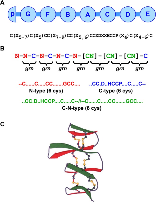
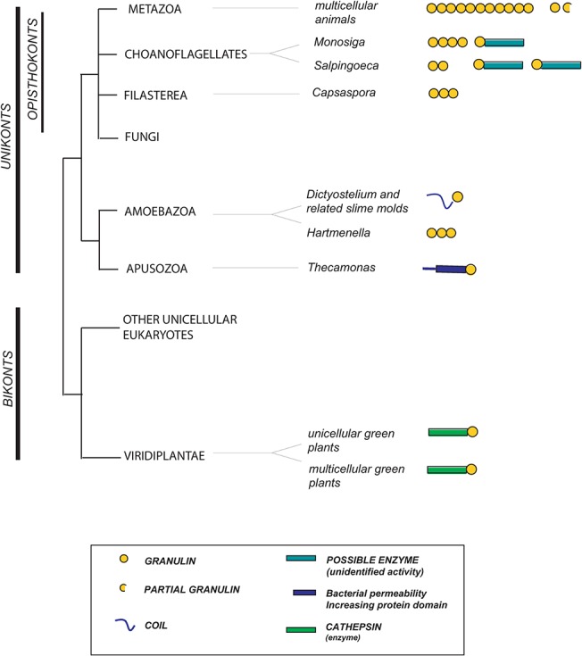
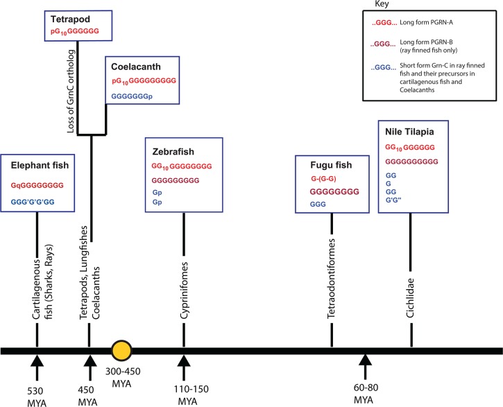
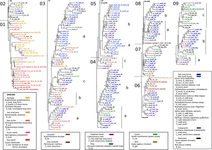
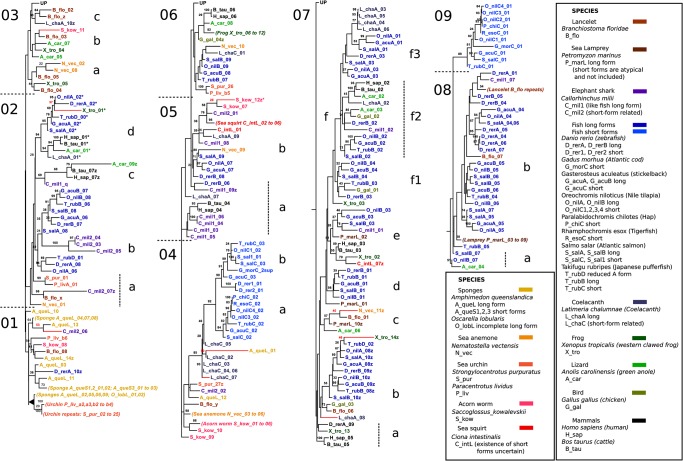
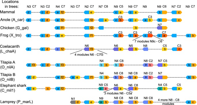
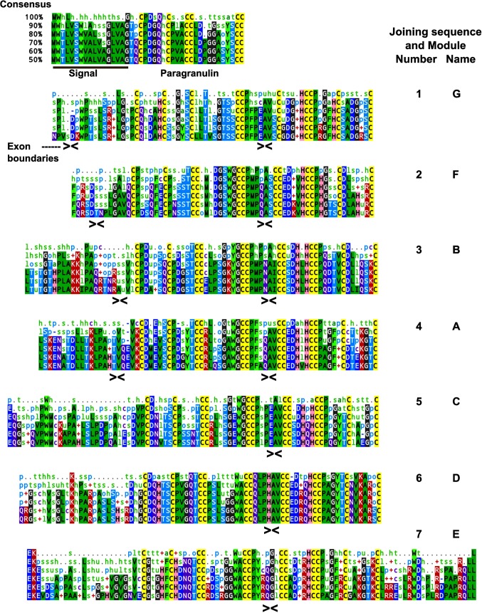
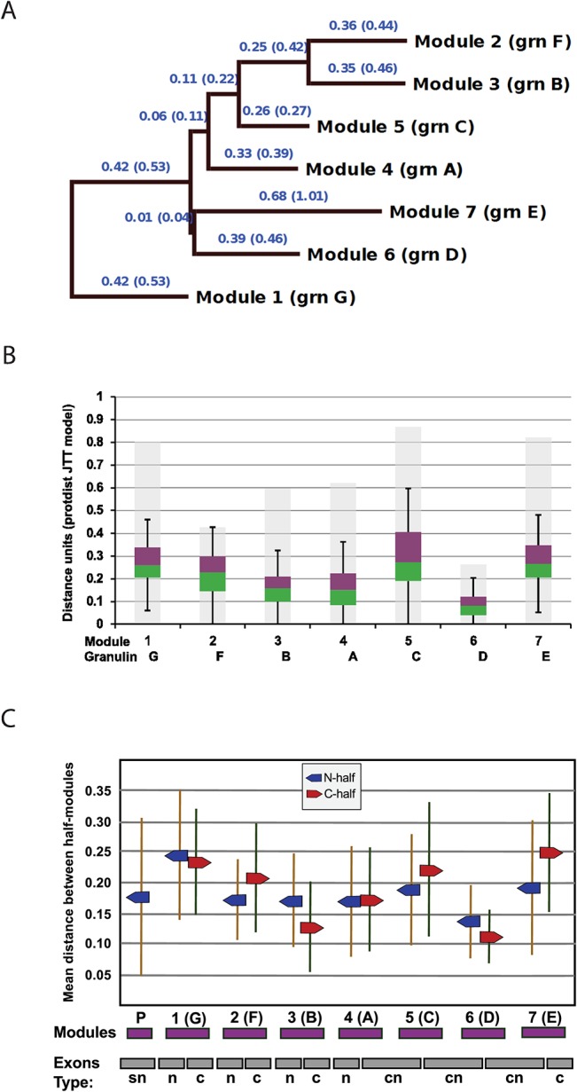
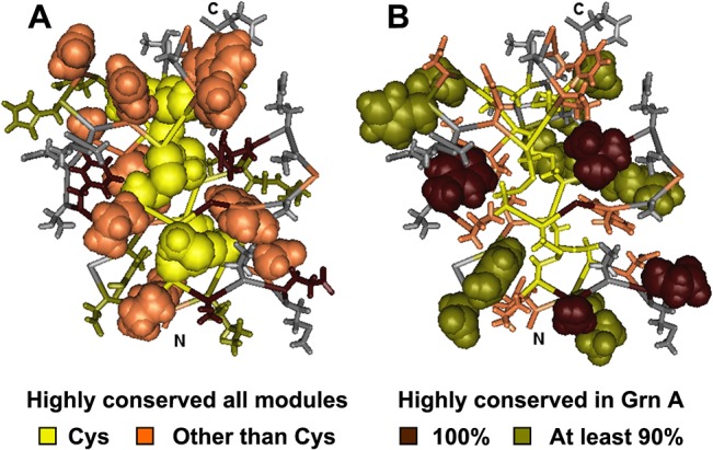
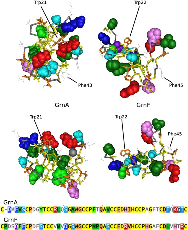
Progranulin is a secreted growth factor that is active in tumorigenesis, wound repair, and inflammation. Haploinsufficiency of the human progranulin gene, GRN, causes frontotemporal dementia. Progranulins are composed of chains of cysteine-rich granulin modules. Modules may be released from progranulin by proteolysis as 6kDa granulin polypeptides. Both intact progranulin and some of the granulin polypeptides are biologically active. The granulin module occurs in certain plant proteases and progranulins are present in early diverging metazoan clades such as the sponges, indicating their ancient evolutionary origin. There is only one Grn gene in mammalian genomes. More gene-rich Grn families occur in teleost fish with between 3 and 6 members per species including short-form Grns that have no tetrapod counterparts. Our goals are to elucidate progranulin and granulin module evolution by investigating (i): the origins of metazoan progranulins (ii): the evolutionary relationships between the single Grn of tetrapods and the multiple Grn genes of fish (iii): the evolution of granulin module architectures of vertebrate progranulins (iv): the conservation of mammalian granulin polypeptide sequences and how the conserved granulin amino acid sequences map to the known three dimensional structures of granulin modules. We report that progranulin-like proteins are present in unicellular eukaryotes that are closely related to metazoa suggesting that progranulin is among the earliest extracellular regulatory proteins still employed by multicellular animals. From the genomes of the elephant shark and coelacanth we identified contemporary representatives of a precursor for short-from Grn genes of ray-finned fish that is lost in tetrapods. In vertebrate Grns pathways of exon duplication resulted in a conserved module architecture at the amino-terminus that is frequently accompanied by an unusual pattern of tandem nearly identical module repeats near the carboxyl-terminus. Polypeptide sequence conservation of mammalian granulin modules identified potential structure-activity relationships that may be informative in designing progranulin based therapeutics.


Conflict of interest statement

Competing Interests: The authors have declared that no competing interests exist.

Figures

Fig 1
Fig 1. The protein and gene structure of a mammalian progranulin.
(A). Progranulin is composed of multiple repeats of a highly conserved 12-cysteine granulin motif [1, 39]. Mammalian progranulins possess 7.5 granulin modules labeled A to G (circles) as well as a half-granulin motif called paragranulin (half circle, p) that contains the N-terminal six cyteines of the full granulin motif. The modules are separated by short joining sequences and can be released as individual granulin peptides of about 6kDa by proteolysis of PGRN (B) Each granulin module is encoded by two exons [40]. These are referred to as N-type exons (red), C-type exons (blue) that encode the first six N-terminal and last six C-terminal cysteines respectively or CN exons (green) that span the final C-terminally located six cysteines and the first N-terminally located six cysteines of adjacent granulin modules. The corresponding granulin polypeptide modules are bracketed. (C) The spatial conformation of carp granulin, determined by 2-dimension nuclear magnetic resonance spectroscopy [41] reveals that the granulin modules adopt a compact fold of beta turns (red and green ribbons) with the 6 disulfide bridges shown as yellow bonds.
Fig 2
Fig 2. The nature and distribution of genes containing granulin modules in unicellular organisms, plants, fungi, and metazoa.
Granulin modules are represented as yellow circles. Metazoans possess only progranulins, linear chains of granulin modules that may contain multiple modules or as few as 1.5. Similar progranulin like proteins are found in organisms closely related to metazoans including Choanoflagellates and Filasterea. Granulin-modules are encountered in other unikonts including slime molds and an Apusozoa but appear to be absent from fungi. In Dictyostelium and related slime molds the Grn genes contain one granulin module and a short N-terminal polypeptide tail. Among bikonts, genes containing granulin modules are reliably detected only in green plants (viridiplantae). In plants and some unikonts (Choanoflagellates and Apusozoa), but not in metazoa, the granulin modules are associated with non-granulin proteins (identified in the figure key). The distances between taxa are arbitrary.
Fig 3
Fig 3. Syntenic conservation between the genomic regions around GrnC gene from the coelacanth, Zebrafish short-form Grn genes and a human Chr7 region.
The genomic context of the Coelacanth GrnC (L_ChaC) based on an Ensembl scaffold (vertical line) aligns with the short-form Grn genes of the zebrafish, and a region of human chromosome 7. Genes in color are orthologues. Gene names are as assigned in the NCBI databases (Grn1 and Grn2 are D_rer1 and D_rer2 in Figs 5 and 6). *This gene was unassigned by NCBI and was identified by pBlast analysis (Ensembl:ENSDARG00000035821, NCBI accession for translated protein XP_005159758.1). An additional predicted protein coding sequence for piggyBac transposable element-derived protein 3-like (XP_005159764.1, not shown) is contained within the DNA sequence for XP_005159758.1.
Fig 4
Fig 4. The structural variability of vertebrate GRN genes is built upon three conserved GRN orthology groups.
Vertebrates have three Grn gene orthology groups which are represented here for the elephant shark Callorhinchus milii, the coelacanth Latimeria chalumnae, land vertebrates (tetrapods), and three representative modern ray-finned fish [Danio rerio, (zebrafish), Takifugu rubripes (fugu fish), and Oreochromis niloticus (Nile tilapia)]. GrnA orthologues (red) are characterized by a variant Cys10 granulin motif in the second granulin module. The coelacanth and tetrapod orthologues have lost part of the first granulin domain which is now represented by a six-cysteine paragranulin sequence (p). The gold circle represents a tetraploidization event in fish genomes between 450 and 300 Mya (gold circle,[71]). The GrnB orthology group (brown), found only in ray-finned fish probably arose during this genome duplication. The third orthology group, GrnC (blue) is absent in tetrapods, occurs in elephant sharks and coelacanths as long-form Grn genes, and in ray-finned fish as short-form Grn genes. In fugu fish the ‘long-form’ PGRN-A gene (red) has been reduced to a short-form with three modules (two modules are shown in parenthesis as they were identified from EST sequences). The positions along the horizontal axis represent the order of evolutionary precedence from early (elephant shark) to late (tilapia) with the time line based on reference [71]. Variations from the canonical 12-cysteine module are indicated as G’.
Fig 5
Fig 5. N-half module relationships illustrated by a DNA-based maximum likelihood tree.
Letters and numbers are used to label some sub-trees and groups of sub-trees to facilitate reference from the text. Abbreviations for species and the specific Grn gene if there two or more are shown in the “species” boxes in the figure. In half-module labelling, this abbreviation is followed by a number based on the sequential numbering of whole modules encoded within the gene. For visual clarity, a "z" follows the number of the last module in a long-form of progranulin. When a repetitive module sequence or a lack of data make the number of modules uncertain, as in the lancelet (B_flo), the last letters of the alphabet replace numbering for the last sequential modules. N-half modules which are unpaired with C-half modules, paragranulins, are labeled "p", and followed by a number if there is more than one in the progranulin. An asterisk (*) indicates a 5-Cys form of half-module. (See also Fig 6 for a note on the cod module nomenclature). Bipartition support values of ≥20 are included. These were based upon data for trees from which rogue taxa had been pruned. The rogue taxa are indicated by red branches, and the support values for their placement in this topology are in red.
Fig 6
Fig 6. C-half module relationships illustrated by a DNA-based maximum likelihood tree.
The Fig 5 legend provides most of the explanation. In addition, by analogy with labelling of paragranulins, a C-half with no paired N-half is labelled "q". The small form from Atlantic cod, G_morC, has the module form gpp, but if the second position stop codon in coding exon 4 is suppressed, the read completes an otherwise good C-half. This was included in the analysis and the virtual module called G_morC_2sup. Thus the third G_morC N-half module is properly a second paragranulin, G_morC_p2 in Fig 5, but is also the N-half of the G_morC_2sup virtual module.
Fig 7
Fig 7. Schematic alignments of long-form progranulins from representative vertebrates summarizing positional half-module similarities and differences.
The upper line, location in trees, provides the location of half-modules in the trees of Figs 5 and 6 and refers to the Nsub- or Csub- sections where they are placed. Thus, N3 is short for Nsub03, etc. Where the tree location differs from the column heading, it is written immediately above the half-module. The letters on half-module shapes refer to the labelled tree location within the sub-section in the trees of Figs 5 and 6. Colours are intended as a visual guide. Repeats of similar modules are shown as one module with the number of repeats written below (contraction of lamprey repeats was unnecessary).
Fig 8
Fig 8. Consensus sequences for mammalian progranulin.
The mammalian polypeptide sequences were aligned at five levels of identity form 100% to 50%. Cysteine residues are highlighted in yellow. Amino acids are in capital letters on a background colour. Other letters and symbols are the default Mview notation for residue categories, (see methods for details). The colour scheme was adjusted to facilitate coordination with colours used in 3D models. The signal peptide is underlined. J indicates a joining sequence between modules, the granulin modules are identified by number and letter designations and the positions of exon boundaries are indicated by arrowheads. Paragranulin is a half granulin module at the N-terminus of mammalian progranulin.
Fig 9
Fig 9. Interspecies variability within mammalian granulin modules.
(A) To calculate distance relationships between mammalian consensus granulin modules the aligned consensus sequences for the 7 full modules in mammalian progranulin were analyzed with PROTDIST followed by FITCH. The JTT matrix was used, and global rearrangements applied. Branch lengths in parentheses result from removing the 12 conserved Cys residue positions from the analysis. (B) To determine the degrees of sequence variability for each granulin module the sequence distances between 37 species were calculated for each module using the Phylip Protdist program (JTT matrix). The data are represented by a box plot. The median divides the box with green down to the 1st quartile and purple up to the 3rd quartile. The background grey bars encompass all distances including outliers. (C). Diversity at the nucleotide level was determined by calculating the pairwise distances between DNA sequences from 37 species for each half module using DNAdist with the LogDet method. The means and standard deviations are plotted for each N-half, including the paragranulin, p, and each C-half in a way which shows their relationship to the modular structure of the progranulin and to its underlying exon structure.
Fig 10
Fig 10. Mapping conserved and variable residues onto the granulin fold.
The 3D structure of the well-folded granulin A (MMDB ID: 63884; PDB ID: 2JYE) was used as a model for illustrating conserved and variable residues in mammalian granulin modules. A: Highly conserved residues in all granulin modules are rendered with space-fill side chains. With no side chain, conserved glycine shows only as tube backbone. B: The residues highly conserved within granulin A in 37 mammalian species are shown with space fill rendition of side chains. Colouring is specified in the key. More variable residue positions are shown in grey. (Amino acids depicted as coloured tube backbones in A correspond to the space-filled residues in B, and vice-versa).
Fig 11
Fig 11. Comparison of the spatial placement of conserved residues in GrnA and GrnF.
The GrnA (right) and GrnF (left) modules are shown in two orientations comparing the conformation around the common tryptophan (Trp21, GrnA and Trp22, GrnF) is shown in brown tube style. The space fill side chains are those which show module-specific high conservation. Residues conserved in all granulin modules are shown only as tube backbones coloured yellow for cysteine and black for those highly conserved in all granulin modules. The most variable residues are coloured grey. The colour coding for the space filling side chains follows the conventions in Fig 8. Red side-chains are basic, dark blue residues are acidic.

Similar articles

Cited by

References

    1. Bhandari V, Palfree RG, Bateman A. Isolation and sequence of the granulin precursor cDNA from human bone marrow reveals tandem cysteine-rich granulin domains. Proceedings of the National Academy of Sciences of the United States of America. 1992;89(5):1715–9. Epub 1992/03/01. PubMed PMID: - PMC - PubMed
    1. Plowman GD, Green JM, Neubauer MG, Buckley SD, McDonald VL, Todaro GJ, et al. The epithelin precursor encodes two proteins with opposing activities on epithelial cell growth. The Journal of biological chemistry. 1992;267(18):13073–8. Epub 1992/06/25. PubMed PMID: . - PubMed
    1. Toh H, Chitramuthu BP, Bennett HP, Bateman A. Structure, function, and mechanism of progranulin; the brain and beyond. J Mol Neurosci. 2011;45(3):538–48. Epub 2011/06/22. 10.1007/s12031-011-9569-4 . - DOI - PubMed
    1. Tolkatchev D, Malik S, Vinogradova A, Wang P, Chen Z, Xu P, et al. Structure dissection of human progranulin identifies well-folded granulin/epithelin modules with unique functional activities. Protein science: a publication of the Protein Society. 2008;17(4):711–24. Epub 2008/03/25. 10.1110/ps.073295308 - DOI - PMC - PubMed
    1. Baker M, Mackenzie IR, Pickering-Brown SM, Gass J, Rademakers R, Lindholm C, et al. Mutations in progranulin cause tau-negative frontotemporal dementia linked to chromosome 17. Nature. 2006;442(7105):916–9. Epub 2006/07/25. 10.1038/nature05016 . - DOI - PubMed

Publication types

Substances

Grants and funding

This study was supported by grants from Canadian Cancer Society Research Institute (CCSRI 700379) and Natural Sciences and Engineering Grant number (RGPIN/327185-2011). The funders had no role in study design, data collection and analysis, decision to publish, or preparation of the manuscript.

LinkOut - more resources