. 2015 Jul 30;162(3):505-15.
doi: 10.1016/j.cell.2015.07.006. Epub 2015 Jul 23.

Cross-Generational Reproductive Fitness Enforced by Microchimeric Maternal Cells

Affiliations

Cross-Generational Reproductive Fitness Enforced by Microchimeric Maternal Cells

Jeremy M Kinder et al. Cell. .

Abstract

Exposure to maternal tissue during in utero development imprints tolerance to immunologically foreign non-inherited maternal antigens (NIMA) that persists into adulthood. The biological advantage of this tolerance, conserved across mammalian species, remains unclear. Here, we show maternal cells that establish microchimerism in female offspring during development promote systemic accumulation of immune suppressive regulatory T cells (Tregs) with NIMA specificity. NIMA-specific Tregs expand during pregnancies sired by males expressing alloantigens with overlapping NIMA specificity, thereby averting fetal wastage triggered by prenatal infection and non-infectious disruptions of fetal tolerance. Therefore, exposure to NIMA selectively enhances reproductive success in second-generation females carrying embryos with overlapping paternally inherited antigens. These findings demonstrate that genetic fitness, canonically thought to be restricted to Mendelian inheritance, is enhanced in female placental mammals through vertically transferred maternal cells that promote conservation of NIMA and enforce cross-generational reproductive benefits.


Figures

Figure 1
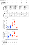
Figure 1. Developmental exposure to maternal tissue primes expanded NIMA-specific FOXP3+ Tregs
(A) Representative plots showing the gating strategy used to identify I-Ab:2W1S specific among CD4+ T cells (top), FOXP3+ Tregs among I-Ab:2W1S specific CD4+ T cells (middle), and composite data (bottom) for percent FOXP3+ among CD4+ T cells with I-Ab:2W1S specificity (filled) compared with bulk CD4+ T cells (open) in the spleen plus peripheral lymph nodes of naive (blue), NIMA-2W1S (red), NIPA-2W1S (green), or 2W1S-self (gray) 8 week old adult mice. (B) Total number of I-Ab:2W1S specific FOXP3+ Tregs (top) and CD4+ T cells (bottom) for each group of mice described in panel A. (C) Percent Helioshi among I-Ab:2W1S specific (red line) or bulk (gray shaded) FOXP3+ CD4+ T cells for each group of mice described in panel A. (D) Mating strategy for generating genetically identical NIMA-2W1S offspring born to either MHC class II I-Ab/b (I-Ab:2W1S55-68 peptide presented by cells of both maternal and fetal origin) or I-Ad/d (I-Ab:2W1S55-68 peptide only presented by cells of fetal origin) haplotype mothers, and composite data for percent FOXP3+ among I-Ab:2W1S specific CD4+ T cells and Helioshi among I-Ab:2W1S specific FOXP3+ cells for each group of NIMA-2W1S (red) compared with naive (blue) mice. (E) Percent FOXP3+ among I-Ab:2W1S specific CD4+ T cells, and Helioshi among I-Ab:2W1S specific FOXP3+ cells for each group of cross-fostered offspring exposed to 2W1S-OVA in utero and/or postnatally through breastfeeding by 2W1S-OVA+ mothers. Each point represents the result from an individual female mouse, and these data are representative of at least three separate experiments each with similar results. Bars, mean ± 95% confidence interval. ** P < 0.01, *** P < 0.001. See also Figure S1.
Figure 2
Figure 2. NIMA-specific Treg expansion requires persistent postnatal exposure to microchimeric maternal cells
(A) Maternal 2W1S-OVA+ microchimeric cell encoding DNA levels in each tissue of naive (blue filled), NIMA-2W1S (red filled), or NIMA-2W1S mice treated with anti-OVA depleting antibody (red open). (B) Cell surface OVA expression levels among splenocytes from 2W1S-OVA+ compared with naive control mice after staining with anti-OVA (red line) or rabbit IgG isotype (gray shaded) antibodies. (C) Representative plots and composite data for FOXP3+ Tregs among I-Ab:2W1S specific CD4+ T cells, and Helios expression among I-Ab:2W1S specific FOXP3+ cells for each group of mice described in panel A. Each point represents the result from an individual female mouse at 8 weeks of age, and these data are representative of at least three separate experiments each with similar results. Bars, mean ± 95% confidence interval. * P < 0.05, ** P < 0.01, *** P < 0.001. See also Figure S2.
Figure 3
Figure 3. Expanded NIMA-specific Treg accumulation in female offspring parallels discordant maternal cell microchimerism in gender-specific reproductive tissue
(A) 2W1S-OVA+ encoding DNA levels in each tissue among NIMA-2W1S female (red circle), littermate 2W1S-NIMA male (red triangle), naive female (blue circle) and naive male (blue triangle) mice. (B) Representative plots and composite data showing I-Ab:2W1S specific CD4+ T cells (top), FOXP3+ Tregs among I-Ab:2W1S specific CD4+ T cells (middle), and Helios expression among Tregs with I-Ab:2W1S specificity (red line) or bulk specificity (gray shaded) among NIMA-2W1S female compared with NIMA-2W1S littermate male mice. Each point represents the result from an individual mouse at 8 weeks of age, and these data are representative of at least three separate experiments each with similar results. Bars, mean ± 95% confidence interval. * P < 0.05, ** P < 0.01
Figure 4
Figure 4. NIMA-specific Treg expansion accelerated during pregnancy with NIMA-matched fetal antigen stimulation
Representative plots and composite data showing I-Ab:2W1S specific CD4+ T cells (top), FOXP3+ Tregs among I-Ab:2W1S specific CD4+ T cells (middle), and composite data for percent and number of FOXP3+ CD4+ T cells with I-Ab:2W1S specificity in virgin and midgestation (E11.5) naive female (blue) compared with NIMA-2W1S (red) female mice after mating with 2W1S-OVA+ transgenic male mice or non-transgenic controls. Each point represents the result from an individual mouse, these data are representative of at least three separate experiments each with similar results. Bars, mean ± 95% confidence interval. *** P < 0.001
Figure 5
Figure 5. Overlap between NIMA and paternal-fetal alloantigen protects against fetal resorption and in utero bacterial invasion following prenatal infection
(A) Percent fetal resorption (top) and average recoverable bacterial CFUs from each concepti per litter (bottom) five days following L. monocytogenes intravenous maternal infection initiated midgestation (E11.5) for naive female (blue) compared with NIMA-H-2d-(2W1S-OVA) (red) female mice during allogeneic pregnancy sired by H-2d or third party H-2k males, or depletion of microchimeric 2W1S-OVA+ maternal cells with anti-OVA antibody prior to mating. (B) Percent fetal resorption (top) and average recoverable bacterial CFUs from each concepti per litter (bottom) five days following L. monocytogenes intravenous maternal infection initiated midgestation (E11.5) for naive female (blue) compared with NIMA-H-2k-(2W1S-OVA) (red) female mice during allogeneic pregnancy sired by H-2k or third party H-2d males, or depletion of microchimeric 2W1S-OVA+ maternal cells with anti-OVA antibody prior to mating. Each point represents the result from an individual mouse, and these data are representative of at least three separate experiments each with similar results. Bars, mean ± 95% confidence interval. *** P < 0.001. See also Figure S3
Figure 6
Figure 6. Overlap between NIMA and paternal-fetal antigen protects against fetal wastage triggered by partial depletion of maternal FOXP3+ regulatory T cells
(A) Percent fetal resorption for naive (blue) FOXP3WT/WT and FOXP3DTR/WT female mice compared with each group of NIMA-H-2d FOXP3DTR/WT (red) female mice five days after initiating diphtheria toxin during allogeneic pregnancy sired by H-2d or third party H-2k males, or depletion of microchimeric 2W1S-OVA+ maternal cells with anti-OVA antibody prior to mating. (B) Percent fetal resorption for naive (blue) FOXP3WT/WT and FOXP3DTR/WT female mice compared with each group of NIMA-H-2k FOXP3DTR/WT (red) female mice five days after initiating diphtheria toxin during allogeneic pregnancy sired by H-2k or third party H-2d males, or depletion of microchimeric 2W1S-OVA+ maternal cells with anti-OVA antibody prior to mating. Each point represents the result from an individual mouse, these data are representative of at least three separate experiments each with similar results. Bars, mean ± 95% confidence interval. * P < 0.05, ** P < 0.01, *** P < 0.001. See also Figure S4.
Figure 7
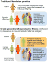
Figure 7. Cross-generational reproductive fitness enforced by vertically transferred microchimeric maternal cells in eutherian placental mammals
In traditional Mendelian genetics (top), pregnancies among female offspring are equally susceptible to fetal wastage or other complications stemming from disruptions in fetal tolerance regardless of paternal MHC haplotype specificity. Comparatively, persistent postnatal maintenance of tolerogenic microchimeric maternal cells in female offspring promotes cross-generational reproductive fitness (bottom) by selectively protecting against fetal wastage during next generation pregnancies sired by males with shared overlapping NIMA specificity.

Comment in

Similar articles

Cited by

References

    1. Akiyama Y, Caucheteux SM, Vernochet C, Iwamoto Y, Tanaka K, Kanellopoulos-Langevin C, Benichou G. Transplantation tolerance to a single noninherited MHC class I maternal alloantigen studied in a TCR-transgenic mouse model. Journal of immunology. 2011;186:1442–1449. - PMC - PubMed
    1. Andrassy J, Kusaka S, Jankowska-Gan E, Torrealba JR, Haynes LD, Marthaler BR, Tam RC, Illigens BM, Anosova N, Benichou G, et al. Tolerance to noninherited maternal MHC antigens in mice. Journal of immunology. 2003;171:5554–5561. - PubMed
    1. Araki M, Hirayama M, Azuma E, Kumamoto T, Iwamoto S, Toyoda H, Ito M, Amano K, Komada Y. Prediction of reactivity to noninherited maternal antigen in MHC-mismatched, minor histocompatibility antigen-matched stem cell transplantation in a mouse model. Journal of immunology. 2010;185:7739–7745. - PubMed
    1. Bakkour S, Baker CA, Tarantal AF, Wen L, Busch MP, Lee TH, McCune JM. Analysis of maternal microchimerism in rhesus monkeys (Macaca mulatta) using real-time quantitative PCR amplification of MHC polymorphisms. Chimerism. 2014;5:6–15. - PMC - PubMed
    1. Belkaid Y, Piccirillo CA, Mendez S, Shevach EM, Sacks DL. CD4+CD25+ regulatory T cells control Leishmania major persistence and immunity. Nature. 2002;420:502–507. - PubMed

Publication types

LinkOut - more resources