. 2015 Sep 29;6(29):27440-60.
doi: 10.18632/oncotarget.4578.

Galeterone and VNPT55 induce proteasomal degradation of AR/AR-V7, induce significant apoptosis via cytochrome c release and suppress growth of castration resistant prostate cancer xenografts in vivo

Affiliations

Galeterone and VNPT55 induce proteasomal degradation of AR/AR-V7, induce significant apoptosis via cytochrome c release and suppress growth of castration resistant prostate cancer xenografts in vivo

Andrew K Kwegyir-Afful et al. Oncotarget. .

Abstract

Galeterone (Gal) is a first-in-class multi-target oral small molecule that will soon enter pivotal phase III clinical trials in castration resistant prostate cancer (CRPC) patients. Gal disrupts androgen receptor (AR) signaling via inhibition of CYP17, AR antagonism and AR degradation. Resistance to current therapy is attributed to up-regulation of full-length AR (fAR), splice variants AR (AR-Vs) and AR mutations. The effects of gal and VNPT55 were analyzed on f-AR and AR-Vs (AR-V7/ARv567es) in LNCaP, CWR22Rv1 and DU145 (transfected with AR-Vs) human PC cells in vitro and CRPC tumor xenografts. Galeterone/VNPT55 decreased fAR/AR-V7 mRNA levels and implicates Mdm2/CHIP enhanced ubiquitination of posttranslational modified receptors, targeting them for proteasomal degradation. Gal and VNPT55 also induced significant apoptosis in PC cells via increased Bax/Bcl2 ratio, cytochrome-c release with concomitant cleavage of caspase 3 and PARP. More importantly, gal and VNPT55 exhibited strong in vivo anti-CRPC activities, with no apparent host toxicities. This study demonstrate that gal and VNPT55 utilize cell-based mechanisms to deplete both fAR and AR-Vs. Importantly, the preclinical activity profiles, including profound apoptotic induction and inhibition of CRPC xenografts suggest that these agents offer considerable promise as new therapeutics for patients with CRPC and those resistant to current therapy.

Keywords: androgen receptors (AR/AR-V7); galeterone (gal); gal’s analog VNPT55; mechanisms of AR/AR-V7 degradation; prostate cancer.


Conflict of interest statement

CONFLICTS OF INTEREST

Vincent C. O. Njar is the lead inventor of galeterone (VN/124-1 or TOK-001) and VNPT55, patents and technologies thereof owned by the University of Maryland, Baltimore, and licensed to Tokai Pharmaceuticals, Inc. Puranik Purushottamachar and Andrew K. Kwegyir-Afful are co-inventors of VNPT55 and related compounds. A patent application to protect VNPT55 and related novel compounds has been filed. The other authors declare no potential conflict of interest.

Figures

Figure 1
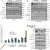
Figure 1. Effects of gal and VNPT55 on AR/AR-V7 mRNA and protein expression
A. LNCaP cells treated with increasing concentrations of gal and VNPT55 (1, 5, 10, 20 μM) for 24 h and mRNA collected, qRT-PCR analysis of fAR mRNA; *p < 0.01, **p < 0.001. CWR22Rv1 cells were treated with increasing concentrations of gal and VNPT55 (1, 5, 10, 20 μM) for 24 h and mRNA collected, qRT-PCR of analysis of AR-V7 mRNA levels. B. (left panel) Effects of gal and VNPT55 on fAR/AR-V7 in CWR22Rv1 cells, B. (right panel) DU145 cells transfected with 0.5 μg of AR-V7 expression plasmid for 16 h and Cells subsequently treated with gal and VNPT55 (5 and 10 μM) for 24 h. C. (left panel), DU145 cells were transfected with ARv567es expression plasmid as in B and treated with gal and VNPT55 (5 and 10 μM) for 24 h, C. (right panel) densitometry for three (3) replicates of experiments done in B (CWR22Rv1 Cells) **p < 0.001. D. (left panel) LNCaP and (right panel) CWR22Rv1 cells treated with gal at 20 μM for 16 h, thereafter, MG132 (5 μM) was added for an extra 8 h. Immunoblot analysis was carried out on fAR and AR-V7 protein expression. E. (left panels) LNCaP and (right panels) CWR22Rv1 were treated as in D with VNPT55. F. Densitometric analysis of D and E (fAR-L = full length AR in LNCaP cells, fAR-R = full length AR in CWR22Rv1 cells). G. LNCaP (left panel) and CWR22Rv1 (right panel) cells were treated as in D and E and 1 mg of total cell lysate used in immunoprecipitation and probed with ubiquitin antibodies.
Figure 2
Figure 2. Significance of Akt and Mdm2 phosphorylation in gal/VNPT55 induced fAR/AR-V7 degradation
A. and B. LNCaP and CWR22Rv1 cells, serum starved for 12 h were treated with increasing concentrations (5, 10 and 20 μM) of gal or VNPT55 for 24 h. C. Densitometry was done for phosphorylated levels of AR, normalizing p-AR to the total AR expression after gal and VNPT55 treatment. D. and E. LNCaP cells were serum starved for 12 h and pre-treated with 20 μM LY294002 (PI3k inhibitor) or 20 μM U0126 (MAPK inhibitor) for 1 h and then treated with gal/VNPT55 (20 μM). F. CWR22Rv1 cells were serum starved in phenol red free media supplemented with charcoal dextran FBS for 12 h and treated with gal (20 μM) for 24 h after pretreating with LY294002 (20 μM) or U0126 (20 μM) for 1 h. Samples were immunoblotted for full length AR (fAR), AR-V7, phospho Akt, phospho Mdm2 and phospho p42/44, using Gapdh as loading control.
Figure 3
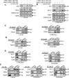
Figure 3. Gal enhanced AR/AR-V7 degradation implicates E3 ligase Mdm2 and CHIP
A. LNCaP cells seeded at 60% confluency were transfected with Mdm2 or CHIP siRNAs (100 nM each), with scrambled siRNA as control. Cells were then exposed to 20 μM gal for 24 h. B. CWR22Rv1 cells were subjected to similar transfection and treatment conditions as in A, separated proteins were probed for fAR, AR-V7, Mdm2 CHIP. C. LNCaP (left panel) and CWR22Rv1 (right panel) cells were serum starved for 12 h and treated with 10 μM of gal for 14 h; 1 mg of total cell lysate was subjected to immunoprecipitation with Mdm2 rabbit polyclonal antibodies. D. LNCaP (left panel) and CWR22Rv1 (right panel) cells were treated as in C and subjected to immunoprecipitation analysis with Hsp90 rabbit polyclonal antibodies. Samples were immunoblotted for Mdm2, Hsp90 and AR with mouse monoclonal antibodies. E. (left and right panels) 1 mg of total cell lysates from CWR22Rv1 cells treated with 10 μM of gal for 11 h was subjected to immunoprecipitation with CHIP and Hsp70 polyclonal antibodies, respectively, to analyze Hsp70-CHIP-AR-V7 interaction. F. Co-immunoprecipitation assays in LNCaP and CWR22Rv1 cells with AR (N20) polyclonal and AR-V7 mouse monoclonal antibodies were carried out with 1 mg cell lysates after treating with gal (10 μM) for 14 h (LNCaP cells) and 11 h (CWR22Rv1 cells). Membranes were probed for AR, AR-V7, Mdm2, Hsp90 and Hsp70 mouse monoclonal antibodies.
Figure 4
Figure 4. Gal and VNPT55 induce apoptosis in LNCaP and CWR22Rv1 cells
A. (left panel) Apoptotic induction was analyzed with the acridine orange/ethidium bromide (AO/EB) staining assay in LNCaP (top panel) and CWR22Rv1 (bottom panel) cells. Indicated cells lines were treated with 2.5 μM of gal or VNPT55 for 72 h. Cells were washed 1X gently with warm PBS and incubated with a 1:2 ratio of ethidium bromide: acridine orange in PBS for 30 minutes. Cells were washed 1X again and images taken with a fluorescence microscope Nikon TE2000. Live, apoptotic and necrotic cells stain green, orange and red respectively. Arrows next to L indicate live cells; arrows pointing to A indicate apoptotic cells; and arrows pointing N indicate necrotic cells. A. (right panel), CWR22Rv1 cells treated with gal and VNPT55 at 5 and 10 μM after 24 h were collected and washed 2X with PBS. Cells were then stained with annexin-v and PI. FACS analysis was performed for apoptotic cells after gal and VNPT55 treatment at 5 and 10 μM (*p < 0.01, **p < 0.001). B. LNCaP (left panel) and CWR22Rv1 (right panel), cells were serum starved in phenol-red free media supplemented with charcoal dextran cFBS for 12 h and treated with increasing doses of gal or VNPT55 (5, 10 and 20 μM) for 24 h. C. LNCaP cells were treated with gal at 20 μM for different time points (0, 3, 6, 12, 18, 24 h). D. & E. LNCaP cells were exposed to gal or VNPT55 at 20 μM in combination with calpeptin (40 μM) or ZVAD-fmk (20 μM) for 24 h. F. LNCaP cells serum starved for 12 h were treated with gal (5, 10 and 20 μM) for 24 h and immunoblotted for Bax and Bcl2. G. Densitometry analysis of Bax/Bcl2 ratio show an increase with increase in gal concentration (**p < 0.001). H. (top panel) Bax co-localizes with mitochondria and enhances mitochondrial permeability. LNCaP cells were treated with 10 μM galeterone or VNPT55 for 24 hours. Cells were then stained with mitotracker (RED CMXRos) and Bax antibody. (bottom panel) CWR22Rv1 cells were treated as in LNCaP cells and subsequently stained with mitotracker and Bax antibody. Images were taken and merged. Areas of co-localization are yellow. I. CWR22Rv1 cells were seeded in an 8-chamber slide and treated with gal or VNPT55 (5 and 10 μM) for 24 h. Cells were then incubated with 125 nM of mitotracker at 37°C for 15 minutes after which cells were fixed with 3.7% paraformaldehyde and stained with cytochrome c at 4°C for 12 h.
Figure 5
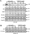
Figure 5. Effects of gal/VNPT55 on AR in epithelial prostate cells and nuclear receptors in LNCaP cells
A. 12 h serum starved LNCaP cells were treated with gal (5, 10, 20 μM) or VNPT55 (5, 10, 20 μM) for 24 h, using DMSO as control. B. Immortalized untransformed prostate epithelial cells (PWR-1E cells) were incubated in phenol red free charcoal dextran FBS for 12 h and treated with gal or VNPT55 at 5, 10 and 20 μM for an additional 24 h.
Figure 6
Figure 6. Antitumor activity of galeterone and VNPT55 in a CWR22Rv1 xenograft
A. Representative tumors from the 2 groups. Effect of gal and VNPT55 was evaluated in castrated CWR22Rv1 xenograft-bearing mice. Mice (n = 5) were administered with gal (0.15 mmol/kg/twice daily) and VNPT55 (0.15 mmol/kg/twice daily), by intraperitoneal injection, 5 days per week for 34 days. Tumors were measured twice a week. B. Mean body weights of mice were weighed once a week for the duration of the study. C. Hematoxylin and eosin staining of normal organs, formalin fixed and paraffin embedded tissues to show in vivo and off-target toxicity or not of compounds. D. Representative images of full length AR, AR-3 and PCNA immunostaining in vehicle and treated groups. E. ImageJ was used to quantify Immunohistochemical staining in D. E. ImageJ was used to quantify Immunohistochemical staining in D. F. Xenograft tumor tissues were harvested and analyzed by western blotting. Effects on fAR, AR-V7, cyclin D1, Bcl2 and Bax were analyzed in both galeterone and VNPT55 groups compared to controls. G. Densitometry analysis of protein expression from western blot analysis was plotted to quantify the effects seen in vivo.
Figure 7
Figure 7. Schematic representation for proposed mechanisms of Gal on full length AR and splice variant AR-V7 degradation
1) Major pathway for gal-induced fAR degradation (left). Gal treatment of PC cells implicates PI3K/Akt pathway: Akt phosphorylates AR at Ser210. Active Akt also phosphorylates Mdm2, which enhances ubiquitination of AR. Ubiquitinated AR is degraded by the 26S proteasome. 2) Major pathway for gal-induced AR-V7 degradation (right)). Gal treatment of PC cells stimulates increased interaction between CHIP-AR-V7, which then enhances ubiquitination of AR-V7 and targets it to degradation via the 26S proteasome.

Similar articles

Cited by

References

    1. Miyamoto H, Messing EM, Chang C. Androgen deprivation therapy for prostate cancer: current status and future prospects. The Prostate. 2004;61:332–353. - PubMed
    1. Vasaitis TS, Bruno RD, Njar VC. CYP17 inhibitors for prostate cancer therapy. The Journal of steroid biochemistry and molecular biology. 2011;125:23–31. - PMC - PubMed
    1. Vasaitis TS, Njar VC. Novel, potent anti-androgens of therapeutic potential: recent advances and promising developments. Future medicinal chemistry. 2010;2:667–680. - PubMed
    1. Osanto S, Van Poppel H. Emerging novel therapies for advanced prostate cancer. Therapeutic advances in urology. 2012;4:3–12. - PMC - PubMed
    1. Shapiro D, Tareen B. Current and emerging treatments in the management of castration-resistant prostate cancer. Expert review of anticancer therapy. 2012;12:951–964. - PubMed

Publication types

MeSH terms