. 2015 Jul 16;6(7):e1827.
doi: 10.1038/cddis.2015.193.

Shear stress regulates endothelial cell autophagy via redox regulation and Sirt1 expression

Affiliations

Shear stress regulates endothelial cell autophagy via redox regulation and Sirt1 expression

J Liu et al. Cell Death Dis. .

Abstract

Disturbed cell autophagy is found in various cardiovascular disease conditions. Biomechanical stimuli induced by laminar blood flow have important protective actions against the development of various vascular diseases. However, the impacts and underlying mechanisms of shear stress on the autophagic process in vascular endothelial cells (ECs) are not entirely understood. Here we investigated the impacts of shear stress on autophagy in human vascular ECs. We found that shear stress induced by laminar flow, but not that by oscillatory or low-magnitude flow, promoted autophagy. Time-course analysis and flow cessation experiments confirmed that this effect was not a transient adaptive stress response but appeared to be a sustained physiological action. Flow had no effect on the mammalian target of rapamycin-ULK pathway, whereas it significantly upregulated Sirt1 expression. Inhibition of Sirt1 blunted shear stress-induced autophagy. Overexpression of wild-type Sirt1, but not the deacetylase-dead mutant, was sufficient to induce autophagy in ECs. Using both of gain- and loss-of-function experiments, we showed that Sirt1-dependent activation of FoxO1 was critical in mediating shear stress-induced autophagy. Shear stress also induced deacetylation of Atg5 and Atg7. Moreover, shear stress-induced Sirt1 expression and autophagy were redox dependent, whereas Sirt1 might act as a redox-sensitive transducer mediating reactive oxygen species-elicited autophagy. Functionally, we demonstrated that flow-conditioned cells are more resistant to oxidant-induced cell injury, and this cytoprotective effect was abolished after inhibition of autophagy. In summary, these results suggest that Sirt1-mediated autophagy in ECs may be a novel mechanism by which laminar flow produces its vascular-protective actions.


Figures

Figure 1
Figure 1
Laminar shear stress enhanced autophagic response in cultured endothelial cells. (a) Fluorescence images showing shear stress (20 dyn/cm2 for 8 h)-induced: (from top to bottom) realignment of actin fibers in human umbilical vein endothelial cells (HUVECs); accumulation of LC3 puncta (arrows) in HUVECs (asterisks indicate punctate LC3- cells); puncta accumulation of GFP-LC3 (arrows) in transfected telomerase-immortalized human microvascular endothelium cell (TIME) cells; acidification of autolysosomes detected with acridine orange staining (red fluorescence represents acidic vesicles); flow cessation decreased the abundance of GFP-LC3 in shear-adapted TIME cells. (b) The average number of LC3 puncta per cell and proportion of punctate LC3+ cells under static (S) and laminar flow (F) conditions. (c) Comparison of the proportion of cells containing different numbers of LC3 puncta under static and flow conditions. (d) Time course of laminar flow-induced autophagic response in HUVECs. (e) Effects of flow cessation (FC) on punctate LC3 in cells adapted to flow for 8 h. (f) Semi-quantitative data showing the effect of laminar flow on GFP-LC3 puncta accumulation in transfected TIME cells. (g) LC3 immunofluorescence showing that low-magnitude flow (LowF; 4 dyn/cm2) or oscillatory flow (OF; ±5 dyn/cm2 at 1 Hz) did not have the same effect as laminar flow (12 dyn/cm2) on autophagy induction. Data are mean±standard error of the mean (S.E.M.). *P<0.05, unpaired t-test, n=3–5. LF, laminar flow
Figure 2
Figure 2
Effects of laminar flow (20 dyn/cm2) on (a) LC3 level, (b) p62 level, and (c) mRNA levels of LC3A, beclin-1, and Atg5. In a, autophagy induction by amino-acid starvation (by treating cells with Hank's balanced salt solution for 4 h) was used as a positive control. Data are mean±S.E.M. *P<0.05, unpaired t-test, n=3–4. C, control; S, static; F, flow; NS, no significance; AS, amino-acid starvation
Figure 3
Figure 3
Sirt1 was critical in mediating shear stress-induced autophagy in endothelial cells. (a) Effects of laminar flow on the phosphorylation levels of mTOR and ULK1, and the expression level of Sirt1 (representative images from four experiments). (b) Time course of the effect of laminar flow on the mRNA expression of Sirt1. (c) Effect of laminar flow on the promoter activity of human Sirt1 as measured by luciferase reporter assay. (d) Effects of the Sirt1 activator resveratrol (10 μM) on the protein levels of Sirt1 and LC3. (e) Effects of overexpression of Flag-tagged wild-type Sirt1 and Sirt1-H363Y mutant on LC3 expression. (f) Effects of Sirt1 and Sirt1-H363Y overexpression on accumulation of LC3 puncta. (g) Effects of wild-type Sirt1 and Sirt1-H363Y overexpression on the mRNA expression levels of various autophagy-related genes as indicated. (h) Effects of the Sirt1 inhibitor EX-527 (10 μM) on flow-induced autophagy. (i) Effects of EX-527 on flow-induced upregulation of Atg5, beclin-1, and LC3A. (j) Effects of Sirt1 gene silencing with siRNA on flow-induced LC3 puncta accumulation. Data are mean±S.E.M. *P<0.05, unpaired t-test or one-way analysis of variance, n=3–4. Res, resveratrol
Figure 4
Figure 4
Shear stress-induced autophagy in EC was redox dependent. (a) ROS production measured with Amplex Red Hydrogen Peroxide Assay in cells maintained under laminar flow and static conditions, in the absence and presence of the NADPH oxidase inhibitors diphenyleneiodonium (DPI; 10 μM) and diapocynin (100 μM). (b) Effects of EUK-134 (10 μM) and N-acetyl cysteine (NAC; 1 mM) on flow-induced LC3 puncta accumulation. (c) Effects of EUK-134 (E) and NAC (N) on the expression levels of Atg5, beclin-1, and LC3A in cells maintained under flow condition. (d) Effects of EUK-134 on the expression levels of Atg5, beclin-1, and LC3A in cells maintained under static condition. (e) Western blot and densitometry data showing the effects of laminar flow on protein levels of Sirt1 and LC3 in the absence and presence of EUK-134 or NAC. Data are mean±S.E.M. *P<0.05, unpaired t-test or one-way analysis of variance as appropriate (n=3–5). S, static; F, flow; NS, no significance; Apo, diapocynin; NAC, N-acetyl cysteine
Figure 5
Figure 5
Sirt1 was a redox-sensitive regulator of reactive oxygen-induced autophagy. (a) Concentration-dependent effects of exogenous H2O2 on the expression level of Sirt1. (b) Effects of H2O2 (300 μM) on Sirt1 mRNA expression at 6 and 12 h. Serum-free treatment was used as a control. (c) Effects of H2O2 and serum free on Sirt1 protein expression. (d) Effects of H2O2 on phosphorylation of mTOR and ULK1. (e) Effects of H2O2 and serum-free treatment on the protein level of LC3. (f) H2O2 increased the expression levels of Atg5, beclin-1, and LC3A, which were blocked by pretreatment with EX-527 (10 μM). (g) Western blot and quantitative densitometry data showing the effects of control and Sirt1 siRNA on the autophagic response induced by H2O2 (300 μM). Data are mean±S.E.M. *P<0.05, one-way analysis of variance, n=3–4. The western blots were representative examples from three experiments. NS, no significance; SF, serum free
Figure 6
Figure 6
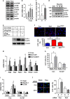
Role of FoxO1 in shear stress- and Sirt1-mediated autophagic response. (a) Immunofluorescence images showing the effects of laminar flow and Sirt1 overexpression on nuclear retention of FoxO1. Nuclei were counter-stained with DAPI (blue). (b) Effects of laminar flow, resveratrol (Res; 10 μM), and H2O2 (300 μM) on the acetylation level of FoxO1 in normal cells (upper) or cells expressing ectopic Flag-FoxO1 (lower). Cell lysates were immunoprecipitated with an anti-FoxO1 or anti-Flag antibody and detected by western blotting with an anti-acetyl lysine (Ac-Lys) antibody. (c) Effects of overexpressing wild type (WT) or the constitutively active form (AAA) of FoxO1 on the expression levels of glutathione peroxidase (GPx), Atg5, beclin-1, and LC3A, in the absence and presence of Res. (d) Overexpression of FoxO1-AAA enhanced the protein level of LC3-II in the presence of Res. (e) Effects of FoxO1 gene silencing with different siRNA sequences on laminar flow-induced autophagic response detected by LC3 immunofluorescence. (f) Effects of FoxO1 siRNA on Sirt1 overexpression (pSirt1)-induced upregulation of Atg5, beclin-1, and LC3A. (g) Laminar flow decreased the acetylation levels of Atg5 and Atg7 detected by immunoprecipitation (IP) with anti-acetyl lysine antibody and western blotting with anti-Atg5 and Atg7 antibodies. Data are mean±S.E.M. *P<0.05, one-way analysis of variance or unpaired t-test, n=4–6. S, static; F, flow; pCon, control plasmid; Res, resveratrol; IP, immunoprecipitation; WT, wild type; GPx, glutathione peroxidase
Figure 7
Figure 7
Autophagic response had cytoprotective actions in endothelial cells. (a) Flow-adapted cells were pretreated with the autophagy inhibitor 3-MA (10 μM) or the Sirt1 inhibitor EX-527, and acutely challenged with a high concentration (600 μM) of H2O2 for 2 h. (b) Effects of laminar flow adaptation on H2O2-induced cell death in the absence and presence of 3-MA. (c) Effects of autophagy induction by amino-acid starvation (by incubating cells in Hank's balanced salt solution for 4 h) in static cells on H2O2-induced cell death. (d) Effects of Atg5 gene silencing with different siRNA sequences on the cytoprotective effects of resveratrol (10 μM) assessed by serum deprivation-induced cell apoptosis. (e) Effects of Atg5 gene silencing on the cytoprotective effects of laminar flow assessed by staurosporine (100 nM)-induced cell apoptosis. Data are mean±S.E.M. *P<0.05, one-way analysis of variance, n=4–5. H, H2O2; F, flow; AS, amino-acid starvation; R, resveratrol; Stauro, staurosporine; C, control; NS, no significance

Similar articles

Cited by

References

    1. 1Davies PF. Hemodynamic shear stress and the endothelium in cardiovascular pathophysiology. Nat Clin Pract Cardiovasc Med 2009; 6: 16–26. - PMC - PubMed
    1. 2Chiu JJ, Chien S. Effects of disturbed flow on vascular endothelium: pathophysiological basis and clinical perspectives. Physiol Rev 2011; 91: 327–387. - PMC - PubMed
    1. 3Mizushima N. Autophagy: process and function. Genes Dev 2007; 21: 2861–2873. - PubMed
    1. 4Ravikumar B, Sarkar S, Davies JE, Futter M, Garcia-Arencibia M, Green-Thompson ZW et al. Regulation of mammalian autophagy in physiology and pathophysiology. Physiol Rev 2010; 90: 1383–1435. - PubMed
    1. 5Glick D, Barth S, Macleod KF. Autophagy: cellular and molecular mechanisms. J Pathol 2010; 221: 3–12. - PMC - PubMed

Publication types

MeSH terms

Substances