Left frontal glioma induces functional connectivity changes in syntax-related networks
- PMID: 26155456
- PMCID: PMC4491091
- DOI: 10.1186/s40064-015-1104-6
Left frontal glioma induces functional connectivity changes in syntax-related networks
Abstract
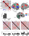
Background: A glioma leads to a global loss of functional connectivity among multiple regions. However, the relationships between performance/activation changes and functional connectivity remain unclear. Our previous studies (Brain 137:1193-1212; Brain Lang 110:71-80) have shown that a glioma in the left lateral premotor cortex or the opercular/triangular parts of the left inferior frontal gyrus causes agrammatic comprehension accompanied by abnormal activations in 14 syntax-related regions. We have also confirmed that a glioma in the other left frontal regions does not affect task performances and activation patterns.
Results: By a partial correlation method for the time-series functional magnetic resonance imaging data, we analyzed the functional connectivity in 21 patients with a left frontal glioma. We observed that almost all of the functional connectivity exhibited chaotic changes in the agrammatic patients. In contrast, some functional connectivity was preserved in an orderly manner in the patients who showed normal performances and activation patterns. More specifically, these latter patients showed normal connectivity between the left fronto-parietal regions, as well as normal connectivity between the left triangular and orbital parts of the left inferior frontal gyrus.
Conclusions: Our results indicate that these pathways are most crucial among the syntax-related networks. Both data from the activation patterns and functional connectivity, which are different in temporal domains, should thus be combined to assess any behavioral deficits associated with brain abnormalities.
Keywords: Agrammatic comprehension; Frontal regions; Functional MRI; Functional connectivity; Glioma; Neural network.
Figures

Similar articles
-
Task-Induced Functional Connectivity of the Syntax-Related Networks for Patients with a Cortical Glioma.Cereb Cortex Commun. 2020 Sep 1;1(1):tgaa061. doi: 10.1093/texcom/tgaa061. eCollection 2020. Cereb Cortex Commun. 2020. PMID: 34296124 Free PMC article.
-
Differential reorganization of three syntax-related networks induced by a left frontal glioma.Brain. 2014 Apr;137(Pt 4):1193-212. doi: 10.1093/brain/awu013. Epub 2014 Feb 11. Brain. 2014. PMID: 24519977
-
Diffuse glioma-induced structural reorganization in close association with preexisting syntax-related networks.Cortex. 2023 Oct;167:283-302. doi: 10.1016/j.cortex.2023.07.005. Epub 2023 Jul 24. Cortex. 2023. PMID: 37586138
-
Differential intrinsic functional connectivity changes in semantic variant primary progressive aphasia.Neuroimage Clin. 2019;22:101797. doi: 10.1016/j.nicl.2019.101797. Epub 2019 Mar 27. Neuroimage Clin. 2019. PMID: 31146321 Free PMC article.
-
[Lateralization of the Frontal Association Cortex: Syntax-related Networks].Brain Nerve. 2018 Oct;70(10):1075-1085. doi: 10.11477/mf.1416201139. Brain Nerve. 2018. PMID: 30287693 Japanese.
Cited by
-
Correlation between brain functional connectivity and neurocognitive function in patients with left frontal glioma.Sci Rep. 2022 Nov 8;12(1):18302. doi: 10.1038/s41598-022-22493-6. Sci Rep. 2022. PMID: 36347905 Free PMC article.
-
Disrupted functional connectivity affects resting state based language lateralization.Neuroimage Clin. 2016 Oct 24;12:910-927. doi: 10.1016/j.nicl.2016.10.015. eCollection 2016. Neuroimage Clin. 2016. PMID: 27882297 Free PMC article.
-
Task-Induced Functional Connectivity of the Syntax-Related Networks for Patients with a Cortical Glioma.Cereb Cortex Commun. 2020 Sep 1;1(1):tgaa061. doi: 10.1093/texcom/tgaa061. eCollection 2020. Cereb Cortex Commun. 2020. PMID: 34296124 Free PMC article.
-
Functional Mapping before and after Low-Grade Glioma Surgery: A New Way to Decipher Various Spatiotemporal Patterns of Individual Neuroplastic Potential in Brain Tumor Patients.Cancers (Basel). 2020 Sep 13;12(9):2611. doi: 10.3390/cancers12092611. Cancers (Basel). 2020. PMID: 32933174 Free PMC article. Review.
-
Determining priority signs and symptoms for use as clinical outcomes assessments in trials including patients with malignant gliomas: Panel 1 Report.Neuro Oncol. 2016 Mar;18 Suppl 2(Suppl 2):ii1-ii12. doi: 10.1093/neuonc/nov267. Neuro Oncol. 2016. PMID: 26989127 Free PMC article. Review.
References
LinkOut - more resources
Full Text Sources
Other Literature Sources

