. 2015 Jul 6;107(9):djv184.
doi: 10.1093/jnci/djv184. Print 2015 Sep.

M-Trap: Exosome-Based Capture of Tumor Cells as a New Technology in Peritoneal Metastasis

Affiliations

M-Trap: Exosome-Based Capture of Tumor Cells as a New Technology in Peritoneal Metastasis

Alexandre de la Fuente et al. J Natl Cancer Inst. .

Abstract

Background: Remodeling targeted tissues for reception of tumor cells metastasizing from primary lesions is a consequence of communication between the tumor and the environment that governs metastasis. This study describes a novel approach that aims to disrupt the process of metastasis by interfering with this intense dialogue.

Methods: Proteomics and adhesion assays identified exosomes purified from the ascitic fluid of ovarian cancer patients (n = 9) as intermediaries of tumor cell attachment. A novel tumor cell capture device was fabricated by embedding exosomes onto a 3D scaffold (metastatic trap [M-Trap]). Murine models of ovarian metastasis (n = 3 to 34 mice per group) were used to demonstrate the efficacy of M-Trap to capture metastatic cells disseminating in the peritoneal cavity. Kaplan-Meier survival curves were used to estimate cumulative survival probabilities. All statistical tests were two-sided.

Results: The exosome-based M-Trap device promoted tumor cell adhesion with a nonpharmacological mode of action. M-Trap served as a preferential site for metastasis formation and completely remodeled the pattern of peritoneal metastasis in clinically relevant models of ovarian cancer. Most importantly, M-Trap demonstrated a statistically significant benefit in survival outcomes, with mean survival increasing from 117.5 to 198.8 days in the presence of M-Trap; removal of the device upon tumor cell capture further improved survival to a mean of 309.4 days (P < .001).

Conclusions: A potent artificial premetastatic niche based on exosomes is an effective approach to impair the crosstalk between metastatic cells and their environment. In the clinical setting, the capacity to modulate the pattern of dissemination represents an opportunity to control the process of metastasis. In summary, M-Trap transforms a systemic, fatal disease into a focalized disease where proven therapeutic approaches such as surgery can extend survival.


Figures

Figure 1.
Figure 1.
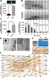
Exosomes as components of ascitic fluid involved in cell communication. A) Short-term calcein-labeled SKOV3 cell adhesion assay performed under Basal (nontreated) and Ascites (ascites-pretreated) conditions. Upper panels illustrate representative fluorescence images of adhered cells, while quantification of fluorescence intensity is represented in the histogram. Data are means from three independent experiments. Error bars represent standard deviation (P = .0012, two-sided Student’s t test). B) Representative electrophoresis gel performed under denaturant conditions showing protein distribution upon HPLC fractionation of total cell-free ascites (upper panel) and ascites depleted of exosomes (middle panel) by ultracentrifugation (see the Supplementary Methods, available online). SKOV3 cell adhesion assay performed upon pretreatment with each HPLC fraction demonstrated a statistically significant reduction in adhesion ability of ascites after depletion of exosomes (data are means from three independent experiments). Error bars represent standard deviation (P = .0035, Student’s t test; lower panel). C) Adhesion assay performed as in panel (A) under basal (nontreated) and exosomes (pretreated with 50 μg of purified exosomes), demonstrated statistically significant adhesion mediated by purified exosomes (data are means from three independent experiments). Error bars represent standard deviation (P < .001, Student’s t test). D) Electron microscopy images of exosomes purified from ascites of ovarian cancer patients (see the Supplementary Methods, available online): SEM, left panel; scale bar = 500nm; representative electron microscopy, right panel; scale bar = 300nm; inserts showing a 10-fold magnification of a representative vesicle. E) Immuno blot of 40 μg of total protein extracted from exosomes purified from the ascitic fluid of ovarian cancer patients, demonstrated human CD9 and CD81 as markers of exosomes (upper panel) (see the Supplementary Methods, available online). Coomassie staining of total extract is shown as loading control (lower panel). F) Ingenuity pathway analysis (IPA) upon proteome analysis by Maldi TOF/TOF of exosomes purified from ascitic fluid of ovarian cancer patients (see the Supplementary Methods, available online). Comprehensive protein network analysis pointed to cell adhesion as a main biological process associated with exosome proteome, characterized by integrins and extracellular matrix proteins and a central role for the ERK 1/2 and Akt pathways (insert).
Figure 2.
Figure 2.
Metastatic trap (M-Trap) design and characterization of mode of action. A) Exosomes purified from ascites of ovarian cancer patients (25 µL at 2 µg/µL) were embedded into the 3D scaffold; representative electron microscopy (TEM) of a fiber of the 3D nanomesh scaffold with adhered exosomes, in comparison with the surface of a fiber of the scaffold in the absence of exosomes (insert; scale bar = 5 μm). B) Representative electron microscopy (TEM) images showing immunogold staining of exosomes adhered to M-Trap device, revealed by TEM as vesicles containing CD9 (left panel) and CD81 (right panel) colloidal gold particles (scale bar = 500nm for both panels) (see the Supplementary Methods, available online). C) Substrate-bound nonpharmacological mode of action of M-Trap as demonstrated by release experiments. DiD-labeled exosomes (25 µl at 2 µg/µl) were immersed in phosphate-buffered solution, and supernatants and devices (scaffold) were collected at indicated times and fluorescent imaged (in vivo image system). D) Representative fluorescent images of SKOV3 cells adhered to a fiber of the 3D scaffold in the presence (M-Trap; right panel) or not (Scaffold; left panel) of exosomes under dynamic orbital rotation conditions (scale bar = 100 μm) (see the Supplementary Methods, available online). E) Luminometer quantification of calcein-labeled SKOV3 cells captured under dynamic conditions to bare 3D scaffolds (scaffold); bare 3D scaffolds decorated with tetraspanins CD9 (scaffold + CD9) or CD81 (scaffold + CD81); bare 3D scaffold pretreated with Poly-Hema (scaffold Poly-Hema); M-Trap device (M-Trap); M-Trap device pretreated with blocking antibodies against CD9 (M-Trap + anti-CD9); and CD81 (M-Trap + anti-CD81) (see the Supplementary Methods, available online). Modulation of cell adhesion resulted in a gradual capacity to capture SKOV3 cells with a maximal effect by M-Trap (data are means from three independent experiments). Error bars represent standard deviation (P = .001, M-Trap versus bare scaffold, two-sided Student’s t test). SN = supernatant.
Figure 3.
Figure 3.
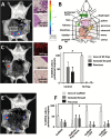
Capture of metastatic tumor cells by metastatic trap (M-Trap) technology in an in vivo model of ovarian cancer dissemination. A) Representative bioluminescent distribution of metastasis at pancreatic fat and gonadal fat pad as natural pattern of dissemination one week after intraperitoneal injection of SKOV3 cells, stably transfected with luciferase gene reporter. Representative haematoxylin-eosin sections of pancreatic fat and gonadal fat pad revealing the presence of tumor infiltrates (right panels; scale bar for HE images = 100 μm). B) Schematic peritoneal anatomy of mice indicating M-TRAP implantation at the inner wall of the peritoneum. C) Representative distribution of peritoneal metastasis in the presence of M-Trap, one week after intraperitoneal injection of SKOV3 cells (left panel). Maximum projection of confocal microscopy images from M-Trap device after death and surgical removal, showing fluorescent DiD-labeled SKOV3 cells aligned along the fibers of M-Trap (upper right panel; scale bar = 100 μm). Representative haematoxylin-eosin sections confirming the absence of tumor implants at the natural sites of metastasis (middle and lower right panels; scale bar for HE images = 100 μm). D) Distribution of SKOV3 cell implants represented as percentage of total biolumiscence signal per mouse quantified with the in vivo image system, in the presence (M-Trap; n = 24) or absence (control; n = 34) of M-Trap device. Error bars represent standard deviation (P < .0001, Student′s t test). E) Representative bioluminescence pattern of SKOV3 cell implants in the presence of an empty scaffold, resulting in a partial metastasis remodeling effect. F) Distribution of SKOV3 cell implants under modulated adhesion properties: empty 3D scaffold (n = 15); empty 3D scaffold pretreated with Poly-Hema (P < .0001; n = 8); empty 3D scaffold decorated with CD9 (P = .003; n = 3); or CD81. Error bars represent standard deviation (P =.004, one-way analysis of variance; n = 3). M-TRAP = metastatic trap.
Figure 4.
Figure 4.
Efficiency of metastatic trap (M-Trap) technology in clinically relevant models of sustained ovarian peritoneal metastasis. A) A first scenario of sustained release of tumor cells into the peritoneal cavity was achieved by intraperitoneal injection of SKOV3 cells generating implants at natural sites of metastasis (pancreas and gonadal fat pad; first left panel). M-Trap device decorated with DiD-fluorescent exosomes was surgically implanted 60 days after SKOV3 cell injection (second panel). SKOV3 cells sustainably released by the pancreas and gonadal fat pad lesions into the peritoneal cavity were captured by M-Trap device 120 days after SKOV3 cell injection (third panel). Peritoneal pattern of SKOV3 cell implants upon death revealed the presence of primary lesions at gonadal fat pad and pancreatic fat. An additional and focalized implant within M-Trap device resulting from the efficient capture of SKOV3 cells metastasizing into the peritoneal cavity from the primary lesions during the four months of the experiment (fourth panel; n = 5) was found in comparison with the natural pattern of massive peritoneal carcinomatosis at four months shown in control mice in the absence of M-Trap device (fifth panel; n = 5). Quantification of global peritoneal bioluminescence signal in control and M-Trap groups revealed no statistical difference in tumor burden (histogram showing similar global peritoneal SKOV3 cell signal for control (mean = 27.2, 95% confidence interval [CI] = 22.1 to 36.9) and M-Trap (mean 28.3, 95% CI = 20.1 to 39.6) groups. Error bars represent standard deviation). B) Second scenario of sustained tumor cell release into the peritoneum was achieved by orthotopic injection of SKOV3 cells (first left panel), resulting in a massive peritoneal dissemination after two months, as evidenced both in vivo (second panel) and upon death (third panel; n = 5). Implantation of M-Trap device with DiD-fluorescent labeled exosomes two weeks after generation of orthotopic tumors (fourth panel) resulted in an efficient capture of tumor cells sustainably disseminating from the primary tumor into the peritoneal cavity, as evidenced both by in vivo imaging showing a focalized disease (fifth panel) and a completely remodeled pattern of dissemination with two lesions at ovary and M-Trap device upon death (sixth panel; n = 5). M-TRAP = metastatic trap.
Figure 5.
Figure 5.
Preclinical trial with metastatic trap (M-Trap) technology demonstrates a benefit in survival. A) Schema with the three arms of the study: 1) control group: mice intraperitoneally injected with 2.5x10⁶ luciferase-expressing SKOV3 cells and follow-up (n = 7; upper panel); 2) M-Trap group: mice surgically implanted with M-Trap device one week before SKOV3 cells intraperitoneal injection and follow-up (n = 7; middle panel); 3) re-operated group: mice implanted with M-Trap device one week before SKOV3 cell intraperitoneal injection and surgically removed one month after injection, and follow-up (n = 4; lower panel). In vivo images showing SKOV3 cells (bioluminescence signal) and M-Trap device (fluorescence signal) confirmed the eradication of peritoneal disease upon surgical removal of M-Trap. Follow-up was performed with in vivo image system until endpoint, defined as a drop in 25% maximum weight. Kaplan-Meier survival curves show the cumulative survival probabilities for the three groups in the study, with subject at risk and survival proportions (re-operated group with three out of four mice still alive at one year follow-up; mean survival for control group was 117.5 days (95% confidence interval [CI] = 107.6 to 127.4); M-Trap group was 198.8 days (95% CI = 170.4 to 209.2); re-operated group was 309.4 days (95% CI = 249.4 to 369.4); P <.001, two-sided log-rank test). B) Extensive peritoneal carcinomatosis shown by bioluminescence in control group at endpoint. C) Representative immunohistological haematoxylin-eosin section of tissues targeted of metastasis at endpoint in control group. Scale bar = 100 µm. D) Bioluminiscence examination of M-Trap group at endpoint. Disseminated peritoneal carcinomatosis in control group has been transformed into a focalized disease, with a tumor mass growing within the device. E) Immunohistological haematoxylin-eosin section of M-Trap device at endpoint showing the tumor mass growing within M-Trap scaffold implanted at the inner wall of the peritoneum. Scale bar = 100 µm. M-TRAP = metastatic trap.

Similar articles

Cited by

References

    1. Hanahan D, Weinberg RA. Hallmarks of cancer: the next generation. Cell. 2011;144 (5):646–674. - PubMed
    1. Sleeman JP, Christofori G, Fodde R, et al. Concepts of metastasis in flux: the stromal progression model. Semin Cancer Biol. 2012;22 (3):174–186. - PubMed
    1. Nguyen DX, Bos PD, Massague J. Metastasis: from dissemination to organ-specific colonization. Nat Rev Cancer. 2009;9 (4):274–284. - PubMed
    1. Psaila B, Lyden D. The metastatic niche: adapting the foreign soil. Nat Rev Cancer. 2009;9 (4):285–293. - PMC - PubMed
    1. Peinado H, Lavotshkin S, Lyden D. The secreted factors responsible for pre-metastatic niche formation: old sayings and new thoughts. Semin Cancer Biol. 2011;21 (2):139–146. - PubMed

Publication types