. 2015 Nov;3(11):1257-68.
doi: 10.1158/2326-6066.CIR-15-0044. Epub 2015 Jul 2.

CTLA-4 Blockade Synergizes Therapeutically with PARP Inhibition in BRCA1-Deficient Ovarian Cancer

Affiliations

CTLA-4 Blockade Synergizes Therapeutically with PARP Inhibition in BRCA1-Deficient Ovarian Cancer

Tomoe Higuchi et al. Cancer Immunol Res. 2015 Nov.

Abstract

Immune checkpoint blockade has shown significant therapeutic efficacy in melanoma and other solid tumors, but results in ovarian cancer have been limited. With evidence that tumor immunogenicity modulates the response to checkpoint blockade, and data indicating that BRCA-deficient ovarian cancers express higher levels of immune response genes, we hypothesized that BRCA(-) ovarian tumors would be vulnerable to checkpoint blockade. To test this hypothesis, we used an immunocompetent BRCA1-deficient murine ovarian cancer model to compare treatment with CTLA-4 or PD-1/PD-L1 antibodies alone or combined with targeted cytotoxic therapy using a PARP inhibitor. Correlative studies were performed in vitro using human BRCA1(-) cells. We found that CTLA-4 antibody, but not PD-1/PD-L1 blockade, synergized therapeutically with the PARP inhibitor, resulting in immune-mediated tumor clearance and long-term survival in a majority of animals (P < 0.0001). The survival benefit of this combination was T-cell mediated and dependent on increases in local IFNγ production in the peritoneal tumor environment. Evidence of protective immune memory was observed more than 60 days after completion of therapy. Similar increases in the cytotoxic effect of PARP inhibition in the presence of elevated levels of IFNγ in human BRCA1(-) cancer cells support the translational potential of this treatment protocol. These results demonstrate that CTLA-4 blockade combined with PARP inhibition induces protective antitumor immunity and significant survival benefit in the BRCA1(-) tumor model, and support clinical testing of this regimen to improve outcomes for women with hereditary ovarian cancer.


Conflict of interest statement

Disclosure of Potential Conflicts of Interest.

No potential conflicts of interest were disclosed.

Figures
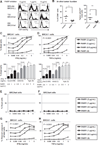
Figure 1
Figure 1
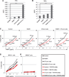
Th1-type cytokines enhance the cytotoxic effect of PARP inhibition in BRCA1 ovarian cancer cells. A, BRCA1 (BR5-Akt) and BRCAwt (T22, ID8) cell lines were cultured in 24-well plates at a starting concentration of 1 104 cells per well with or without the PARP inhibitor (PARPi) at the indicated dose, and analyzed at 72 hours for cell viability by flow cytometry. B, peritoneal cells were harvested by PBS wash on day 7 from BRCA1 tumor–bearing mice treated with the PARPi (40 mg/kg/day; days 3–6) and analyzed by flow cytometry for the percentage of CD45 tumor cells (left). In addition, total ascites cells were plated at serial dilutions for 72 hours, and the number of tumor colonies among total ascites cells was counted as an estimate of viable tumor burden (right). (Left, P 1/4 0.0271; right, P 1/4 0.0032, Student t test.) C and D, BRCA1 cells were cultured in 24-well plates for 72 hours in the presence of 0, 0.5, 1, or 2 µg/mL of PARPi and either IFNγ (C, top, PARPi dose effect, P < 0.0001; IFNγ dose effect, P < 0.0001; interaction, P < 0.0001) or TNFα (D, top, PARPi dose effect, P < 0.0001; TNFα dose effect, P < 0.0001, interaction, P < 0.0001) at the concentrations indicated. Cells were then stained with a fixable cell viability dye and analyzed by flow cytometry for the percentage of dead cells. Top dose-response curves show the interaction dose effect between cytokine concentration and PARP inhibitor treatment. Bottom bar graphs illustrate the dose effect independent of interaction. E and F, BRCAwt (T22) cells were assayed for viability as in C and D and show no treatment effect with increasing concentrations of cytokines or the PARP inhibitor. G and H, the fixable viability dye allowed for analysis of intracellular staining for active caspase-3. BRCA cells treated with PARPi and IFNγ (G) or TNFα (H) were intracellularly stained and cells positive for active caspase-3 are presented as a percentage of total cells (PARPi dose effect, P < 0.0001; IFNγ dose effect, P < 0.0001; interaction, P < 0.0001). Assays were repeated a minimum of three times. 0, P < 0.05; 00, P < 0.025; 000, P < 0.005; 0000, P < 0.0001 by ANOVA and Tukey procedure for multiple comparisons.
Figure 2
Figure 2
Checkpoint blockade with CTLA-4 antibody combined with PARP inhibition enhances T-cell effector function in the peritoneal tumor environment. Using the protocol depicted in Supplementary Fig. S1, BRCA1 tumor–bearing mice were sacrificed on day 21 for analysis (n 1/4 5/group). Peritoneal (A) and splenic (B) CD4+ and CD8+ T cells were analyzed for CD44 and CD62L expression, and percentage of CD62Llow/CD44hi cells was gated to determine the percentage of effector/memory CD4+ and CD8+ T cells of total CD4+ or CD8+ T cells, respectively. Peritoneal (C) cells and splenocytes (D) were restimulated in 24- well plates with PMA and ionomycin for 5 hours in the presence of Golgi transport inhibitors. CD4+ and CD8+ T cells were then analyzed by flow cytometry for intracellular cytokine expression. The percentages of T cells from treated animals producing IFNγ (top), TNFα (middle), or both (bottom) were compared with untreated controls. Data are representative of two experiments: * , P < 0.05; ** , P < 0.025; **** , P < 0.0001 by ANOVA and Tukey procedure for multiple comparisons.
Figure 3
Figure 3
Increases in IFNγ production in response to combined CTLA-4 blockade and PARP inhibition in vivo are sufficient to enhance tumor cell cytotoxicity. Mice were treated as in Supplementary Fig. S1 and euthanized on day 21 followed by retrieval of peritoneal cells (n 1/4 5/group). A total of 5 106 peritoneal cells were restimulated ex vivo with 10 µg/mL of anti-CD3 for 18 hours. Cell-free supernatants were harvested and pooled by treatment group, and levels of IFNγ (A) and TNFα (B) were determined by ELISA. C–E, supernatants were added to BRCA cells at a 4 dilution with DMEM-C in the presence of PARPi at indicated concentrations and cultured 72 hours in the absence (black lines) or presence (red lines) of IFNγ and TNFα neutralizing mAbs (10 µg/mL). C, cells were analyzed for viability by flow cytometry, and values shown are the percentages of dead cells with or without neutralizing antibody treatment. D, overlay of data in C. E, the same as D using BRCAwt cells (T22). *, P < 0.05; **, P < 0.025; ***, P < 0.005; ****, P < 0.0001 by ANOVA and Tukey procedure for multiple comparisons.
Figure 4
Figure 4
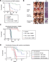
Combined treatment with a PARP inhibitor and CTLA-4 antibody promotes IFNγ-mediated tumor rejection in a BRCA1 tumor model. A, mice were treated as depicted in Supplementary Fig. S1 (40 mg/kg/day PARPi), and survival was compared with that of untreated controls (n 1/4 10/group). B, on day 21, the peritoneal cavity was exposed for evaluation of macroscopic solid tumor burden, and histologic sections of omentum were examined for microscopic tumor implants. Representative samples are shown. C, combination therapy with PARP inhibition (40 mg/kg/day) and antibody blockade of CTLA-4, PD-1, or PD-L1 as in Supplementary Fig. S1 (n 1/4 5–10 per group). D, survival of mice treated with PARPi (20 mg/kg/day) and CTLA-4 mAb combination therapy and neutralizing antibodies to TNFα , IFNγ, or both every 4 days beginning on day 7 (n 1/4 5–15/group); survival comparisons are the Kaplan–Meier curves. Differences among groups were determined with the log-rank (Mantel–Cox) test: *, P < 0.05;***, P < 0.005. Each experiment was repeated at least twice, with representative data shown.
Figure 5
Figure 5
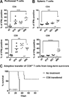
Combined treatment with the PARP inhibitor and CTLA-4 antibody induces protective immunity. Peritoneal cells (A) and splenocytes (B) were retrieved on days 7, 14, and 21 from combination therapy— treated mice, and from long-term survivors on day 90, and restimulated ex vivo with PMA and ionomycin for analysis by flow cytometry for intracellular IFNγ production by CD4+ and CD8+ T cells. * , P < 0.05; ** , P < 0.025, Tukey multiple comparisons test. C, adoptive transfer of CD8+ T cells from long-term survivors protects recipients from tumor development. CD8+ T cells were isolated by MACS-negative selection from long-term combination therapy survivors, pooled, and 2×10⁵ cells were adoptively transferred to recipient mice 12 hours prior to challenge with 2×10⁵ BRCA- tumor cells. Mice were monitored for survival (n 1/4 5/group) log-rank (Mantel–Cox) test.
Figure 6
Figure 6
IFNγ enhances cytotoxicity in human BRCA1 tumor cells exposed to a PARP inhibitor in vitro. BRCA1 UWB1.289 cells or UWB1.289 cells transfected with competent BRCA1 were cultured with titrated doses of the PARP inhibitor in the presence of recombinant human IFNγ or TNFα as indicated. After 72 hours, cells were analyzed for viability by flow cytometry. A, BRCA1 UWB1.289 cells exposed to IFNγ and PARP inhibition (top, PARPi dose effect, P < 0.0001; IFNγ dose effect, P < 0.0001; interaction, P 1/4 0.3872 based on two-way ANOVA; bottom, * , P < 0.05; **, P < 0.025 by Tukey multiple comparisons test). B, BRCA1 UWB1.289 cells exposed to TNFα and PARP inhibition (top, TNFα dose effect, P 1/4 0.0499; interaction, P 1/4 0.9655 by two-way ANOVA; bottom, *, P < 0.05, Tukey multiple comparisons test). C and D, BRCA1- transfected UWB1.289 cells exposed to IFNγ or TNFα and PARP inhibition (no statistically significant dose effect for IFNγ or TNFα).

Similar articles

Cited by

References

    1. Pardoll DM. The blockade of immune checkpoints in cancer immuno-therapy. Nat Rev Cancer. 2012;12:252–264. - PMC - PubMed
    1. Hodi FS, O'Day SJ, McDermott DF, Weber RW, Sosman JA, Haanen JB, et al. Improved survival with ipilimumab in patients with metastatic melanoma. N Engl J Med. 2010;363:711–723. - PMC - PubMed
    1. Lavou eV, Th edrez A, Lev^eque J, Foucher F, Henno S, Jauffret V, et al. Immunity of human epithelial ovarian carcinoma: the paradigm of immune suppression in cancer. J Transl Med. 2013;11:147. - PMC - PubMed
    1. Ai M, Curran MA. Immune checkpoint combinations from mouse to man. Cancer Immunol Immunother. 2015;64:885–892. - PMC - PubMed
    1. Yang YF, Zou JP, Mu J, Wijesuriya R, Ono S, Walunas T, et al. Enhanced induction of antitumor T-cell responses by cytotoxic T lymphocyte-associated molecule-4 blockade: the effect is manifested only at the restricted tumor-bearing stages. Cancer Res. 1997;57:4036–4041. - PubMed

Publication types

MeSH terms