. 2015 Jun 18;161(7):1633-43.
doi: 10.1016/j.cell.2015.06.002.

Crystal Structure of Antagonist Bound Human Lysophosphatidic Acid Receptor 1

Affiliations

Crystal Structure of Antagonist Bound Human Lysophosphatidic Acid Receptor 1

Jill E Chrencik et al. Cell. .

Abstract

Lipid biology continues to emerge as an area of significant therapeutic interest, particularly as the result of an enhanced understanding of the wealth of signaling molecules with diverse physiological properties. This growth in knowledge is epitomized by lysophosphatidic acid (LPA), which functions through interactions with at least six cognate G protein-coupled receptors. Herein, we present three crystal structures of LPA1 in complex with antagonist tool compounds selected and designed through structural and stability analyses. Structural analysis combined with molecular dynamics identified a basis for ligand access to the LPA1 binding pocket from the extracellular space contrasting with the proposed access for the sphingosine 1-phosphate receptor. Characteristics of the LPA1 binding pocket raise the possibility of promiscuous ligand recognition of phosphorylated endocannabinoids. Cell-based assays confirmed this hypothesis, linking the distinct receptor systems through metabolically related ligands with potential functional and therapeutic implications for treatment of disease.


Figures

Figure 1
Figure 1
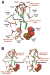
The overall structure of LPA1 colored by region and compound structure in the bioactive conformation. (A) The ligand ONO-9780307, shown with green carbons, demarcates the binding pocket, which is partially occluded by an N-terminal alpha helix (red) packing closely against the extracellular loops, ECL1 and ECL2 (orange). There are three native disulfide bonds in the extracellular region, one of which constrains the N-terminal capping helix to ECL2. (B) The bioactive conformation of ONO-9780307 in green carbons with high torsional strain energy required for positioning the indane ring adjacent to the dimethoxy phenyl ring. Release of this strain with an optimal torsion angle would result in a clash with the binding pocket.
Figure 2
Figure 2
Generation of the disulfide engineered LPA1 (dsLPA1-bRIL) crystallization construct. (A). Prediction of cysteine mutants replacing Asp2045.37 and Val2826.59 at the tip of TMV and VI respectively. (B). Functional analysis through a calcium mobilization assay measured in response to the LPA agonist dsLPA1 compared to full-length wild-type LPA1. The EC50 value of the stabilized receptor (994 nM) is about ten-fold higher compared to wild-type (153 nM). Data are presented as a mean (± SEM, n=3). (C). Thermal stability analysis of dsLPA1-bRIL compared to the non-engineered crystallization construct LPA1-bRIL. The engineered disulfide bond increases the apparent Tm by about 5°C in the presence of ONO-9780307, which was the ligand used for stability characterization of new constructs. (D). Crystal images for dsLPA1-bRIL in the presence of ONO-3080573. See also Figure S1.
Figure 3
Figure 3
Comparison of S1P1 and LPA1 structural features. (A). Top-view of LPA1 with structurally divergent regions of S1P1 (PDB ID: 3V2Y) overlayed (cyan). The configuration of the extracellular region is constrained by two intraloop disulfide bonds in ECL2 and ECL3 and a disulfide bond connecting ECL2 with the N-terminus. Although the electron density for the ECL3 loop is poor compared to the rest of the structure, the approximate position could be properly determined and is further from the N-terminal helix compared to S1P1. In S1P1 Lys285 in this region forms a cation dipole interaction with the C-terminal end of the capping helix, an interaction that is missing in LPA1. TMI is shifted 3 Å closer to TMVII at the tip compared to S1P1 and ECL0 is a loop in LPA1. Neither ECL0 nor ECL3 are involved in crystal contacts. (B). The shape of the LPA1 binding pocket is spherical in nature (solid surface) compared to the more linear binding pocket of S1P1 (blue mesh surface). This difference is caused by three changes in the amino acid composition between the two receptors. The first at position 3.33 is an aspartate in LPA1 and a phenylalanine in S1P1. The second at position 5.43 is a tryptophan in LPA1 and a cysteine in S1P1. Finally at position 6.51 LPA1 has a glycine whereas S1P1 has a leucine. The reduction in side chain bulk at this position opens a sub-pocket occupied by the indane ring of the antagonist series.
Figure 4
Figure 4
Analysis of the potential ligand entry for S1P1 and LPA1. (A). The position of TMI in S1P1 is shifted away from TMVII with a distance of 11 Å as measured between the Cα of Thr481.35 and Glu2947.36, this helical position allows access of ligand (ML056 shown in green carbon sticks) in the plasma membrane to the binding pocket of S1P1. (B). The position of TMI in LPA1 is closer to TMVII closing access of ligand (ONO-9780307 shown in green carbon sticks) in the plasma membrane to the binding pocket. The closer packing of TMI relative to S1P1 may be driven by the presence of Glu3017.43 which forms a hydrogen bond interaction with the backbone of Gly561.39 and Val591.42. (C). Molecular dynamics analysis of the distance between TMI and TMVII measured at four locations represented by a box plot for each measurement with the minimum and maximum indicated by vertical lines. The measurements correspond to the distances in shown in A monitored over the course of the simulation. The distance between the two helices is consistently larger and more variable for S1P1 than LPA1 both with and without ML056 and ONO-9780307 ligands respectively.
Figure 5
Figure 5
Analysis of the ligand binding pocket of LPA1. (A). Detailed binding interactions for ONO-9780307 in the LPA1 pocket. Polar interactions are represented by dashed yellow lines, and calculated positions of polar hydrogens are shown to more accurately represent the hydrogen bonding network. The indane binding pocket is formed by Gly2746.51 and Leu2756.52 which are represented by van Der Waals spheres. Areas for improving ligand binding are indicated in red text, and red spheres indicate positions for additional polar interactions. (B). Binding pocket interactions for additional co-crystal structures of ONO-9910539 and ONO-3080573. ONO-9910539 has improved interactions due to the introduction of an acetyl group in the para position of the phenyl ring which interacts with Trp2105.43. ONO-3080573 reduces the torsional strain induced by the indane position in ONO-9780307 through the replacement of a methylene linker with an ether linkage. In addition replacement of the pyrrole ring with a para substituted phenyl ring alters the interactions of the acid group away from the phosphate binding pocket toward Lys2947.36. See also Figure S2, Table S1 and Table S2.
Figure 6
Figure 6
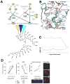
Pharmacologically pleitropic signaling lipids overlap through common acyl chain binding pockets in the LPA and cannabinoid receptors and enzymatic interconversion of polar head group. (A). The LPA, S1P and cannabinoid receptors are grouped in the same clade based on a sequence alignment of the transmembrane region. LPA and cannabinoid receptors are proposed to overlap in ligand specificity when binding polyunsaturated acyl chains delimited by different head group requirements. The biosynthetic map details the conversion of standard phospholipids to both LPA and CB receptor agonists through common pathways. Both known agonists for the cannabinoid system (2-AG and AEA) are one enzymatic step away from LPA ligands via phosphorylation of the head group (2-ALPA and pAEA). (B). Molecular modeling of the endogenous agonist lysophosphatidic acid with an 18 carbon acyl chain (LPA). In the presence of agonist, both Trp2716.48 and Trp2105.43 are predicted to change rotameric states to increase the volume of the binding pocket and direct the respective aromatic π clouds toward the bound ligand. The predicted agonist binding pocket is capable of docking 2-ALPA and pAEA which are generated through enzymatic action from endogenous cannabinoids. (C). Conformational root mean square fluctuation of LPA over the course of a 100 ns molecular dynamics simulation. The orientation of the ligand is consistent throughout the calculation indicating a reasonable starting point for modeling agonist interactions. (D). Analysis of intracellular signaling and cellular functions in B103 cells heterologously expressing full-length wild-type LPA1. The data are presented as mean normalized values (± SEM) to maximum responses induced by 3 μM 1-oleoyl-LPA (n =3) for Ca2+ mobilization assay, and by 10 μM forskolin (n =3) for cAMP assay. For the Ca2+ mobilization assay EC50 values for LPA 18:1 sn1 (n=3), 1-ALPA (n=3), 2-ALPA (n=2) and pAEA (n=2) are 146 ± 74 nM, 1125 ± 1484 nM, 98 ± 143 nM and 2120 ± 3885 nM respectively. For the cAMP assay EC50 values for LPA 18:1 sn1 (n=2), 2-ALPA (n=2) and pAEA (n=2) are 0.345 ± 0.202 nM, 0.302 ± 0.54 nM and 1.50 ± 2.04 nM respectively. Neurite retraction was quantified under microscopic observation. Filamentous (F)-actin stress fibers, produced by Rho GTPase activation like neurite retraction, were also produced by pAEA stimulation. The representative cell morphologies were shown (bars, 50 μm). The F-actin stress fiber formation was observed after 15 min treatment with LPA and pAEA (bars, 10 μm).

Similar articles

Cited by

References

    1. Alexandrov AI, Mileni M, Chien EY, Hanson MA, Stevens RC. Microscale fluorescent thermal stability assay for membrane proteins. Structure. 2008;16:351–359. - PubMed
    1. Aoki J. Mechanisms of lysophosphatidic acid production. Semin Cell Dev Biol. 2004;15:477–489. - PubMed
    1. Ballesteros JA, Weinstein H. Integrated methods for the construction of three dimensional models and computational probing of structure-function relations in G-protein coupled receptors. Methods Neurosci. 1995;25:366–428.
    1. Beard H, Cholleti A, Pearlman D, Sherman W, Loving KA. Applying physics-based scoring to calculate free energies of binding for single amino acid mutations in protein-protein complexes. PloS one. 2013;8:e82849. - PMC - PubMed
    1. Benito C, Romero JP, Tolon RM, Clemente D, Docagne F, Hillard CJ, Guaza C, Romero J. Cannabinoid CB1 and CB2 receptors and fatty acid amide hydrolase are specific markers of plaque cell subtypes in human multiple sclerosis. J Neurosci. 2007;27:2396–2402. - PMC - PubMed

Publication types