. 2015 Jul 1;125(7):2781-94.
doi: 10.1172/JCI80950. Epub 2015 Jun 15.

Molecular pharmacodynamics of emixustat in protection against retinal degeneration

Molecular pharmacodynamics of emixustat in protection against retinal degeneration

Jianye Zhang et al. J Clin Invest. .

Abstract

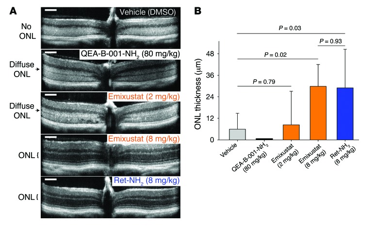
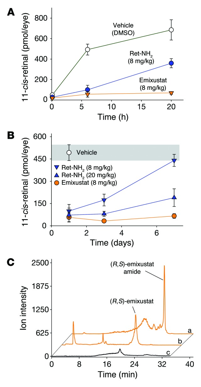
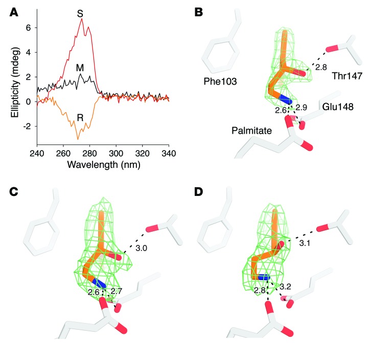
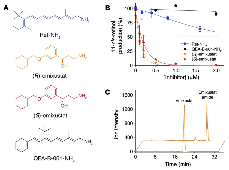
Emixustat is a visual cycle modulator that has entered clinical trials as a treatment for age-related macular degeneration (AMD). This molecule has been proposed to inhibit the visual cycle isomerase RPE65, thereby slowing regeneration of 11-cis-retinal and reducing production of retinaldehyde condensation byproducts that may be involved in AMD pathology. Previously, we reported that all-trans-retinal (atRAL) is directly cytotoxic and that certain primary amine compounds that transiently sequester atRAL via Schiff base formation ameliorate retinal degeneration. Here, we have shown that emixustat stereoselectively inhibits RPE65 by direct active site binding. However, we detected the presence of emixustat-atRAL Schiff base conjugates, indicating that emixustat also acts as a retinal scavenger, which may contribute to its therapeutic effects. Using agents that lack either RPE65 inhibitory activity or the capacity to sequester atRAL, we assessed the relative importance of these 2 modes of action in protection against retinal phototoxicity in mice. The atRAL sequestrant QEA-B-001-NH2 conferred protection against phototoxicity without inhibiting RPE65, whereas an emixustat derivative incapable of atRAL sequestration was minimally protective, despite direct inhibition of RPE65. These data indicate that atRAL sequestration is an essential mechanism underlying the protective effects of emixustat and related compounds against retinal phototoxicity. Moreover, atRAL sequestration should be considered in the design of next-generation visual cycle modulators.


Figures

Figure 9
Figure 9. Impact of treatment with retinoid cycle modulators on the quantity of enlarged photoreceptors in BALB/c mice.
Four- to five-week old male and female BALB/c mice were exposed to bright light at 20,000 lux for 2 hours on the day after they were gavaged with oil (control, 100 μl), emixustat (racemic, 40 mg/kg), or MB-002 (80 mg/kg). TPM images of intact BALB/c mouse eyes were obtained on day 2 after exposure to light, and representative 3D reconstructions for each group are shown in A. In each 3D reconstruction, the RPE is at the top, and the section through the photoreceptor cell layer is shown 9–12 μm below the RPE. Treatments are indicated in each image. Enlarged photoreceptor cells, indicated with red arrowheads, were noted only in animals that were exposed to light and treated with oil or MB-002. Enlarged photoreceptor cells were counted for animals in each treatment group (B). Error bars indicate SDs; n = 8, 5, 8, and 10 for the vehicle, no light–, emixustat-, and MB-002–treated mice, respectively. P values shown in the graph were calculated by ANOVA and represent the statistical significance for each treated group versus control animals exposed to light and treated by gavage with oil.
Figure 8
Figure 8. Crystal structure of RPE65 in complex with MB-002.
The bound MB-002 molecules are shown as orange sticks. The σA-weighted Fo-Fc omit electron density is shown as green mesh contoured at 3 σ. Residues within 4.5 Å of the bound ligands are shown as gray sticks.
Figure 7
Figure 7. Effects of the emixustat derivative MB-002 on RPE65 activity and protection from light-induced retinopathy.
(A) Chemical structures of emixustat and MB-002. (B) Inhibitory effect of MB-002 on retinoid isomerase activity in vitro. (C) Inhibitory effects of visual cycle modulators on 11-cis-retinal regeneration in C57BL6J mice. Post-bleaching recovery of ocular 11-cis-retinal was measured after a a 6-hour dark incubation period. (D) OCT images of retinae from pigmented Abca4–/– Rdh8–/– mice treated with MB-002 or retinylamine. The images, obtained 3 days after light exposure, show a lack of protective effects with MB-002. Scale bars: 100 μm. (E) Representative 3D TPM image of the retinal photoreceptor and RPE cell layers from an albino Abca4–/– Rdh8–/– mouse treated with MB-002 and exposed to intense light showing numerous swollen photoreceptor outer segments (red arrowheads). RPE cells are at the top of the section, and the photoreceptor cell layer section is shown at 10 μm below the RPE. Mice were exposed to bright light 1 day after they were gavaged with either oil (control), Ret-NH2 (2 mg/mouse), or MB-002 (2 mg/mouse). TPM images of intact mouse eyes were obtained on day 2 after light exposure. Enlarged photoreceptors were counted in each group at a distance of 8 to 10 μm from the RPE. The mean numbers (± SD) of enlarged photoreceptors for each treatment group are as follows: 252 ± 33 for the oil-treated group; none in the no-light group; 5 ± 5 in Ret-NH2–treated animals; and 233 ± 45 in MB-002–treated animals. n = 4.
Figure 6
Figure 6. Emixustat-atRAL Schiff base formation in vitro and in vivo.
(A) Chromatographic separation and detection of primary amines, atRAL, and their Schiff base conjugates. Emixustat or Ret-NH2 was incubated with atRAL for 2 hours, and the mixture was separated on a C-18 column. Representative HPLC chromatograms indicate the formation of an emixustat Schiff base (peak “a”) and Ret-NH2 Schiff base (peak “b”) at an aldehyde/amine ratio of 1:1. Peak “c” corresponds to unreacted atRAL. (B) Quantification of Schiff bases formed upon incubation of increasing concentrations of atRAL with either 0.2 mM emixustat (inverted triangles) or Ret-NH2 (circles). (C) The chromatogram illustrates separation of an emixustat Schiff base standard. Peak 1, between 6 and 7 minutes, corresponds to the emixustat retinylidene Schiff base as indicated by its MS spectrum (D) with a dominant ion of m/z = 530.5 corresponding to [MH]+ of the emixustat-retinylidene Schiff base. Peak 2 corresponds to atRAL. Inset in D shows the MS/MS fragmentation pattern of the m/z = 530.5 parent ion. (E) Detection of emixustat-retinylidene Schiff base in vivo; 8 mg/kg emixustat was administered to Abca4–/– Rdh8–/– mice 2 hours before light bleaching. After bleaching, mice were euthanized, and eye extracts were analyzed by LC-MS. Peak 1′ observed in the chromatogram corresponds to an emixustat Schiff base. Its identity was confirmed by its UV/Vis spectrum with a characteristic absorbance maximum of 462 nm (inset) and by MS (F), in which fragmentation of the m/z = 530.5 ion yielded the product ion at m/z = 438.5, identical to that shown in D. mAU, milliabsorbance units.
Figure 5
Figure 5. Impact of treatment with retinoid cycle modulators on the quantity of enlarged photoreceptors in Abca4–/– Rdh8–/– mice.
Abca4–/–Rdh8–/– mice were exposed to bright light on the day after they were gavaged with oil (control), Ret-NH2 (80 mg/kg), emixustat (racemic, 40 mg/kg), or QEA-B-001-NH2 (80 mg/kg). TPM images of intact double-KO mouse eyes were obtained on day 2 after exposure to light, and representative images for each group are shown. In each TPM 3D section, RPE cells are at the top of the section, and the photoreceptor cell layer section is shown 9–12 μm below the RPE. Treatments are indicated in each image. Enlarged photoreceptor cells, indicated with red arrowheads, were noted only in animals that were exposed to light and treated with oil or QEA-B-001-NH2. Enlarged photoreceptor cells were counted (graph) for animals in each treatment group; mean values are displayed. Error bars indicate SD; (n = 4); P values were calculated by ANOVA and represent the statistical significance for each treated group versus control animals exposed to light and treated by gavage with oil.
Figure 4
Figure 4. Protective effects of primary amines against light-induced retinal degeneration in Abca4–/– Rdh8–/– mice.
Four-week-old Abca4–/– Rdh8–/– mice treated with the tested amines were kept in the dark for 24 hours, bleached with 10,000 lux light for 1 hour, and then kept in the dark for 3 days. (A) Representative OCT images of mice treated with QEA-B-001-NH2 (80 mg/kg), emixustat (2 mg/kg and 8 mg/kg), and Ret-NH2 (8 mg/kg). A dramatic decrease in the ONL indicates advanced retinal degeneration. Scale bars: 100 μm. (B) Quantification of the protective effects of QEA-B-001-NH2, emixustat, and Ret-NH2 are shown by measuring the average thickness of the ONL. Only non-scattering ONL layers were measured. For images in which the ONL layer boundary was obscured due to light scattering, the ONL length was recorded as 0 μm. Mean values ± SD are shown; n = 5–6 for each group. P values were calculated by ANOVA.
Figure 3
Figure 3. Inhibition of visual chromophore regeneration by emixustat and Ret-NH2 in C57BL6J mice.
Recovery of 11-cis-retinal was measured in mouse eyes after exposure to bright light. (A) The progress of 11-cis-retinal recovery after light exposure in mice treated with either emixustat or Ret-NH2. Both drugs were administrated at the same dose 24 hours prior to light exposure. (B) Persistence of the inhibitory effect in mice treated with Ret-NH2 or emixustat. Both drugs were administrated 1, 3, or 7 days before light exposure; the recovery of 11-cis-retinal was measured after a 6-hours dark adaptation. (A and B) Mean values ± SD are shown; n = 5–6 for each data point. (C) Detection of emixustat and its amides in mouse eyes by LC-MS. The graph shows extracted ion chromatograms for m/z = 502.3 [MH]+, representing emixustat palmitamide (a), and m/z = 264.3 [MH]+, corresponding to the free amine form of the compound (b). Chromatogram “c” reveals an intensity of m/z = 502.3 ion for an eye extract obtained from an untreated animal.
Figure 2
Figure 2. Analysis of emixustat enantiomer binding to RPE65.
(A) Circular dichroism (CD) spectra of (R)-, (S)-, and racemic (M) emixustat. (B, C, and D) Respective unbiased σA-weighted omit Fo-DFc electron density maps at 3 root mean square deviation (RMSD) contour levels and associated models for the complexes obtained from racemic, (R)-, and (S)-emixustat. Dashed lines indicate hydrogen bonds with bond lengths given in Å. For B, (R)-emixustat is clearly bound (compare with C), despite the addition of equimolar concentrations of (R)- and (S)-emixustat prior to crystallization.
Figure 1
Figure 1. Inhibition of RPE65 by retinol analogs and formation of emixustat amide.
(A) Primary amines used in this study that are structurally similar to retinol (vitamin A). (B) 11-cis-Retinol production in the presence of inhibitors depicted in A. Primary amines were preincubated with bovine RPE microsomes at room temperature for 5 minutes, then all-trans-retinol was added and the mixture incubated at 37°C. All incubation mixtures were quenched by the addition of methanol after 1 hour of incubation. Inhibition of RPE65 enzymatic activity was measured as a decline in 11-cis-retinol production. Note that (S)-emixustat was 10 times more potent than Ret-NH2 in lowering 11-cis-retinol production. Without an inhibitor, typical activity was between 25 and 32 pmol/minute, and this was set as 100% activity. (C) Extracted ion LC-MS chromatograms showing acylation of emixustat (m/z = 264.2 [MH]+, lower trace) by LRAT in RPE microsomes to form corresponding emixustat palmitamide (m/z = 502.3 [MH]+, upper trace).

Similar articles

Cited by

References

    1. Batten ML, et al. Lecithin-retinol acyltransferase is essential for accumulation of all-trans-retinyl esters in the eye and in the liver. J Biol Chem. 2004;279(11):10422–10432. doi: 10.1074/jbc.M312410200. - DOI - PMC - PubMed
    1. Redmond TM, Poliakov E, Yu S, Tsai JY, Lu Z, Gentleman S. Mutation of key residues of RPE65 abolishes its enzymatic role as isomerohydrolase in the visual cycle. Proc Natl Acad Sci U S A. 2005;102(38):13658–13663. - PMC - PubMed
    1. Palczewski K, et al. Crystal structure of rhodopsin: a G protein-coupled receptor. Science. 2000;289(5480):739–745. doi: 10.1126/science.289.5480.739. - DOI - PubMed
    1. Palczewski K. G protein-coupled receptor rhodopsin. Annu Rev Biochem. 2006;75:743–767. doi: 10.1146/annurev.biochem.75.103004.142743. - DOI - PMC - PubMed
    1. Kiser PD, Golczak M, Palczewski K. Chemistry of the retinoid (visual) cycle. Chem Rev. 2014;114(1):194–232. doi: 10.1021/cr400107q. - DOI - PMC - PubMed

Publication types

MeSH terms