. 2015 Jul 16;59(2):321-32.
doi: 10.1016/j.molcel.2015.05.022. Epub 2015 Jun 11.

SIRT5 Regulates both Cytosolic and Mitochondrial Protein Malonylation with Glycolysis as a Major Target

Affiliations

SIRT5 Regulates both Cytosolic and Mitochondrial Protein Malonylation with Glycolysis as a Major Target

Yuya Nishida et al. Mol Cell. .

Abstract

Protein acylation links energetic substrate flux with cellular adaptive responses. SIRT5 is a NAD(+)-dependent lysine deacylase and removes both succinyl and malonyl groups. Using affinity enrichment and label free quantitative proteomics, we characterized the SIRT5-regulated lysine malonylome in wild-type (WT) and Sirt5(-/-) mice. 1,137 malonyllysine sites were identified across 430 proteins, with 183 sites (from 120 proteins) significantly increased in Sirt5(-/-) animals. Pathway analysis identified glycolysis as the top SIRT5-regulated pathway. Importantly, glycolytic flux was diminished in primary hepatocytes from Sirt5(-/-) compared to WT mice. Substitution of malonylated lysine residue 184 in glyceraldehyde 3-phosphate dehydrogenase with glutamic acid, a malonyllysine mimic, suppressed its enzymatic activity. Comparison with our previous reports on acylation reveals that malonylation targets a different set of proteins than acetylation and succinylation. These data demonstrate that SIRT5 is a global regulator of lysine malonylation and provide a mechanism for regulation of energetic flux through glycolysis.


Figures

Figure 1
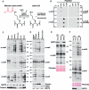
Figure 1. Generation of Malonyl-Lysine Specific Antibodies and Characterization of Malonylation Distribution in Mouse Tissues and Subcellular Liver Compartments
(A) Malonyl-lysine and the demalonylation reaction catalyzed by SIRT5. The malonyl group on the lysine residue is shown in red. (B) Malonyl-lysine antibodies specifically identify malonyl-lysine containing peptide, but not acetyl- or succinyl-lysine in a dotplot assay. (C) Western blot analysis comparing lysine malonylation, succinylation, and acetylation levels in mouse organs derived from WT or Sirt5−/− mice. The absence of SIRT5 in Sirt5−/− mouse tissues was confirmed using anti-SIRT5 antibody. (D) Western blot analysis for lysine malonylation, succinylation and acetylation in whole cell lysates, cytosolic or mitochondrial fractions derived from WT or Sirt5−/− mouse livers. Purity of subcellular fractionation was confirmed by western blot for the cytosolic protein tubulin and the mitochondrial protein VDAC. (E) Global hyper-malonylation is reversibly de-malonylated by SIRT5 overexpression. A lentivirus vector was used either as an empty vector (lane 2) or to overexpress SIRT5 (lane3) in SIRT5 KO primary hepatocytes. Lysine malonylation levels were examined in whole cell lysates by western blotting. (F) Global maK level changes following starvation. Global malonylation levels in whole liver lysates were compared between fed and starved WT or SIRT5 KO mice. Note that malonyl-lysine levels are higher in feeding than fasting conditions both in WT and SIRT5 KO mice whereas acetylation is higher in fasting than feeding and unaffected by SIRT5.
Figure 2
Figure 2. Enrichment and Identification of Whole Liver Lysine Malonylome by Label-Free Quantitation
(A) Whole cell liver lysates were prepared from 5 WT and 5 Sirt5−/− mice (Sv129, n = 5, 10 months of age, fed). Individual samples were digested with trypsin and spiked with 150 fmol of a malonyllysine peptide standard. Lysine malonylated peptides were immunoprecipitated and samples were analyzed by LC-MS/MS in duplicate. Ion intensity chromatograms were extracted using MS1 Filtering and peptide abundance compared between WT and KO animals. (B) Scatterplot shows individual malonylated peptide abundances quantified by MS1 Filtering from WT and Sirt5−/− mice. Dashed lines represent fold change between the two conditions (WT versus Sirt5−/− mice). Peptides showing significant difference in abundance (p value < 0.05) are shown in purple while all other peptides are shown in red. (C) Overlap of the malonyl-lysine sites and proteins targeted by SIRT5 in the mouse liver malonylome (KO:WT ≥ 1.5, p value < 0.05). For peptide information and quantification of individual sites, see Tables S1 and S2.
Figure 3
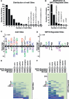
Figure 3. Site Distribution, Sequence Logo, and Conservation Analysis
(A) Distribution of the number of maK sites identified per protein. Most malonylated proteins contain a single site, but some contain multiple sites. (B) Distribution of the number of SIRT5-regulated sites per protein. Abbreviations include CPS1 (Carbamoyl-phosphate synthase, mitochondrial), FDH (Cytosolic 10-formyltetrahydrofolate dehydrogenase), MMSDH (Methylmalonate-semi-aldehyde dehydrogenase, mitochondrial), ARG1 (Arginase-1), ACAT (Acetyl-CoA acetyltransferase, mitochondrial), OSF-3 (Peroxiredoxin-1), L-FABP (Fatty acid-binding protein, liver), EF-1α1 (Elongation factor 1-alpha 1), BHMT (Betaine-homocysteine S-methyltransferase 1), 17-β-HSD5 (Estradiol 17 beta-dehydrogenase 5), LACS1 (Long-chain-fatty-acid-CoA ligase 1), ENO1 (Alpha-enolase), GAPDH (Glyceraldehyde-3-phosphate dehydrogenase), ASS1 (Argininosuccinate synthase), PYGL (Glycogen phosphorylase, liver form), PGK1 (Phosphoglycerate kinase 1), ACBP (Acyl-CoA-binding protein), CA-III (Carbonic anhydrase 3), BHMT (Betaine-homocysteine S-methyltransferase 1), TP-α (Trifunctional enzyme subunit alpha, mitochondrial), and RPS20 (40S ribosomal protein S20). (C and D) Consensus sequence logo plots centered on the lysine of all identified maK sites (±5 amino acids) (C) or all identified SIRT5-regulated maK sites (≥1.5-fold and p < 0.05) (D). (E and F) Heatmap depicting the conservation index of maK (E) or SIRT5-regulated maK sites (F) across seven vertebrate species. The percentage of sites conserved for all sites, target and non-target sites is listed above the heatmap. Lysine (K), glutamine (Q), arginine (R), aspartic acid (D), or glutamic acid (E) and other amino acids are represented by the colors indicated in the heat-map. See Table S5 for individual site conservation details.
Figure 4
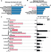
Figure 4. Glycolytic Enzymes Are Hyper-Malonylated in the Absence of SIRT5
(A and B) Ingenuity pathway analysis of malonylated proteins (A) and SIRT5-regulated malonylated proteins (B). (C) Glycolytic enzymes are highlighted next to the reaction they catalyze. The color intensity of boxes (white to red) indicates the the ratio of SIRT5 KO hypermalonylation (Sirt5−/− /WT) with red being high and white corresponding to a value of 1. MaK sites in each enzyme and their regulation by SIRT5 are shown at right as a bar graph of mean MS intensity in the presence and absence of SIRT5.
Figure 5
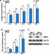
Figure 5. Glycolysis Is Suppressed in the Absence of SIRT5
(A) Lactic acid production is impaired in primary hepatocytes derived from Sirt5−/− versus WT animals. Lactic acid was measured in culture media of primary hepatocytes isolated from WT and Sirt5−/− animals at different time points. Each graph is representative of two independent experiments; n = 3 measurements/sample; mean ± SD. (B) Glycolytic flux measured by deuterium-labeled glucose is suppressed in KO primary hepatocytes. Oxidized glucose was measured in primary hepatocytes isolated from WT and Sirt5−/− animals at different time points. Graph is representative of two independent experiments; n = 3 measurements/sample, mean ± SD. (C) Comparison of Glucose transporter 2 expression between WT and SIRT5 KO primary hepatocytes as assayed by western blotting using tubulin as a loading control.
Figure 6
Figure 6. The Major Site of Malonylation in GAPDH by SIRT5 (K184) Is Important for GAPDH Enzymatic Activity
(A) GAPDH is reversibly de-malonylated by WT but not enzymatically inactive mutant SIRT5 overexpression in cell culture. Flag-tagged GAPDH and SIRT5 were co-overexpressed in WT or SIRT5 KO MEFs, and western blotting against maK was performed following immunoprecipitation by anti-Flag antibody. (B) GAPDH is reversibly de-malonylated by SIRT5 in vitro. After purifying GAPDH and SIRT5 WT or H158Y enzymatically inactive mutant overexpressed in HEK293T cells, respectively, de-malonylation was performed by adding NAD. MaK level of GAPDH was assayed by western blotting. The quantification of maK levels is shown in the right panel, in which WT but not H158Y SIRT5 effectively demalonylated GAPDH in vitro. (C) Sites of malonylation in the GAPDH protein with respect to its two functional domains, NAD binding, and catalytic domains. The ratio of malonylation between WT and Sirt5−/− samples are shown above. (D) Relative malonylation between WT and Sirt5−/− samples is illustrated as a redness color scale, with the most SIRT5-regulated lysine (K184) shown as 100% red. (E) K184E mutant shows defective GAPDH enzymatic activity. Endogenous human GAPDH was knocked down with two different shRNAs in HEK293T cells after a 4-day selection with puromycin. Expression vector for Flag-tagged mouse GAPDH WT, or mutants C150S (enzymatically inactive) and K184E were overexpressed. GAPDH activity was measured in whole-cell lysates 24 hr after transfection of expression vectors by enzymatic assay. (F) Knockdown of endogenous GAPDH and expression of WT, C150S, and K184E mutants was confirmed by western blotting using an antiserum against GAPDH or against Flag.

Similar articles

Cited by

References

    1. Altschul SF, Madden TL, Schäffer AA, Zhang J, Zhang Z, Miller W, Lipman DJ. Gapped BLAST and PSI-BLAST: a new generation of protein database search programs. Nucleic Acids Res. 1997;25:3389–3402. - PMC - PubMed
    1. Baeza J, Dowell JA, Smallegan MJ, Fan J, Amador-Noguez D, Khan Z, Denu JM. Stoichiometry of site-specific lysine acetylation in an entire proteome. J. Biol. Chem. 2014;289:21326–21338. - PMC - PubMed
    1. Bao X, Zhao Q, Yang T, Fung YME, Li XD. A chemical probe for lysine malonylation. Angew. Chem. Int. Ed. Engl. 2013;52:4883–4886. - PubMed
    1. Berger SL. Histone modifications in transcriptional regulation. Curr. Opin. Genet. Dev. 2002;12:142–148. - PubMed
    1. Beysen C, Murphy EJ, McLaughlin T, Riiff T, Lamendola C, Turner HC, Awada M, Turner SM, Reaven G, Hellerstein MK. Whole-body glycolysis measured by the deuterated-glucose disposal test correlates highly with insulin resistance in vivo. Diabetes Care. 2007;30:1143–1149. - PubMed

Publication types

MeSH terms

LinkOut - more resources