. 2015 Jun 12:6:7324.
doi: 10.1038/ncomms8324.

WASH and Tsg101/ALIX-dependent diversion of stress-internalized EGFR from the canonical endocytic pathway

Affiliations

WASH and Tsg101/ALIX-dependent diversion of stress-internalized EGFR from the canonical endocytic pathway

Alejandra Tomas et al. Nat Commun. .

Abstract

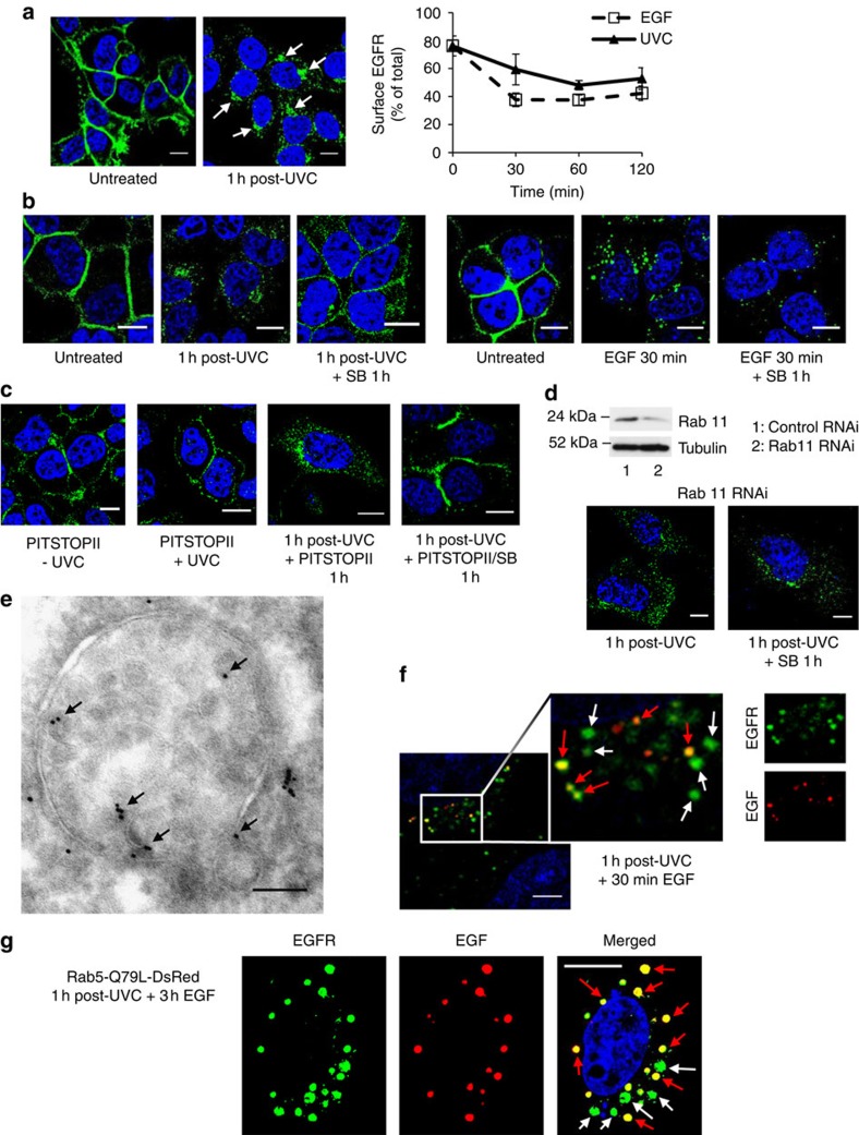
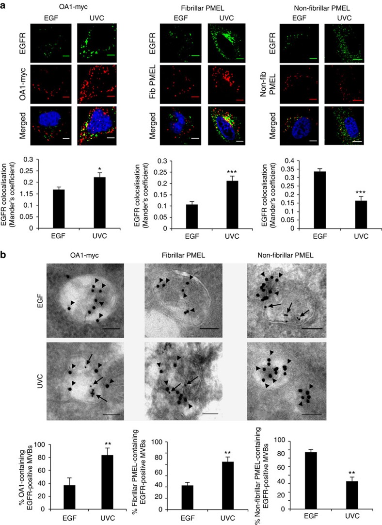
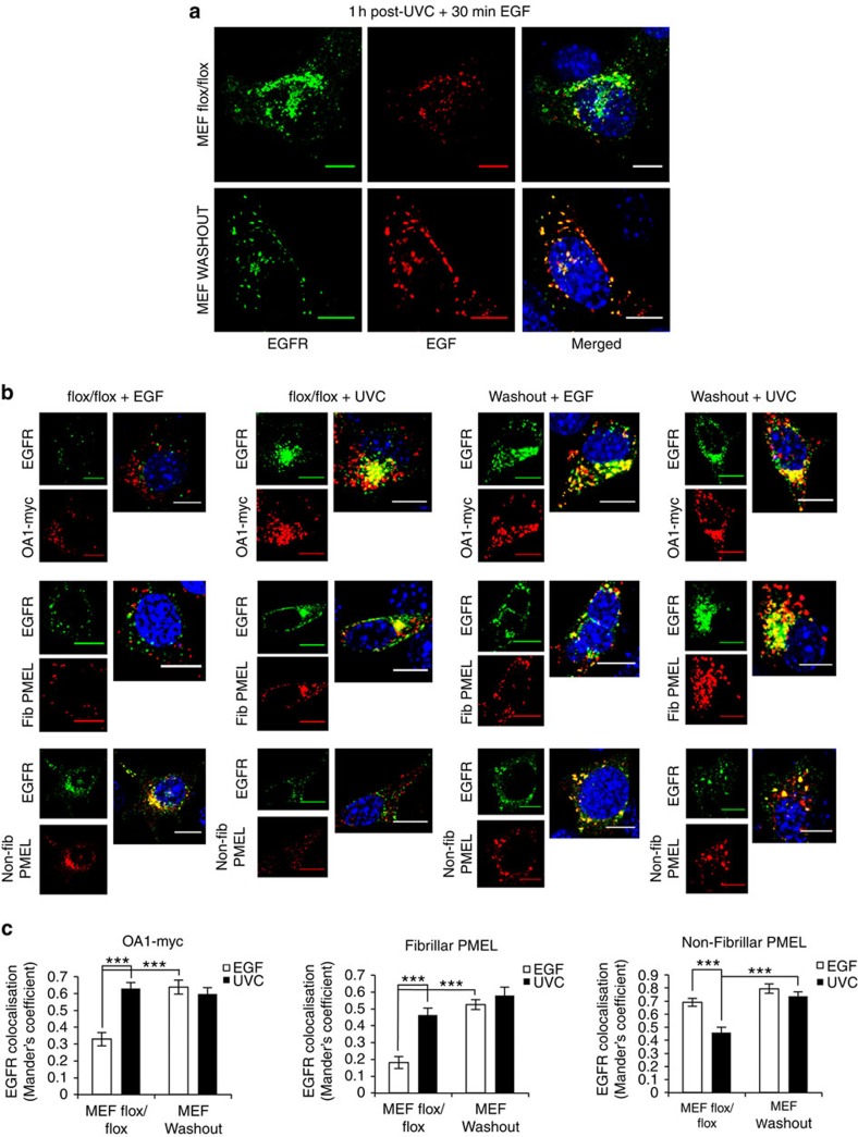
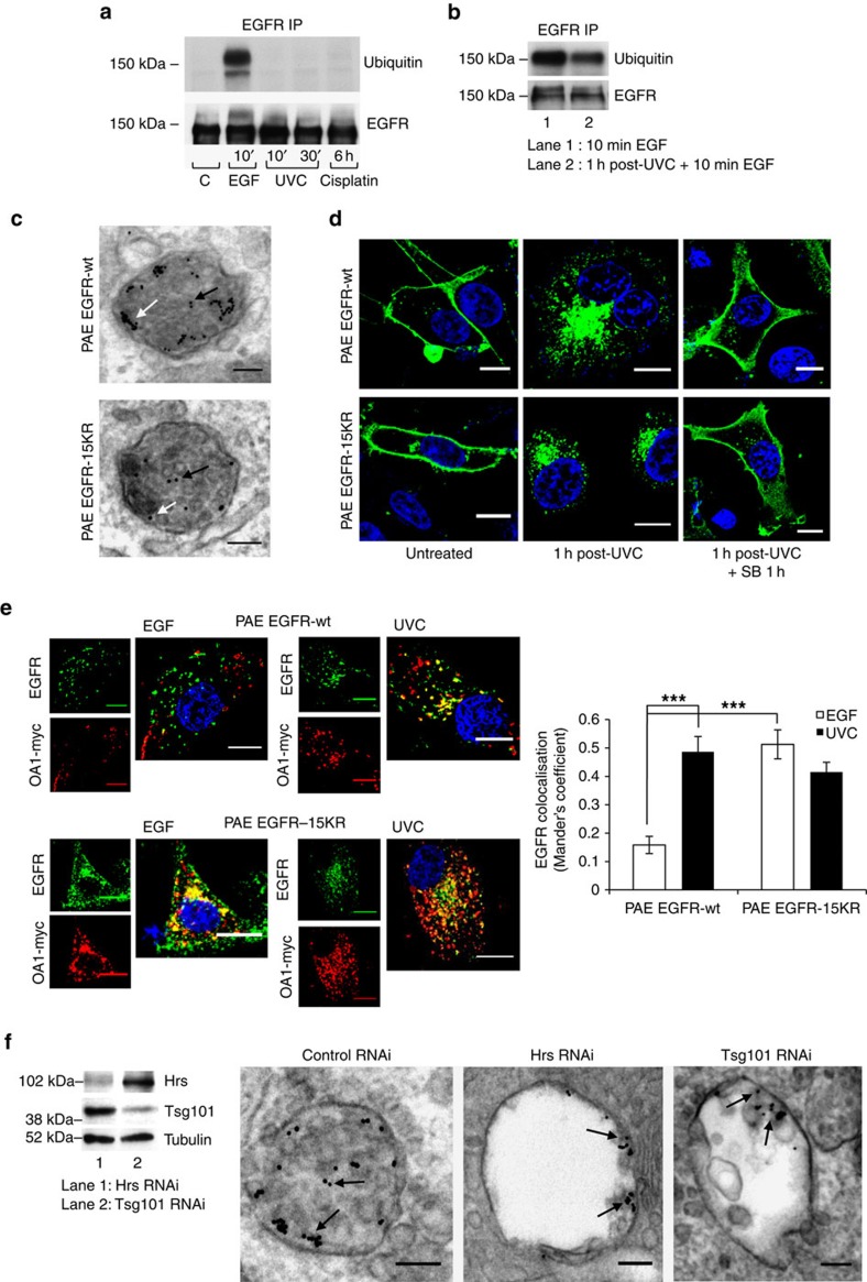
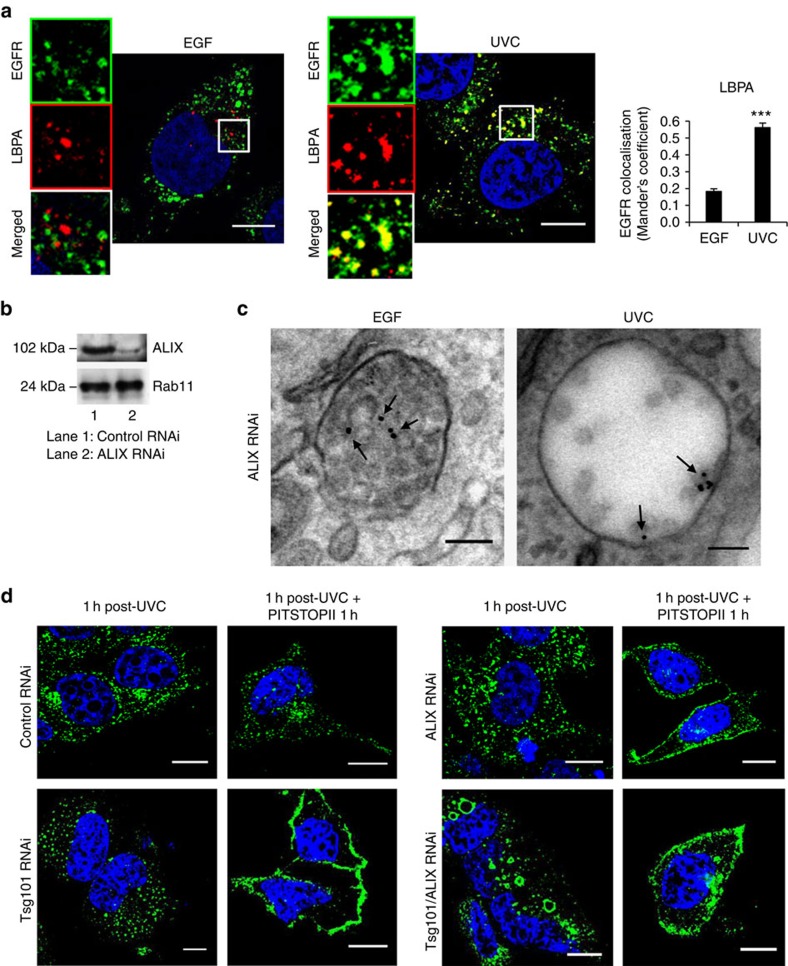
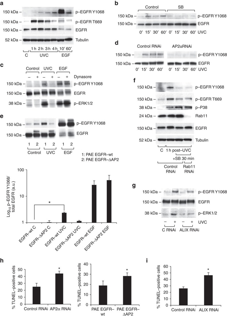
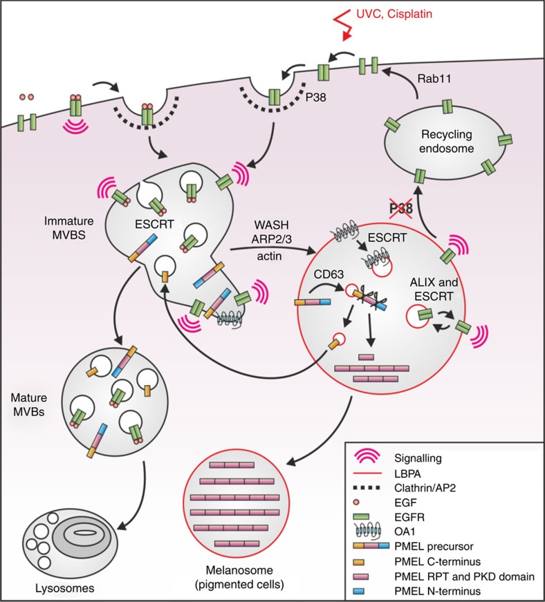
Stress exposure triggers ligand-independent EGF receptor (EGFR) endocytosis, but its post-endocytic fate and role in regulating signalling are unclear. We show that the p38 MAP kinase-dependent, EGFR tyrosine kinase (TK)-independent EGFR internalization induced by ultraviolet light C (UVC) or the cancer therapeutic cisplatin, is followed by diversion from the canonical endocytic pathway. Instead of lysosomal degradation or plasma membrane recycling, EGFR accumulates in a subset of LBPA-rich perinuclear multivesicular bodies (MVBs) distinct from those carrying EGF-stimulated EGFR. Stress-internalized EGFR co-segregates with exogenously expressed pre-melanosomal markers OA1 and fibrillar PMEL, following early endosomal sorting by the actin polymerization-promoting WASH complex. Stress-internalized EGFR is retained intracellularly by continued p38 activity in a mechanism involving ubiquitin-independent, ESCRT/ALIX-dependent incorporation onto intraluminal vesicles (ILVs) of MVBs. In contrast to the internalization-independent EGF-stimulated activation, UVC/cisplatin-triggered EGFR activation depends on EGFR internalization and intracellular retention. EGFR signalling from this MVB subpopulation delays apoptosis and might contribute to chemoresistance.


Figures

Figure 1
Figure 1. Dual role of p38 in ligand-independent stress-triggered EGFR trafficking in HeLa cells.
(a) EGFR immunolocalization in untreated versus UVC-exposed cells 1 h post UVC exposure (left), and quantification of surface downregulation in cells exposed to UVC or treated with EGF for the indicated times (right). Arrows indicate perinuclear accumulation of EGFR (green) after UVC exposure. Data are mean±s.e.m. of three independent experiments. (b) Untreated or UVC-exposed HeLa cells were fixed after 1 h, or further incubated with the p38 inhibitor SB202190 (SB) for 1 h (left). Untreated or EGF-treated cells were fixed after 30 min, or further incubated with SB for 1 h in the continuous presence of EGF (right). p38 inhibition causes EGFR (green) redistribution to the plasma membrane following UVC, but not EGF exposure. (c) Pre-treatment for 30 min with PITSTOPII prevents UVC-induced EGFR (green) internalization (left) but PITSTOPII addition 1 h after UVC-induced EGFR internalization does not affect perinuclear EGFR accumulation or the recycling induced by simultaneous p38 inhibition (right). (d) Cells transfected with control or Rab11 siRNA were immunoblotted after 72 h for Rab11 and tubulin to assess knockdown efficiency (top). Rab11 knockdown did not prevent UVC-induced EGFR (green) internalization but prevented EGFR recycling after subsequent SB treatment (bottom). (e) Cells transfected with EGFR-GFP were fixed 1 h after UVC exposure and ultrathin cryosections were immuno-labelled for EGFR with 8 nm gold. EGFR-GFP (arrows) is on the limiting membrane and ILVs of MVBs. (f) Immunofluorescence analysis of UVC and EGF sequentially exposed HeLa cells (see Methods for experimental details). Red arrows show endosomes containing EGFR (green) and EGF (red). White arrows show EGFR+ve, EGF-ve endosomes, indicating a separate subset of MVBs containing stress-internalized but not EGF-bound EGFR. Scale bar, 5 μm. (g) Cells transfected with constitutively active Rab5-Q79L-DsRed were exposed to UVC and incubated for 1 h before treatment with EGF-AlexaFluor 647 (red) for 3 h. Red and white arrows show EGFR+ve/EGF+ve and EGFR+ve/EGF-ve endosomes, respectively. Scale bars, 10 μm for confocal and 100 nm for EM, unless otherwise indicated; 4,6-diamidino-2-phenylindole (DAPI)-stained nuclei, blue.
Figure 2
Figure 2. Co-segregation of stress-internalized EGFR with markers of pre-melanosomal MVBs.
(a) HeLa cells were transfected with OA1-myc or PMEL, and either treated with EGF for 30 min or exposed to UVC and incubated for 1 h. Cells were co-stained with EGFR and either myc (left), fibrillar PMEL (middle) or non-fibrillar PMEL (right). Quantification of co-localization between EGFR (green) and expressed marker (red) for the different conditions is shown below each set of images. Data are mean±s.e.m. of three independent experiments, *P<0.05 and ***P<0.001 (Student's t-test). (b) HeLa cells were treated as above in the presence of 10 nm anti-EGFR-gold (arrows) before preparation for cryo-immunoEM. Ultrathin cryosections were labelled for myc (left), fibrillar PMEL (middle) or non-fibrillar PMEL (right) with 15 nm-gold (arrowheads). Depicted are typical examples of OA1 and fibrillar PMEL+ve MVBs containing stress-internalized but not EGF-stimulated EGFR, and non-fibrillar PMEL containing EGF-stimulated but not stress-internalized EGFR. Quantification of the percentage of EGFR+ve MVBs containing each of the different markers following EGF versus UVC exposure is shown below each set of images. Data are mean±s.e.m. of ≥10 cells, **P<0.01 (Student's t-test). Scale bars, 10 μm for confocal and 100 nm for EM images; 4,6-diamidino-2-phenylindole (DAPI)-stained nuclei, blue.
Figure 3
Figure 3. Sorting of EGF-bound and stress-internalized EGFR onto separate MVB subsets is WASH dependent.
(a) Control flox/flox and WASH knock-out (WASHOUT) MEFs transfected with human EGFR were exposed to UVC and incubated for 1 h to allow stress-induced EGFR internalization in the presence of anti-EGFR 108 antibody. Cells were washed, incubated with EGF-AlexaFluor 488 for 30 min (red), and processed for immunofluorescence with an AlexaFluor 555 secondary antibody to label EGFR (green). Both EGF+ve and EGF-ve EGFR-containing endosomes are present in control flox/flox, but these are largely merged in WASHOUT MEFs. (b) Control flox/flox and WASHOUT MEFs were co-transfected with human EGFR and OA1-myc or PMEL, and either treated with EGF for 30 min or exposed to UVC and incubated for 1 h. Cells were co-stained for EGFR (green) and the following markers (in red): myc (top panels), fibrillar PMEL (central panels) or non-fibrillar PMEL (bottom panels). (c) Quantification of co-localization between EGFR and expressed markers for the different conditions. Data are mean±s.e.m. of three independent experiments, ***P<0.001 (Student's t-test). Scale bars, 10 μm; 4,6-diamidino-2-phenylindole (DAPI)-stained nuclei, blue.
Figure 4
Figure 4. Ubiquitin-independent, ESCRT-dependent sorting of stress-internalized EGFR onto ILVs of MVBs.
(a) Immunoprecipitation (IP) of EGFR from HeLa cell lysates and immunoblotting for ubiquitin and EGFR showed robust EGFR ubiquitination after EGF stimulation but not after exposure to UVC or cisplatin. (b) Treatment of HeLa cells with EGF 1 h post-UVC exposure induced strong EGFR ubiquitination, measured as in a, that was reduced compared with EGF alone, most likely because only 50% of EGFR was available for EGF stimulation after UVC exposure. (c) Stable PAE cell sublines expressing EGFR-wt or a ubiquitination-defective EGFR (EGFR-15KR) were exposed to UVC and incubated for 1 h with anti-EGFR-gold. Cells were fixed and processed for EM. Representative images of ultrathin sections with gold in ILVs (black arrows) and on the limiting membrane of MVBs (white arrows) from both sublines are shown. (d) Immunofluorescence shows perinuclear EGFR (green) accumulation 1 h after UVC exposure followed by recycling to the plasma membrane on subsequent p38 inhibition in both PAE EGFR-wt and -15KR cells, consistent with no role for ubiquitination in stress-induced EGFR traffic. (e) PAE EGFR-wt and -15KR cells were transfected with OA1-myc and either treated with EGF for 30 min or exposed to UVC and incubated for 1 h. Ubiquitination-deficient EGFR-15KR (green) showed increased co-staining with OA1-myc (red) following EGF stimulation compared with EGFR-wt, to a similar level to that shown by UVC-internalized EGFR (-wt or -15KR). Data are mean±s.e.m. of three independent experiments, ***P<0.001 (Student's t-test). (f) Lysates from HeLa cells transfected with Hrs or Tsg101 siRNA were immunoblotted after 72 h for Hrs, Tsg101 and tubulin to assess knockdown efficiency (left). RNAi-treated cells were exposed to UVC, incubated for 1 h with anti-EGFR-gold (arrows) and processed for EM. Ultrathin sections (right) show enlarged MVBs containing reduced numbers of EGFR-positive ILVs in Hrs and Tsg101 siRNA-treated compared with control RNAi cells. Scale bars, 10 μm for confocal and 100 nm for EM images; 4,6-diamidino-2-phenylindole (DAPI)-stained nuclei, blue.
Figure 5
Figure 5. ALIX is required for sorting into ILVs and retention in MVBs of stress-internalized, but not EGF-bound EGFR.
(a, left) Co-staining of EGFR (green) with LBPA (red) showed little co-localization in serum-starved HeLa cells treated with EGF for 30 min, but considerable overlap in cells 1 h after UVC exposure. Note that, in serum-free conditions, LBPA does not accumulate in lysosomes, facilitating the detection of MVB-specific labelling. (a, right) Quantification of co-localization of EGFR with LBPA in EGF-treated versus UVC-exposed serum-starved HeLa cells. Data are mean±s.e.m., ***P<0.001 (Student's t-test). (b) Lysates from HeLa cells transfected with control or ALIX siRNA were immunoblotted after 72 h for ALIX and Rab11 (as a loading control) to assess knockdown efficiency. (c) ALIX siRNA-treated cells were stimulated with EGF for 30 min, or exposed to UVC and incubated for 1 h, in the presence of anti-EGFR-gold, before EM processing. Ultrathin sections show gold (arrows) on ILVs of densely packed MVBs after EGF stimulation, but mainly on the limiting membrane of enlarged MVBs containing few ILVs in UVC-exposed cells. (d) Control, Tsg101, ALIX or Tgs101+ALIX siRNA-treated HeLa cells were exposed to UVC, incubated for 1 h and fixed, or further treated with PITSTOPII for 1 h. Immunostaining for EGFR (green) shows that depletion of Tsg101 or ALIX inhibits perinuclear EGFR accumulation, whereas EGFR redistributes to the plasma membrane upon PITSTOPII treatment, indicating that Tsg101 and ALIX are required for intracellular retention of EGFR. EGFR is found in very large vacuoles in double Tsg101+ALIX knocked-down cells after UVC exposure, before redistribution to the plasma membrane upon PITSTOPII treatment. Scale bars, 10 μm for confocal and 100 nm for EM pictures; 4,6-diamidino-2-phenylindole (DAPI)-stained nuclei, blue.
Figure 6
Figure 6. EGFR internalization and intracellular retention in a specific subset of MVBs is required for EGFR TK activation and delays onset of stress-induced apoptosis.
(a) Immunoblotting HeLa lysates showed transient, strong EGFR-T669 phosphorylation and gradually increased EGFR-Y1068 phosphorylation after UVC exposure, compared with weak T669 and rapid, strong Y1068 signal after EGF stimulation. (b) Immunoblotting HeLa lysates pre-incubated for 30 min with SB, exposed to UVC and incubated in the continuous presence of SB, showed that EGFR-Y1068 phosphorylation requires p38 activity. (c) Immunoblotting HeLa lysates showed that 30 min pre-incubation with dynasore prevented EGFR-Y1068 and ERK1/2 phosphorylation induced 1 h post UVC exposure, but not that induced by 30 min exposure to EGF. (d) Immunoblotting HeLa lysates showed that AP2α siRNA treatment inhibited EGFR-Y1068 phosphorylation up to 1 h post UVC exposure. (e, top) Immunoblotting PAE lysates showed that EGF-stimulated Y1068-phosphorylation is similar in PAE EGFR-ΔAP2 and -wt cells. However, although EGFR-wt showed increased Y1068-phosphorylation 15 min post UVC exposure, −ΔAP2 did not. Note that exposure time for p-EGFR Y1068 in EGF-stimulated samples has been reduced to avoid film saturation. (e, bottom) Quantification of EGFR-Y1068 phosphorylation from above. Data were normalized to control (untreated) EGFR-wt and are mean±s.e.m. of three independent experiments, *P<0.05 (Student's t-test). (f) Immunoblotting HeLa lysates showed that 30 min SB treatment after UVC-induced EGFR internalization reduced EGFR-Y1068 phosphorylation that was not rescued by Rab11 siRNA treatment. (g) Immunoblotting HeLa lysates showed that ALIX siRNA treatment reduced EGFR-Y1068 phosphorylation and prevented ERK1/2 phosphorylation 1 h post UVC exposure. (h, left) AP2α RNAi results in increased percentage of TUNEL-positive HeLa cells 2 h post UVC exposure compared with control RNAi. (h, right) PAE EGFR-ΔAP2 showed increased TUNEL-positive cells 8 h post UVC exposure compared with EGFR-wt cells. Data are mean±s.e.m. of three independent experiments, *P<0.05 (Student's t-test). (i) ALIX RNAi causes a similar increase compared with control RNAi to that caused by AP2α RNAi in the percentage of TUNEL-positive HeLa cells 2 h post UVC exposure. Data are mean±s.e.m. of three independent experiments, *P<0.05 (Student's t-test).
Figure 7
Figure 7. A working model for stress- versus EGF-induced EGFR trafficking.
EGF stimulation triggers plasma membrane receptor activation and ubiquitination before internalization, whereas stress exposure induces p38-dependent EGFR-T669 phosphorylation and internalization into CCPs via interaction with AP2. EGF-bound and stress-internalized EGFR are then sorted from early endosomes onto separate MVB subsets, with stress-internalized EGFR undergoing WASH-dependent co-segregation with pre-melanosomal markers OA1 and fibrillar PMEL, whereas EGF-bound EGFR is retained in degradative MVBs by ubiquitin/ESCRT-dependent sorting onto ILVs and transported to lysosomes for degradation. Stress-exposed EGFR becomes activated post-internalization, and is largely retained in non-degradative MVBs from where it signals by the continued action of p38 in a mechanism that involves ALIX- and ESCRT-dependent receptor sorting onto ILVs, and may include cycles of internalization and back-fusion of ILVs with MVB-limiting membranes.

Comment in

Similar articles

Cited by

References

    1. Jones S. & Rappoport J. Z. Interdependent epidermal growth factor receptor signalling and trafficking. Int. J. Biochem. Cell Biol. 51, 23–28 (2014). - PubMed
    1. Sorkin A. & Goh L. K. Endocytosis and intracellular trafficking of ErbBs. Exp. Cell Res. 315, 683–696 (2009). - PubMed
    1. Murphy J. E., Padilla B. E., Hasdemir B., Cottrell G. S. & Bunnett N. W. Endosomes: a legitimate platform for the signaling train. Proc. Natl Acad. Sci. USA 106, 17615–17622 (2009). - PMC - PubMed
    1. Scaltriti M. & Baselga J. The epidermal growth factor receptor pathway: a model for targeted therapy. Clin. Cancer Res. 12, 5268–5272 (2006). - PubMed
    1. Chong C. R. & Janne P. A. The quest to overcome resistance to EGFR-targeted therapies in cancer. Nature Med. 19, 1389–1400 (2013). - PMC - PubMed

Publication types

MeSH terms