

An Endocrine Genetic Signal Between Blood Cells and Vascular Smooth Muscle Cells: Role of MicroRNA-223 in Smooth Muscle Function and Atherogenesis
- PMID: 26065992
- PMCID: PMC4498487
- DOI: 10.1016/j.jacc.2015.03.570
An Endocrine Genetic Signal Between Blood Cells and Vascular Smooth Muscle Cells: Role of MicroRNA-223 in Smooth Muscle Function and Atherogenesis
Abstract
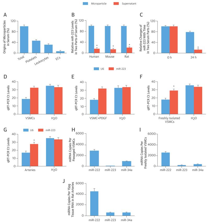
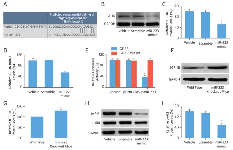
Background: MicroRNA-223 (miR-223) is a hematopoietic lineage cell-specific microRNA. However, a significant amount of miR-223 has been identified in vascular smooth muscle cells (VSMCs) and vascular walls that should not have endogenous miR-223.
Objectives: This study sought to determine the sources of miR-223 in normal and atherosclerotic arteries and the role of miR-223 in atherogenesis.
Methods: The levels and sources of miR-223 in blood cells (leukocytes and platelets), serum, blood microparticles, VSMCs, and vascular walls were determined. Both in vivo and in vitro studies were conducted to evaluate miR-223 secretion by blood cells and the ability of miR-223 to enter VSMCs and vascular walls. Subsequent changes in and the effects of miR-223 levels on serum and arteries in atherosclerotic animals and patients were investigated.
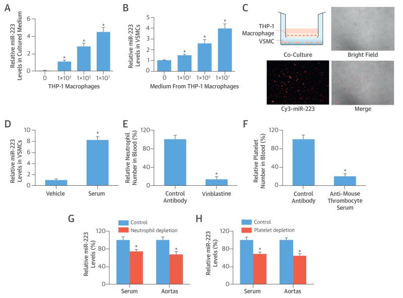
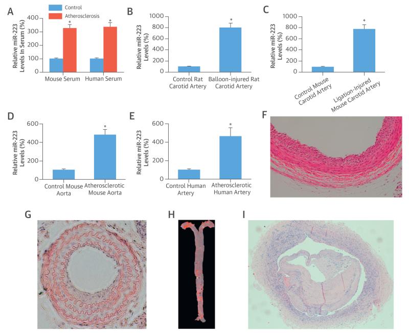
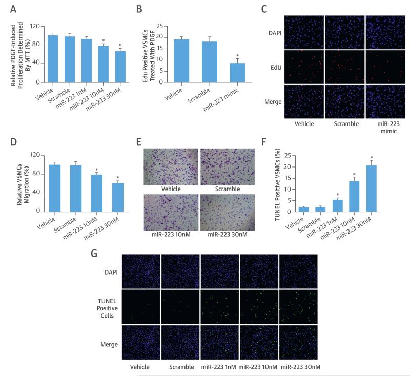
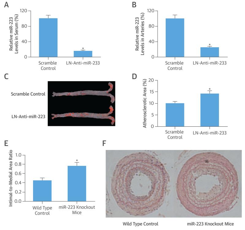
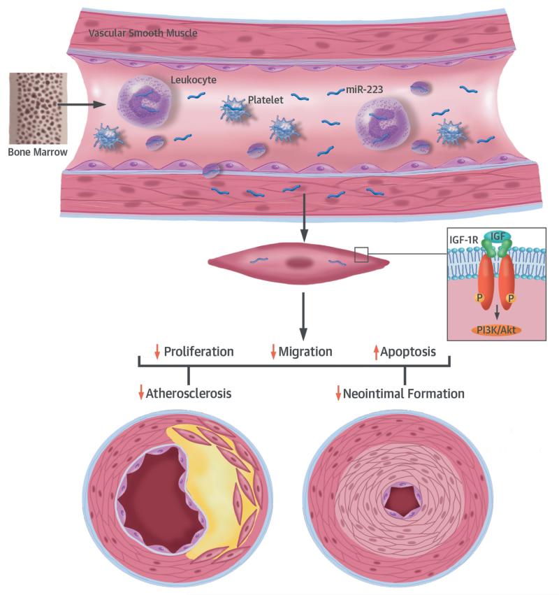
Results: Blood cells were able to secrete miR-223 into serum. MicroRNA-223 from blood cells was the most abundant cell-free miRNA in blood. Blood cell-secreted miR-223 could enter VSMCs and vascular walls, which produced strong biological effects via its target genes. In both atherosclerotic apolipoprotein-E knockout mice and patients with atherosclerosis, miR-223 levels were significantly increased in serum and atherosclerotic vascular walls. The atherosclerotic lesions in apolipoprotein-E knockout mice were exacerbated by miR-223 knockdown. The effect of miR-223 on atherogenesis was verified using miR-223 knockout mice.
Conclusions: Blood cell-secreted miR-223 enters vascular cells and walls, and appears to play important roles in VSMC function and atherogenesis. As a novel endocrine genetic signal between blood cells and vascular cells, miR-223 may provide a novel mechanism and new therapeutic target for atherosclerosis.
Keywords: atherosclerosis; bone marrow; insulin-like growth factor 1 receptor; microRNAs.
Copyright © 2015 American College of Cardiology Foundation. Published by Elsevier Inc. All rights reserved.
Figures
Comment in
-
Unlocking the Many Secrets of Noncoding RNA.J Am Coll Cardiol. 2015 Jun 16;65(23):2538-41. doi: 10.1016/j.jacc.2015.04.014. J Am Coll Cardiol. 2015. PMID: 26065993 No abstract available.
Similar articles
-
Bone Marrow-Derived MicroRNA-223 Works as an Endocrine Genetic Signal in Vascular Endothelial Cells and Participates in Vascular Injury From Kawasaki Disease.J Am Heart Assoc. 2017 Feb 14;6(2):e004878. doi: 10.1161/JAHA.116.004878. J Am Heart Assoc. 2017. PMID: 28196816 Free PMC article.
-
MicroRNA-31 controls phenotypic modulation of human vascular smooth muscle cells by regulating its target gene cellular repressor of E1A-stimulated genes.Exp Cell Res. 2013 May 1;319(8):1165-75. doi: 10.1016/j.yexcr.2013.03.010. Epub 2013 Mar 19. Exp Cell Res. 2013. PMID: 23518389
-
Phenotypic switching of vascular smooth muscle cells in the 'normal region' of aorta from atherosclerosis patients is regulated by miR-145.J Cell Mol Med. 2016 Jun;20(6):1049-61. doi: 10.1111/jcmm.12825. Epub 2016 Mar 15. J Cell Mol Med. 2016. PMID: 26992033 Free PMC article.
-
MicroRNA-126, -145, and -155: a therapeutic triad in atherosclerosis?Arterioscler Thromb Vasc Biol. 2013 Mar;33(3):449-54. doi: 10.1161/ATVBAHA.112.300279. Epub 2013 Jan 16. Arterioscler Thromb Vasc Biol. 2013. PMID: 23324496 Review.
-
MicroRNA-155 in the pathogenesis of atherosclerosis: a conflicting role?Heart Lung Circ. 2013 Oct;22(10):811-8. doi: 10.1016/j.hlc.2013.05.651. Epub 2013 Jul 1. Heart Lung Circ. 2013. PMID: 23827206 Review.
Cited by
-
MicroRNA-223-3p inhibits vascular calcification and the osteogenic switch of vascular smooth muscle cells.J Biol Chem. 2021 Jan-Jun;296:100483. doi: 10.1016/j.jbc.2021.100483. Epub 2021 Feb 26. J Biol Chem. 2021. PMID: 33647318 Free PMC article.
-
Inflammation, Senescence and MicroRNAs in Chronic Kidney Disease.Front Cell Dev Biol. 2020 Aug 6;8:739. doi: 10.3389/fcell.2020.00739. eCollection 2020. Front Cell Dev Biol. 2020. PMID: 32850849 Free PMC article.
-
Exosomes from mesenchymal stem cells expressing miR-125b inhibit neointimal hyperplasia via myosin IE.J Cell Mol Med. 2019 Feb;23(2):1528-1540. doi: 10.1111/jcmm.14060. Epub 2018 Nov 28. J Cell Mol Med. 2019. PMID: 30484954 Free PMC article.
-
microRNAs Associated with Carotid Plaque Development and Vulnerability: The Clinician's Perspective.Int J Mol Sci. 2022 Dec 9;23(24):15645. doi: 10.3390/ijms232415645. Int J Mol Sci. 2022. PMID: 36555285 Free PMC article. Review.
-
Enhanced extracellular vesicle production and ethanol-mediated vascularization bioactivity via a 3D-printed scaffold-perfusion bioreactor system.Acta Biomater. 2019 Sep 1;95:236-244. doi: 10.1016/j.actbio.2018.11.024. Epub 2018 Nov 22. Acta Biomater. 2019. PMID: 30471476 Free PMC article.
References
-
- Libby P, Ridker PM, Hansson GK. Progress and challenges in translating the biology of atherosclerosis. Nature. 2011;473:317–25. - PubMed
Publication types
MeSH terms
Substances
Grants and funding
LinkOut - more resources
Full Text Sources
Other Literature Sources
Medical
Research Materials
