. 2015 Jun 10:5:11220.
doi: 10.1038/srep11220.

Ubiquitin-specific Protease 15 Negatively Regulates Virus-induced Type I Interferon Signaling via Catalytically-dependent and -independent Mechanisms

Affiliations

Ubiquitin-specific Protease 15 Negatively Regulates Virus-induced Type I Interferon Signaling via Catalytically-dependent and -independent Mechanisms

Huan Zhang et al. Sci Rep. .

Abstract

Viral infection triggers a series of signaling cascades, which converge to activate the transcription factors nuclear factor-κB (NF-κB) and interferon regulatory factor 3 (IRF3), thereby inducing the transcription of type I interferons (IFNs). Although not fully characterized, these innate antiviral responses are fine-tuned by dynamic ubiquitination and deubiquitination processes. In this study, we report ubiquitin-specific protease (USP) 15 is involved in regulation of the retinoic acid-inducible gene I (RIG-I)-dependent type I IFN induction pathway. Knockdown of endogenous USP15 augmented cellular antiviral responses. Overexpression of USP15 inhibited the transcription of IFN-β. Further analyses identified histidine 862 as a critical residue for USP15's catalytic activity. Interestingly, USP15 specifically removed lysine 63-linked polyubiquitin chains from RIG-I among the essential components in RIG-I-like receptor-dependent pathway. In addition, we demonstrated that in contrast to USP15 de-ubiquitinating (DUB) activity, USP15-mediated inhibition of IFN signaling was not abolished by mutations eliminating the catalytic activity, indicating that a fraction of USP15-mediated IFN antagonism was independent of the DUB activity. Catalytically inactive USP15 mutants, as did the wild-type protein, disrupted virus-induced interaction of RIG-I and IFN-β promoter stimulator 1. Taken together, our data demonstrate that USP15 acts as a negative regulator of RIG-I signaling via DUB-dependent and independent mechanisms.


Figures
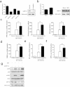
Figure 1
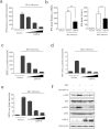
Figure 1. Knockdown of USP15 results in upregulation of type I IFN.
(a) Knockdown effect of the siRNAs at the mRNA and protein level. HEK293T cells were transfected with USP15-specific siRNAs and the endogenous USP15 mRNA and protein were assayed by real-time RT–PCR and immunoblotting. (b) The mRNA and protein level of USP15 after viral infection. HEK293T cells were infected with SEV or mock infected for 16 h. And the endogenous USP15 mRNA and protein were assayed by real-time RT–PCR and immunoblotting. (c) Effects of USP15 knockdown on SEV-induced activation of the IFN-β promoter, IFN-β mRNA and protein levels. HEK293T cells were transfected with the IFN-β–Luc reporter plasmid (0.1 μg) and pRL-TK plasmid (0.02 μg), together with plasmid encoding the USP15-specific siRNAs. At 24 h after transfection, the cells were further infected with SEV or mock infected for 16 h before luciferase assays were performed. Meanwhile, the supernatants were collected for ELISA, and the total RNA was then extracted and the expression of IFNB1 was evaluated with SYBR Green real-time RT–PCR. Data are means ± SD from three independent experiments. ** indicates p ≤ 0.01. (d–f) Effects of USP15 knockdown on the activation of ISRE and the NF-κB and IRF3 promoters. The experiments were performed as in (c), except that the ISRE–Luc, NF-κB–Luc, and IRF3–Luc reporter plasmids were used. Data are means ± SD from three independent experiments. (g) Effects of USP15 knockdown on SEV-induced phosphorylation of NF-κB subunit p65 and IRF3. HEK293T cells were transfected with USP15-specific siRNAs or control siRNAs for 24 h. Cells were further infected with SEV or mock infected for 16 h before immunoblots with the indicated antibodies were performed.
Figure 2
Figure 2. USP15 suppresses the virus-induced type I IFN signaling pathway.
(a) USP15 inhibited the SEV-induced activation of the IFN-β promoter. HEK293T cells grown in 24-well plates were transfected with the IFN-β–Luc reporter plasmid (0.1 μg) and pRL-TK plasmid (0.02 μg) together with increasing quantities (0, 0.1, 0.3, or 0.9 μg) of plasmid encoding USP15. At 24 h after transfection, the cells were further infected with SEV or mock infected for 16 h before luciferase assays were performed. Data are means ± SD from three independent experiments. (b) USP15 inhibited SEV-induced IFN-β mRNA and protein levels. HEK293T cells were transfected with plasmid encoding USP15 (1 μg) or an equivalent amount of empty vector. Then the experiments were performed as in (Fig. 1c). Data are means ± SD from three independent experiments. ** indicates p ≤ 0.01. (c–e) USP15 inhibited SEV-induced activation of ISRE and the NF-κB and IRF3 promoters. The experiments were performed as in (a), except that the ISRE–Luc, NF-κB–Luc, and IRF3–Luc reporter plasmids were used. Data are means ± SD from three independent experiments. (f) USP15 inhibited SEV-induced phosphorylation of NF-κB subunit p65 and IRF3. HEK293T cells were transfected with plasmid encoding USP15 (3 μg) or an equivalent amount of empty vector for 24 h, and then the cells were infected with SEV or mock infected for 16 h. An immunoblotting analysis was performed with the indicated antibodies.
Figure 3
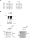
Figure 3. Identification of the DUB activity site His862 in vivo and in vitro.
(a) Putative sites responsible for the DUB activity of USP15. Black boxes indicate the conserved residues tested in this experiment. The sequences were taken from GenBank entries with the following accession numbers: USP4, NM_003363; USP10, NM_001272075; USP13, NM_003940; USP15, NM_006313; USP17, NM_201402 and XM_352721; USP18, NM_017414; USP20, NM_001110303; USP21, NM_001014443; and USP31, NM_020718. (b) H862 is the catalytic site. HEK293T cells were co-transfected with a Flag-tagged Ub expression plasmid (1.0 μg) and expression vectors encoding HA–USP15-WT, HA–USP15-C269A, or HA–USP15-H862A. The immunoblotting analysis was performed with anti-HA and anti-Flag antibodies. (c) Identification of the DUB activity in vitro. The proteins were obtained from USP15/USP15-C269A/USP15-H862A-transfected or mock-transfected HEK293T cells using the HA tagged Protein PURIFICATION KIT. Lys48-/Lys63-linked polyubiquitin was incubated with the protein obtained from mock-transfected (lane 2), USP15-transfected (lane 3), USP15-C269A-transfected (lane 4) or USP15-H862A-transfected (lane 5) HEK293T cells at 37 °C for 30 min, then analyzed by SDS–PAGE. Lane 1, uncleaved Lys48-linked polyubiquitin chain (K48-Ub2–7).
Figure 4
Figure 4. USP15 specifically deconjugates the ubiquitination of RIG-I.
(a–d) HEK293T cells were co-transfected with Flag-tagged RIG-I, IPS-1, TRAF3, TBK1 and HA–USP15-WT or a catalytic mutant (USP15-C269A or USP15-H862A). The cell lysates were immunoprecipitated with an anti-Flag antibody and immunoblotted with an anti-Ub antibody.
Figure 5
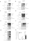
Figure 5. USP15 deconjugates Lys63-linked polyubiquitin chains from RIG-I.
(a–c) HEK293T cells were cotransfected with expression vectors encoding Flag–RIG-I and HA–WT-Ub (a), Flag–RIG-I and HA–K63-Ub (b), or Flag–RIG-I and HA–K48-Ub (c), together with control vectors or expression vectors encoding Myc–USP15-WT, Myc–USP15-C269A, or Myc–USP15-H862A. The cell lysates were immunoprecipitated with an anti-Flag antibody and immunoblotted with an anti-HA antibody. (d–e) HEK293T cells were co-transfected with an expression vector encoding Flag–RIG-I and control vectors or expression vectors encoding HA–USP15-WT or the mutant HA–USP15-C269A or HA–USP15-H862A. The cell lysates were immunoprecipitated with an anti-Flag antibody and immunoblotted with an anti-Ub (K63) antibody (d), or anti-Ub (K48) antibody (e). (f) Knockdown of USP15 potentiated the ubiquitination of RIG-I. HEK293T cells were transfected with USP15-specific siRNAs or control siRNAs and indicated expression plasmids. The cell lysates were immunoprecipitated with an anti-Flag antibody and immunoblotted with an anti-HA antibody. (g) Knockdown of USP15 potentiated SEV-induced ubiquitination of RIG-I. HEK293T cells were transfected with USP15-specific siRNAs or control siRNAs, the cells were then further infected with SEV or mock infected for 16 h. The cell lysates were immunoprecipitated with an anti-RIG-I antibody and immunoblotted with indicated antibodies. (h) USP15 inhibited RIG-I- and RIG-I-N-mediated activation of the IFN-β promoter. HEK293T cells were cotransfected with the IFN-β–Luc reporter plasmid (0.1 μg), pRL-TK plasmid (0.02 μg), and 0.5 μg of plasmid encoding USP15, together with the RIG-I or RIG-I-N expression vector (0.5 μg). Luciferase assays were performed 30 h after transfection. Data are means ± SD from three independent experiments. ** indicates p ≤ 0.01.
Figure 6
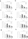
Figure 6. Mutation of the catalytic residues does not abolish USP15 IFN antagonism.
(a-d) HEK293T cells were co-transfected with increasing amounts of plasmid encoding USP15 or the catalytic mutant USP15–C269A or USP15–H862A, together with different reporter plasmids (0.1 μg) and the pRL-TK plasmid (0.02 μg) for 24 h. The cells were then infected with SEV or mock infected for 16 h before the luciferase assays were performed. Data are means ± SD from three independent experiments. (e–h) HEK293T cells were co-transfected with increasing amounts of plasmid encoding USP15 or the USP15-C269A or USP15-H862A catalytic mutant, together with different reporter plasmids (0.1  μg), pRL-TK plasmid (0.02  μg), and plasmid encoding RIG-I-N. Luciferase assays were performed 30  h after transfection. Data are means ± SD from three independent experiments.
Figure 7
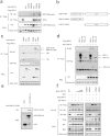
Figure 7. USP15 sequesters the interaction between RIG-I and IPS-1 in a DUB activity-independent manner.
(a) The interaction between USP15 and RIG-I was independent of the DUB activity of USP15. HEK293T cells were co-transfected with expression vectors encoding HA–USP15-WT or the mutant HA–USP15-C269A or HA–USP15-H862A, together with control vectors, or expression vectors encoding Flag–RIG-I. The cell lysates were immunoprecipitated with an anti-Flag antibody and immunoblotted with an anti-HA antibody. (b) Schematic diagram of the deletion mutants of USP15 used in this study. (c) The UCH domain of USP15 was responsible for its interaction with RIG-I. HEK293T cells were co-transfected with plasmid encoding wild-type HA–USP15 or one of its deletion mutants, together with control vectors or expression vectors encoding Flag–RIG-I. The cell lysates were immunoprecipitated with an anti-Flag antibody and immunoblotted with an anti-HA antibody. (d) The N-terminal CARD domain and the C-terminal domain of RIG-I both bind to USP15. HEK293T cells were co-transfected with plasmid encoding wild-type Flag–RIG-I or one of its deletion mutants, together with control vectors or expression vectors encoding HA–USP15. The cell lysates were immunoprecipitated with an anti-Flag antibody and immunoblotted with an anti-HA antibody. (e) GST pulldown assay to confirm the interaction between RIG-I and USP15. Glutathione beads conjugated to GST or GST-RIG-I-N (from recombinant E. coli) were incubated with purified USP15 C250. After washing five times, the beads were boiled and subjected to immunoblotting. (f) USP15 sequestered the interaction between RIG-I and IPS-1. HEK293T cells were co-transfected with plasmid encoding HA–RIG-I, Flag-IPS-1, increasing amounts of wild-type Myc–USP15 or its mutants. 24 h later, cells were further infected with SEV. The cell lysates were immunoprecipitated with an anti-HA antibody and immunoblotted with anti-Flag and anti-Myc antibodies.

Similar articles

Cited by

References

    1. Akira S., Uematsu S. & Takeuchi O. Pathogen recognition and innate immunity. Cell 124, 783–801 (2006). - PubMed
    1. Saito T. & Gale M. Jr. Principles of intracellular viral recognition. Curr. Opin. Immunol. 19, 17–23 (2007). - PubMed
    1. Takeuchi O. & Akira S. MDA5/RIG-I and virus recognition. Curr. Opin. Immunol. 20, 17–22 (2008). - PubMed
    1. Katze M. G., Fornek J. L., Palermo R. E., Walters K. A. & Korth M. J. Innate immune modulation by RNA viruses: emerging insights from functional genomics. Nat. Rev. Immunol. 8, 644–654 (2008). - PMC - PubMed
    1. Zeng W. et al. Reconstitution of the RIG-I pathway reveals a signaling role of unanchored polyubiquitin chains in innate immunity. Cell 141, 315–330 (2010). - PMC - PubMed

Publication types

MeSH terms