. 2015 Jun 23;112(25):7821-6.
doi: 10.1073/pnas.1509744112. Epub 2015 Jun 8.

Amelioration of toxicity in neuronal models of amyotrophic lateral sclerosis by hUPF1

Affiliations

Amelioration of toxicity in neuronal models of amyotrophic lateral sclerosis by hUPF1

Sami J Barmada et al. Proc Natl Acad Sci U S A. .

Abstract

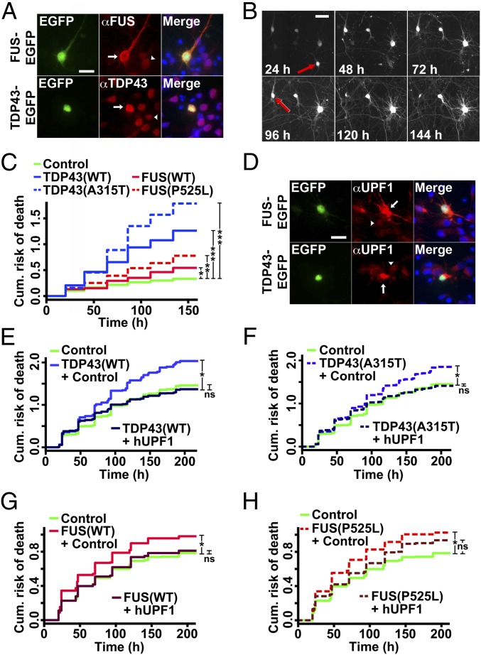
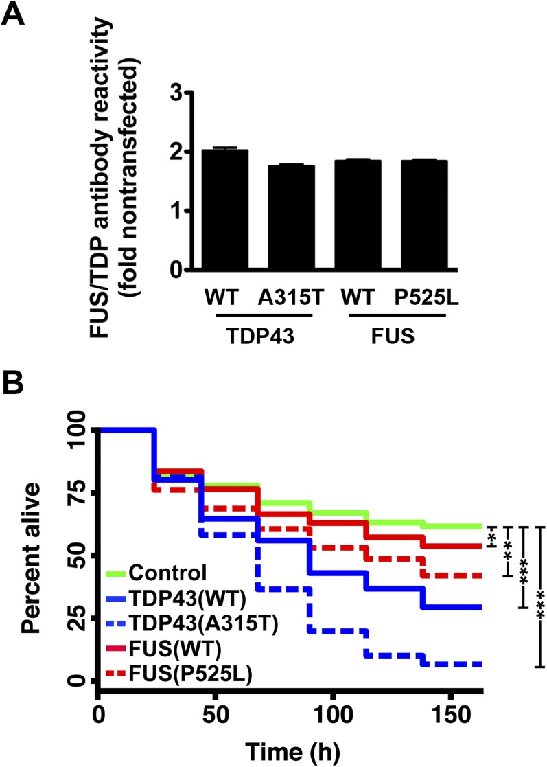
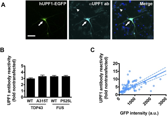
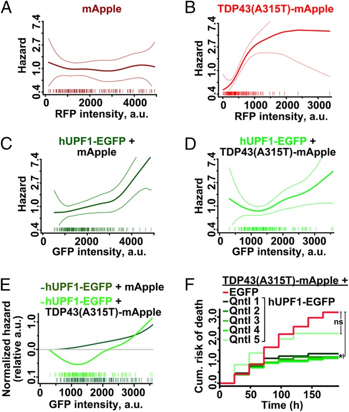
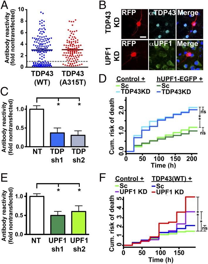
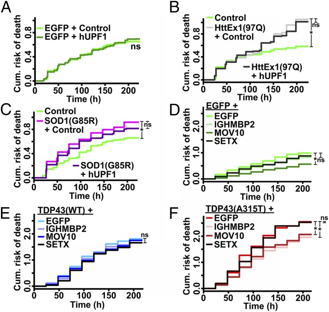
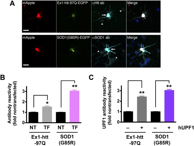
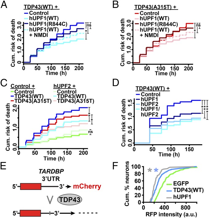
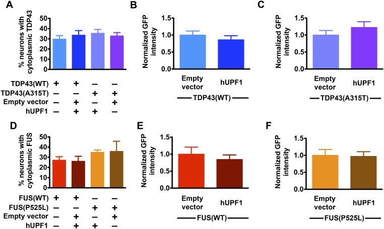
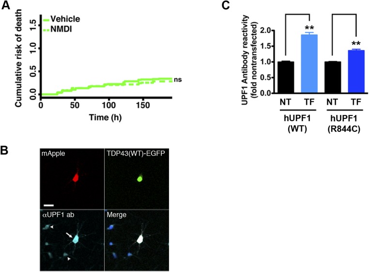
Over 30% of patients with amyotrophic lateral sclerosis (ALS) exhibit cognitive deficits indicative of frontotemporal dementia (FTD), suggesting a common pathogenesis for both diseases. Consistent with this hypothesis, neuronal and glial inclusions rich in TDP43, an essential RNA-binding protein, are found in the majority of those with ALS and FTD, and mutations in TDP43 and a related RNA-binding protein, FUS, cause familial ALS and FTD. TDP43 and FUS affect the splicing of thousands of transcripts, in some cases triggering nonsense-mediated mRNA decay (NMD), a highly conserved RNA degradation pathway. Here, we take advantage of a faithful primary neuronal model of ALS and FTD to investigate and characterize the role of human up-frameshift protein 1 (hUPF1), an RNA helicase and master regulator of NMD, in these disorders. We show that hUPF1 significantly protects mammalian neurons from both TDP43- and FUS-related toxicity. Expression of hUPF2, another essential component of NMD, also improves survival, whereas inhibiting NMD prevents rescue by hUPF1, suggesting that hUPF1 acts through NMD to enhance survival. These studies emphasize the importance of RNA metabolism in ALS and FTD, and identify a uniquely effective therapeutic strategy for these disorders.

Keywords: ALS; FTD; RNA binding proteins; RNA decay; neurodegeneration.


Conflict of interest statement

The authors declare no conflict of interest.

Figures

Fig. 1.
Fig. 1.
hUPF1 rescues neuron loss in ALS models. (A) Primary neurons transfected with TDP43-EGFP and FUS-EGFP were probed using anti-TDP43 and -FUS antibodies, demonstrating selective overexpression in transfected (arrow) vs. untransfected (arrowhead) neurons. (B) Time of death, represented by the last time the cell was observed alive (red arrows), was used to create cumulative hazard plots (C) depicting risk of death over time. *P < 0.05, **P < 0.0001, ***P ≤ 1 × 10⁻¹⁰, by Cox hazards analysis. n = 98–139 neurons per genotype. (D) Immunocytochemistry using an anti-UPF1 antibody confirmed UPF1 overexpression in transfected (arrow) vs. untransfected (arrowhead) neurons. (E–H) The survival of neurons coexpressing hUPF1 and TDP43(WT) (E), TDP43(A315T) (F), FUS(WT) (G), or FUS(P525L) (H) was determined using LFM. In E and F, n = 1,150–1,205 neurons per genotype; *P < 1 × 10⁻⁹. In G and H, n = 312–595 neurons per genotype; *P < 0.01. ns, not significant, by Cox hazards analysis. Results pooled from three or more independent experiments. (Scale bars in A, B, and D, 50 µm.)
Fig. S1.
Fig. S1.
A primary neuron model of ALS. Primary rodent cortical neurons were dissected then transfected with plasmids encoding mApple or mApple-tagged TDP43 or FUS variants on day 4 in vitro. (A) Neurons transfected with TDP43 or FUS displayed an approximate 2-fold mean increase in antibody reactivity, as measured by quantitative fluorescence microscopy. n = 316–433 neurons per genotype. Error bars, ± SEM. (B) Survival of transfected neurons was followed by LFM as described in the text, and survival was plotted using Kaplan–Meier analysis. *P < 0.05, **P < 0.0001, ***P < 1 × 10⁻¹⁰, by Cox hazards analysis. n = 98–139 neurons per genotype, pooled from 8 wells each.
Fig. S2.
Fig. S2.
Quantitative immunocytochemistry of hUPF1-EGFP in transfected neurons. (A) Primary rodent cortical neurons were transfected with hUPF1-EGFP, fixed and probed using antibodies that recognize total UPF1. The amount of UPF1 in transfected neurons (arrow) was compared with endogenous UPF1 in untransfected neurons (arrowhead) to calculate the fold overexpression. (Scale bar, 50 µm.) (B) Primary neurons cotransfected with hUPF1-EGFP and WT and mutant versions of TDP43 or FUS demonstrated a 3-fold increase in UPF1 antibody reactivity by quantitative ICC. n = 316–433 neurons per genotype. (C) The relationship between hUPF1-EGFP intensity and total UPF1 levels was determined for individual neurons using linear regression analysis. Dotted lines, 95% confidence intervals. n = 76 neurons. Data were pooled from eight separate wells.
Fig. 2.
Fig. 2.
The amount of hUPF1 is critical for neuroprotection. (A) mApple levels were unrelated to survival (n = 193, plinear = 0.94, pnonlinear = 0.53), but increasing amounts of TDP43(A315T)-mApple (B) magnified hazard (n = 164, plinear = 1 × 10⁻⁹, pnonlinear = 0.01). In control neurons (C), hUPF1-EGFP levels and hazard were linearly related (n = 193, plinear = 1 × 10⁻⁶, pnonlinear = 0.2), indicative of dose-dependent toxicity. In TDP43(A315T)-mApple transfected neurons, low levels of hUPF1-EGFP (D) reduced hazard, whereas high levels were toxic (n = 164, plinear = 0.005, pnonlinear = 0.3). (E) Splines for hUPF1-EGFP in neurons expressing mApple or TDP43(A315T)-mApple emphasize hUPF1’s neuroprotective properties at low levels. (F) Neurons expressing TDP43(A315T)-mApple and hUPF1-EGFP were separated into quintiles based upon GFP intensity, and survival was analyzed by LFM. *P < 0.04; ns, not significant, Cox hazards analysis. Dotted lines in AD, 95% confidence interval.
Fig. 3.
Fig. 3.
hUPF1 rescues cell death arising from TDP43 overexpression but not knockdown. (A) Expression of TDP43(WT) (n = 136) and TDP43(A315T) (n = 111) down-regulated en-TDP43 in 15–36% of transfected neurons, as judged by anti-TDP43 antibody reactivity. (B and C) Knockdown of en-TDP43 or en-UPF1 in mouse primary neurons using shRNAs. (Scale bar, 25 µm.) (D) en-TDP43 knockdown increased the risk of death in primary neurons, as determined by LFM, but survival was unaffected by hUPF1. Reduction of 40–50% in en-UPF1 (E) exacerbated neurodegeneration due to TDP43 overexpression (F). In C and E, *P < 0.05, ANOVA with Dunnett’s test. In D, n = 305–513 neurons per genotype. In F, n = 249–332 neurons per genotype. For both D and F, ns, P > 0.05; *P < 0.006, **P < 2 × 10⁻⁷, Cox hazards analysis. Results were pooled from 8 to 24 wells per condition, in duplicate.
Fig. 4.
Fig. 4.
SF1 helicases partially rescue TDP43-mediated neurodegeneration. (A) hUPF1 had no effect on the survival of control neurons expressing EGFP alone. n = 862–893 neurons per genotype. (B) Ex1-Htt-97Q increased neuronal risk of death, regardless of hUPF1 expression. n = 197–266 neurons per genotype. (C) hUPF1 expression also failed to improve survival in cells expressing SOD1(G85R). n = 253–1028 neurons per genotype. SF1 helicases were safe in control neurons (D), but none prevent cell death due to TDP43(WT) overexpression (E). In contrast, IGHMBP2 and MOV10 mitigated TDP43(A315T)-mediated toxicity (F). *P < 0.02, ns, not significant, by Cox hazards analysis. In DF, n = 145–198 neurons per genotype. Results were pooled from eight wells per condition, performed in triplicate.
Fig. S3.
Fig. S3.
Quantitative immunocytochemistry of mutant htt and SOD1 in transfected neurons. (A) Primary cortical neurons were dissected, cultured, then transfected with mApple and either mutant htt (Ex1-htt-97Q) or SOD1(G85R) fused to EGFP. Transfected proteins were visualized by immunocytochemistry using antibodies that recognize both endogenous (arrowheads) and exogenous (arrows) htt or SOD1. (Scale bars, 25 µm.) (B) Fold overexpression of htt or SOD1 was calculated by comparing antibody reactivity in nontransfected cells (NT) to that in transfected neurons (TF). htt, 161 neurons each for NT and TF groups; SOD1, 146 neurons each for NT and TF groups. (C) Quantitative immunocytochemistry with antibodies against UPF1 was used to determine the UPF1 level in untransfected (−) cells and those transfected (+) with hUPF1. Ex1-htt-97Q, n = 124 neurons each for (−) and (+) groups; SOD1(G85R), n = 148 neurons each for (−) and (+) groups. In B and C, *P < 0.001, **P < 0.0001, vs. NT by two-tailed t test. Data were pooled from four experiments. Error bars represent ± SEM.
Fig. 5.
Fig. 5.
Neuroprotection by hUPF1 requires helicase activity and involves NMD. hUPF1 improved survival in cells expressing TDP43(WT) (A) and TDP43(A315T) (B), but helicase-null hUPF1(R844C) was ineffective, and rescue was attenuated with the addition of NMDI-1, an inhibitor of NMD. (C) hUPF2 also improved survival in neurons expressing TDP43(WT) and TDP43(A315T). (D) Expression of both hUPF1 and hUPF2 further reduced the risk of death in neurons transfected with TDP43(WT). In A and B, *P < 0.01; in C, *P = 0.02, **P ≤ 1 × 10⁻⁴; in D, *P = 0.02, **P < 0.001, ***P < 1 × 10⁻⁵; ns, not significant by Cox hazards analysis. In A and B, n = 360–686 neurons per genotype. In C and D, n = 87–258 neurons per genotype. Results were pooled from eight wells per condition, in three or more experiments. (E) Binding of TDP43 to the TARDBP 3′UTR triggers splicing and RNA decay of a fluorescent reporter. (F) TDP43(WT) (n = 104) and hUPF1 (n = 56) effectively reduce single-cell reporter intensity, compared with EGFP (n = 130). *P ≤ 0.05, Kolmogorov–Smirnov test. Results pooled from 8–16 wells per experiment, in duplicate.
Fig. S4.
Fig. S4.
Effect of hUPF1 on exogenous TDP43 and FUS. The percentage of cells exhibiting cytoplasmic TDP43-EGFP (A) or FUS-EGFP (D) was unchanged by expression of hUPF1. Likewise, the total levels of TDP43(WT)-EGFP (B), TDP43(A315T)-EGFP (C), FUS(WT)-EGFP (E), and FUS(P525L)-EGFP (F) were unaffected by hUPF1 expression. Data were pooled from eight wells per condition, in triplicate. For AC, n = 319–386 neurons per genotype; for DF, n = 98–112 neurons per genotype.
Fig. S5.
Fig. S5.
Evaluation of NMD inhibitor 1 (NMDI) and mutant hUPF1(R844C) in primary neurons. (A) Primary cortical neurons were treated with NMDI or vehicle (DMSO) at 5 µM, transfected with EGFP, and followed by LFM. Vehicle, n = 170 cells; NMDI, n = 132 cells. ns, not significant (P > 0.05), Cox proportional hazards analysis. Results were combined from eight wells per condition in duplicate. (B) Expression of hUPF1(R844C) in transfected neurons (arrow) was confirmed by immunocytochemistry using antibodies that recognize endogenous (arrowheads) as well as exogenous UPF1. (Scale bar, 25 µm.) (C) Fold overexpression of hUPF1(WT) or hUPF1(R844C) was determined by comparison of antibody reactivity in transfected (TF) vs. nontransfected (NT) neurons. hUPF1(WT), n = 197 neurons each for NT and TF groups; hUPF1(R844C), n = 144 neurons each for NT and TF groups. Error bars, ± SEM. **P < 0.0001 vs NT by 2-tailed t test. Data were pooled from four experiments.

Similar articles

Cited by

References

    1. Huisman MHB, et al. Population based epidemiology of amyotrophic lateral sclerosis using capture-recapture methodology. J Neurol Neurosurg Psychiatry. 2011;82(10):1165–1170. - PubMed
    1. Renton AE, Chiò A, Traynor BJ. State of play in amyotrophic lateral sclerosis genetics. Nat Neurosci. 2014;17(1):17–23. - PMC - PubMed
    1. Barmada SJ, Finkbeiner S. Pathogenic TARDBP mutations in amyotrophic lateral sclerosis and frontotemporal dementia: Disease-associated pathways. Rev Neurosci. 2010;21(4):251–272. - PubMed
    1. Barmada SJ, et al. Autophagy induction enhances TDP43 turnover and survival in neuronal ALS models. Nat Chem Biol. 2014;10(8):677–685. - PMC - PubMed
    1. Bilican B, et al. Mutant induced pluripotent stem cell lines recapitulate aspects of TDP-43 proteinopathies and reveal cell-specific vulnerability. Proc Natl Acad Sci USA. 2012;109(15):5803–5808. - PMC - PubMed

Publication types