. 2015 Jun 4;10(6):e0126240.
doi: 10.1371/journal.pone.0126240. eCollection 2015.

mTORC1 Down-Regulates Cyclin-Dependent Kinase 8 (CDK8) and Cyclin C (CycC)

Affiliations

mTORC1 Down-Regulates Cyclin-Dependent Kinase 8 (CDK8) and Cyclin C (CycC)

Daorong Feng et al. PLoS One. .

Abstract

In non-alcoholic fatty liver disease (NAFLD) and insulin resistance, hepatic de novo lipogenesis is often elevated, but the underlying mechanisms remain poorly understood. Recently, we show that CDK8 functions to suppress de novo lipogenesis. Here, we identify the mammalian target of rapamycin complex 1 (mTORC1) as a critical regulator of CDK8 and its activating partner CycC. Using pharmacologic and genetic approaches, we show that increased mTORC1 activation causes the reduction of the CDK8-CycC complex in vitro and in mouse liver in vivo. In addition, mTORC1 is more active in three mouse models of NAFLD, correlated with the lower abundance of the CDK8-CycC complex. Consistent with the inhibitory role of CDK8 on de novo lipogenesis, nuclear SREBP-1c proteins and lipogenic enzymes are accumulated in NAFLD models. Thus, our results suggest that mTORC1 activation in NAFLD and insulin resistance results in down-regulation of the CDK8-CycC complex and elevation of lipogenic protein expression.


Conflict of interest statement

Competing Interests: The authors have declared that no competing interests exist.

Figures

Fig 1
Fig 1. Hepatic CDK8 and CycC proteins are down regulated in genetically obese and insulin resistant mice.
Control wild-type and db/db (left panels) or ob/ob (right panels) male mice on the C57Bl/6J background were maintained on the normal chow diet. At 4 months of age, these mice were fasted for 16 hours before sacrifice. A, B, C) Liver extracts were prepared and analyzed for the indicated proteins by immunoblotting. TBP or β-actin served as the invariant control. The SREBP-1 precursor (pre-SREBP1) and the nuclear SREBP-1 (nSREBP-1) from liver extracts of the same mice were also analyzed. Each lane represents an independent mouse. The relative protein levels in the livers of db/db (D) or ob/ob (E) mice were further analyzed by densitometry. *p<0.05 and **<0.01 vs. wild-type (n = 6 independent mice).
Fig 2
Fig 2. Hepatic CDK8 protein levels are inversely correlated with nuclear SREBP-1 levels in human NAFLD.
A) Representative immunoblots of indicated proteins in total protein extracts of normal human liver biopsies (<5% fat by ultrasound analysis) or patients with diagnosed with non-alcoholic fatty liver disease, NAFLD (>30% fat). TBP served as the invariant control. B) The ratio of nuclear SREBP-1 (nSREBP1) to CDK8 of densitometry analyses after normalized by TBP from 14 normal and 25 NAFLD liver biopsies.
Fig 3
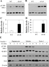
Fig 3. mTORC1 is activated in obese mouse livers, and is involved in nutrient-repletion-induced down-regulation of CDK8 in tissue culture.
Control wild-type and db/db (A) or ob/ob (B) male mice on the C57Bl/6J background were maintained on the normal chow diet. At 4 months of age, the mice were fasted for 16 hours and then re-fed for 4 hours before sacrifice. Liver extracts were prepared and analyzed for the levels of S6K1 phosphorylation on T389 by immunoblotting. Total amount of S6K1 served as the loading control. The relative protein levels in the livers of db/db (C) or ob/ob (D) mice were further analyzed by densitometry. *p<0.05 and **<0.01 vs. wild-type (n = 3 independent mice). E) Effects of nutrient-depletion and nutrient-repletion on the levels of indicated proteins in the presence of mTOR inhibitor rapamycin or Torin 1. FAO cells were cultured in the low-serum DMEM overnight, and then in amino acid-free RPMI for 2 hour. The cells were pre-treated without or with 100nM rapamycin or 250nM Torin 1 for 30 min followed by the addition of regular DMEM containing 100nM insulin for the indicated period of time. Cell extracts were prepared and analyzed for the indicated proteins by immunoblotting. These are representative immunoblots independently performed for four times.
Fig 4
Fig 4. mTORC1 activity negatively regulates the CDK8 and CycC protein levels.
A) Effects of mTOR knockdown on the levels of indicated proteins during nutrient-depletion or nutrient-repletion. HEK293T cells were infected with non-specific (NS) or mTOR-specific shRNA lentiviruses. The cells were nutrient-depleted by placing them in the low serum DMEM medium overnight, and then in amino acid-free RPMI for 2 hour followed by the addition of regular DMEM containing 100 nM insulin for 30 min. The relative protein levels of CDK8 (B) and CycC (C) were further analyzed by densitometry. *p<0.05 vs. control (n = 3). D) Effects of overexpressing a mutant of constitutively active Rheb or Ulk1 on the levels of indicated proteins during nutrient-depletion (ND) or nutrient-repletion (NR) condition. HEK293T cells were transfected with a constitutively active ULK1 mutant cDNA (U, 1 μg), the empty vector (V, 1 μg) or a constitutively active Rheb mutant cDNA (R, 1 μg). Thirty-six hours after transfection, the cells were nutrient-depleted and nutrient-replete as described above. These are representative immunoblots independently performed for five times.
Fig 5
Fig 5. mTORC1 activation is required for feeding-induced down-regulation of CDK8.
A) Representative immunoblots showing the levels of indicated proteins in duplicate from livers of liver-specific Raptor knockout mice or controls. Knockout was achieved by tail-vein injection of AAV-TBG-Cre (or GFP as the control) in Raptor flox/flox mice. One week after the AAV injections, the mice were fasted for 12 hours, and then either sacrificed or re-fed for 5 hours. B) The ratios of CDK8 to β-actin of indicated liver samples as analyzed by densitometry (n = 7 mice for each group). C) Representative immunoblots showing the levels of the indicated proteins in duplicate from livers of liver-specific Raptor knockout mice or controls as indicated. D) The ratios of nSREBP1 to the invariant nucleolin of the indicated liver samples as analyzed by densitometry (n = 4 mice for each group).
Fig 6
Fig 6. Hepatic levels of CDK8 and CycC proteins are down regulated during aging.
C57BL/6J mice were maintained on a normal chow diet and at the ages indicated were fasted for 16 h and liver extracts prepared. A-B) Representative immunoblots showing the levels of indicated proteins in livers of mice with indicated ages, as detected by immunoblotting. β-tubulin or TBP served as the invariant controls. Each lane is from the equal pooling of liver extracts from six mice. The relative protein levels of CDK8 (C), CycC (D) and phosphorylated S6K1 (E) were further analyzed by densitometry. *p<0.05, **p<0.01 and ***p<0.001 vs. control (n = 6).
Fig 7
Fig 7. mTORC1 activation is required for the down-regulation of CDK8 and CycC in aging livers.
Effects of rapamycin treatment in 12-month old mice on the levels of indicated proteins in the livers, as detected by immunoblotting. β-actin was the loading control. Three independent mice were treated by intra-peritoneal injection of rapamycin (2 mg/kg body weight) or vehicle control for three days. Liver extracts were immunoblotted for (A) CDK8, CycC and β- actin, (C) pT389-S6K1 and β-actin, (D) pT402-SREBP1, nSREBP1 and TBP. B) The ratios of CDK8 or CycC to β-actin of indicated treatments were analyzed by densitometry. E) The ratios of pT402-SREBP-1/TBP, nSREBP-1/TBP and pT402-SREBP-1/nSREBP-1 of indicated treatments were analyzed by densitometry. *p<0.05 vs. vehicle (n = 3).

Similar articles

Cited by

References

    1. Ferre P, Foufelle F. Hepatic steatosis: a role for de novo lipogenesis and the transcription factor SREBP-1c. Diabetes Obes Metab. 2010;12 Suppl 2:83–92. 10.1111/j.1463-1326.2010.01275.x - DOI - PubMed
    1. Kawano Y, Cohen DE. Mechanisms of hepatic triglyceride accumulation in non-alcoholic fatty liver disease. J Gastroenterol. 2013;48(4):434–441. 10.1007/s00535-013-0758-5 - DOI - PMC - PubMed
    1. Ferre P, Foufelle F. SREBP-1c transcription factor and lipid homeostasis: clinical perspective. Horm Res. 2007;68(2):72–82. . - PubMed
    1. Filhoulaud G, Guilmeau S, Dentin R, Girard J, Postic C. Novel insights into ChREBP regulation and function. Trends Endocrinol Metab. 2013;24(5):257–268. 10.1016/j.tem.2013.01.003 - DOI - PubMed
    1. Shimomura I, Bashmakov Y, Ikemoto S, Horton JD, Brown MS, Goldstein JL. Insulin selectively increases SREBP-1c mRNA in the livers of rats with streptozotocin-induced diabetes. Proc Natl Acad Sci U S A. 1999;96(24):13656–13661. . - PMC - PubMed

Publication types

MeSH terms

LinkOut - more resources